Abstract Background. Advancing therapeutic approaches to brain metastases (BrMets) is an area of critical need. Preclinical models of BrMets are a rare but much-needed tool to investigate novel therapeutic approaches. We developed a biobank of BrMet patient-derived xenograft (PDX) models established from resected BrMets originating from various solid tumor types. Methods: Resected BrMet tissues were collected. PDX models were established, and ex vivo drug screening was performed using previously published methods (Morikawa et al. Can Res Comm 2023). Molecular profiles of PDXs and matched source tumors were compared, and their correlations with drug response were examined. Results: From Nov 2016 to Sept 2023, 142 surgical cases were collected, of which 126 PDX models were established and maintained growth. Tumor types included common (lung, breast, melanoma) and rare (sarcoma, ovarian, cervical, prostate, renal, and gastrointestinal) tumors. Common mutations across these models included BRCA, ATR, ALK, KMT2C, FAT1, ZFXHX3, MAP3K1, COL6A3, FLT3, MLH1, EGFR, IGFN1, and TP53, though many were not predicted to be pathogenic. In addition, there were less prevalent but potentially targetable alterations, such as PI3K mutations. Copy number variation (CNV) analysis demonstrated a predominance of amplifications over deletions. The observed pathways included those associated with neuronal and structural features such as axonal transport (DNA KEGG database) and extracellular matrix (DNA REACTOME database). The RNA seq analysis revealed clustering mostly based on the primary tumor type. Compared to publicly available metastatic PDX models (NCI database) stratified by primary tumor type, these BrMet models demonstrated differences in the molecular pathway enrichment. PDX models generally exhibited high concordance based on Jaccard Index (JI). The majority of the samples showed JI in the range of 0.4-0.6, with melanoma samples demonstrating JI in the lower 0.2 range. We evaluated the PDX and matched pairs for the selected variants predicted to be pathogenic or possibly functionally impactful. Again, melanoma subtypes showed more divergence, but overall, the majority of the variants were retained in the matched PDX models. Drug sensitivity testing was performed on 13 PDXs using a panel of molecularly targeted agents and chemotherapies. Of 13, nine PDXs had potentially actionable molecular targets. Highly active drugs were identified in these models; however, most drug sensitivities were not predictable based solely on genomic profiles, emphasizing the utility of paired functional characterization. Conclusion: We present a novel biobank of BrMet PDX models. These models provide a valuable resource for probing the biology of BrMet and informing therapeutic strategies. Prospective collection is ongoing to expand the biobank, alongside further studies investigating tumor-microenvironment interactions using immune-competent and organ-on-chip microfluidic blood-brain niche models. AI disclosure: AI was used for language editing only; content was verified by the authors Citation Format: Aki Morikawa, Tusharika Rastogi, Noreen Khan, Peter Ulintz, Derek Nancarrow, Habib Serhan, Xu Cheng, Liwei Bao, Aaron Udager, Matthew Soellner, Jason Heth, Nathan Merrill, Sofia D. Merajver. Developing diverse patient-derived xenograft models of common and rare brain metastases to elucidate molecular landscapes and reveal therapeutic opportunities [abstract]. In: Proceedings of the AACR Special Conference in Cancer Research: Brain Cancer; 2026 Mar 23-25; Philadelphia, PA. Philadelphia (PA): AACR; Cancer Res 2026;86(6_Suppl):Abstract nr B062.
Supplementary Figure S4. Frag J/Jm mediated loss of misfolded Fluc-DM is independent of mutant p53 status.
Supplementary Figure S7. Design and testing of scrambled peptides as negative controls.
Supplementary Figure S3. GRAIL1 is a glycosylated protein that undergoes processing.
Abstract Background: Anaplastic lymphoma kinase-positive non-small cell lung cancer (ALK+ NSCLC) comprises 5-6% of all lung cancers with a median survival rate of 6.8 years. The primary treatment is tyrosine kinase inhibitors (TKI) which have a 30-80% response rate but acquired resistance is inevitable. One area of potential intervention is within the tumor-immune microenvironment (TIME) but the standard immunotherapies, such as immune checkpoint inhibitors, have resulted in low overall response rate (10-15%), toxicity, or inconclusive therapeutic effects in ALK+ NSCLC as a single agent or combined with TKIs in clinical trials. Therefore, it is imperative to gain a more in-depth understanding of the TIME evolution. In particular, there has been increasing evidence showing that macrophage polarization is involved in NSCLC tumorigenesis and drug resistance. The timing, phenotype, and functional state of macrophages within ALK+ NSCLC may offer opportunities for therapeutic targeting. Methods: Three investigations were carried out to investigate ALK+ NSCLC macrophage evolution: (1) a de novo and treatment-naïve study setting where intratracheal instillation of a CRISPR/Cas9 adenoviral system was administered in C57BL/6 mice for chromosomal rearrangement of alk and eml4 leading to spontaneous formation of tumors in the lungs, (2) a heterotopic TKI treatment study setting where ALK+ NSCLC cells syngeneic to C57BL/6 mice were injected subcutaneously, and (3) analysis of ALK+ NSCLC patient legacy and local cohorts. Tumors from the animal studies were harvested at early, intermediate, and late time points for analysis of macrophage phenotypes. Legacy and local patient cohorts were analyzed to investigate and validate macrophage results in a broad and heterogenous population of ALK+ NSCLC patients. Results: Macrophage frequency in ALK+ NSCLC increased during tumor progression with localization occurring primarily at the periphery of the tumor. The addition of Lorlatinib further increased macrophage frequency. Macrophage polarization was also altered by specific TKI treatments. Alectinib increased pro-inflammatory:anti-inflammatory macrophage ratios over time, reaching 0.55 at the late time point, which was significantly higher than Lorlatinib at 0.13. Compared with other oncogene-driven NSCLC, ALK+ patients in legacy and local RNAseq datasets demonstrated a lower frequency of pro-inflammatory macrophages. Conclusions: Differential macrophage polarization was demonstrated using de novo untreated and subcutaneous TKI treated ALK+ NSCLC animal models. Macrophage polarization appears to be TKI-specific with Alectinib treatment increasing pro-inflammatory macrophage subsets and decreasing anti-inflammatory subsets while Lorlatinib shows the opposite despite a smaller tumor size. Macrophage polarization has potential to be a promising avenue for therapeutic intervention. Citation Format: Marisa E. Aikins, Abdullah Saeed, Derek Nancarrow, Peter J. Ulintz, Peggy Hsu, Yusoo Lee, Sofia Merajver, Kiran H. Lagisetty. Macrophage polarization in ALK+ non-small cell lung cancer: Implications for treatment targeting [abstract]. In: Proceedings of the American Association for Cancer Research Annual Meeting 2026; Part 1 (Regular Abstracts); 2026 Apr 17-22; San Diego, CA. Philadelphia (PA): AACR; Cancer Res 2026;86(7 Suppl):Abstract nr 3453.
Cooperativity between mutant p53 and mutant KRAS, although recognized, is poorly understood. In pancreatic cancer, mutant p53 induces splicing factor hnRNPK, causing an isoform switch that produces overexpression of GTPase-activating protein 17 isoform 1 (GAP17-1). GAP17-1 is mislocalized in the cytosol instead of the membrane, owing to the insertion of exon 17 encoding a PPLP motif, thus allowing mutant KRAS to remain in the GTP-bound hyperactive state. However, the role of PPLP in influencing GAP17-1 mislocalization remains unclear. We show that Smad ubiquitination regulatory factor 2 (SMURF2), a known stabilizer of mutant KRAS, interacts with GAP17-1 via the PPLP motif and displaces it from the membrane, facilitating mutant p53-mediated mutant KRAS hyperactivation. We used cell lines with known KRAS and TP53 mutations, characterized SMURF2 expression in multiple pancreatic cancer mouse models (iKras*; iKras*, p53*, and p48-Cre; Kras*), and performed single-cell RNA sequencing and tissue microarray on preclinical and clinical samples. We found that SMURF2 silencing profoundly reduces the survival of mutant TP53; KRAS-driven cells. We show that a GAP17-1 AALA mutant does not bind to SMURF2, stays in the membrane, and keeps mutant KRAS in the GDP-bound state to inhibit downstream signaling. In mouse models, mutant KRAS and SMURF2 upregulation are correlated with pancreatic intraepithelial neoplasia and ductal adenocarcinoma lesions. Furthermore, patients with pancreatic ductal adenocarcinoma who received neoadjuvant therapy and express moderate-to-high SMURF2 show decreased overall survival (P = 0.04). IMPLICATIONS:In TP53 and KRAS double-mutated pancreatic cancer, SMURF2-driven GAP17-1 membrane expulsion facilitates mutant p53-KRAS oncogenic synergy.
Immunosuppression is a common feature of esophageal adenocarcinoma (EAC) and has been linked to poor overall survival (OS). We hypothesized that upstream factors might negatively influence CD3 levels and T cell activity, thus promoting immunosuppression and worse survival. We used clinical data and patient samples of those who progressed from Barrett's to dysplasia to EAC, investigated gene (RNA-Seq) and protein (tissue microarray) expression, and performed cell biology studies to delineate a pathway impacting CD3 protein stability that might influence EAC outcome. We showed that the loss of both CD3-ε expression and CD3+ T cell number correlated with worse OS in EAC. The gene related to anergy in lymphocytes isoform 1 (GRAIL1), which is the prominent isoform in EACs, degraded (ε, γ, δ) CD3s and inactivated T cells. In contrast, isoform 2 (GRAIL2), which is reduced in EACs, stabilized CD3s. Further, GRAIL1-mediated CD3 degradation was facilitated by interferon-stimulated gene 15 (ISG15), a ubiquitin-like protein. Consequently, the overexpression of a ligase-dead GRAIL1, ISG15 knockdown, or the overexpression of a conjugation-defective ISG15-leucine-arginine-glycine-glycine mutant could increase CD3 levels. Together, we identified an ISG15/GRAIL1/mutant p53 amplification loop negatively influencing CD3 levels and T cell activity, thus promoting immunosuppression in EAC.
Abstract Frequent (>70%) TP53 mutations often promote its protein stabilization, driving esophageal adenocarcinoma (EAC) development linked to poor survival and therapy resistance. We previously reported that during Barrett’s esophagus progression to EAC, an isoform switch occurs in the E3 ubiquitin ligase RNF128 (aka GRAIL—gene related to anergy in lymphocytes), enriching isoform 1 (hereby GRAIL1) and stabilizing the mutant p53 protein. Consequently, GRAIL1 knockdown degrades mutant p53. But, how GRAIL1 stabilizes the mutant p53 protein remains unclear. In search for a mechanism, here, we performed biochemical and cell biology studies to identify that GRAIL has a binding domain (315-PMCKCDILKA-325) for heat shock protein 40/DNAJ. This interaction can influence DNAJ chaperone activity to modulate misfolded mutant p53 stability. As predicted, either the overexpression of a GRAIL fragment (Frag-J) encompassing the DNAJ binding domain or a cell-permeable peptide (Pep-J) encoding the above 10 amino acids can bind and inhibit DNAJ-Hsp70 co-chaperone activity, thus degrading misfolded mutant p53. Consequently, either Frag-J or Pep-J can reduce the survival of mutant p53 containing dysplastic Barrett’s esophagus and EAC cells and inhibit the growth of patient-derived organoids of dysplastic Barrett’s esophagus in 3D cultures. The misfolded mutant p53 targeting and growth inhibitory effects of Pep-J are comparable with simvastatin, a cholesterol-lowering drug that can degrade misfolded mutant p53 also via inhibiting DNAJA1, although by a distinct mechanism. Implications: We identified a novel ubiquitin ligase-independent, chaperone-regulating domain in GRAIL and further synthesized a first-in-class novel misfolded mutant p53 degrading peptide having future translational potential.
The advancement of RNAseq and isoform-specific expression platforms has led to the understanding that isoform changes can alter molecular signaling to promote tumorigenesis. An active area in cancer research is uncovering the roles of ubiquitination on spliceosome assembly contributing to transcript diversity and expression of alternative isoforms. However, the effects of isoform changes on functionality of ubiquitination machineries (E1, E2, E3, E4, and deubiquitinating (DUB) enzymes) influencing onco- and tumor suppressor protein stabilities is currently understudied. Characterizing these changes could be instrumental in improving cancer outcomes via the identification of novel biomarkers and targetable signaling pathways. In this review, we focus on highlighting reported examples of direct, protein-coded isoform variation of ubiquitination enzymes influencing cancer development and progression in gastrointestinal (GI) malignancies. We have used a semi-automated system for identifying relevant literature and applied established systems for isoform categorization and functional classification to help structure literature findings. The results are a comprehensive snapshot of known isoform changes that are significant to GI cancers, and a framework for readers to use to address isoform variation in their own research. One of the key findings is the potential influence that isoforms of the ubiquitination machinery have on oncoprotein stability.
Supplementary Figure 2 from Genome-Wide Copy Number Analysis in Esophageal Adenocarcinoma Using High-Density Single-Nucleotide Polymorphism Arrays
ABSTRACT We introduce CoFrEE, a simple python-based approach to extracting copy number data from expression values that works with either RNAseq or array-based expression data. CoFrEE works best in tumor cohorts that include a subset of non-tumor tissues and is applied to processed (RSEM, RPKM or TPM) expression, rather than raw data. Experiments with real public data suggest CoFrEE can provide copy number estimations comparable to existing RNAseq-based approaches, with the advantage of also being applicable to the multitude of older expression-array cohorts.
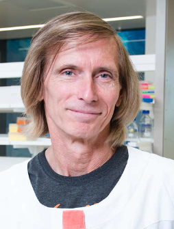 Nicholas Hayward合作论文数Oncogenomics Laboratory, QIMR Berghofer Medical Research Institute48
 David G. Beer合作论文数Department of Surgery, Medical School, University of Michigan27
 Dipankar Ray合作论文数Dipankar Ray Laboratory, Department of Radiation Oncology, Medical School, University of Michigan20
 Bryan J Mowry合作论文数20
 M K Walters合作论文数18Whiteman David C合作论文数18
 Daysha Ferrer-Torres合作论文数18
 Zhuwen Wang (王祝文)合作论文数18
 Theodore S Lawrence合作论文数Department of Radiation Oncology, Medical School, University of Michigan17
 Kiran H Lagisetty合作论文数Department of Surgery, University of Michigan16
 Paramita Ray合作论文数Department of Radiation Oncology, Medical School, University of Michigan16
 Thomas D. Wang合作论文数Department of Biomedical Engineering and Mechanical Engineering, University of Michigan13Srimathi Kasturirangan合作论文数12May San Martinho合作论文数12
 Jason Spence合作论文数Department of Biomedical Engineering, University of Michigan;Department of Internal Medicine, University of Michigan;Department of Cell & Developmental Biology, Medical School, University of Michigan;The Spence Laboratory, University of Michigan10
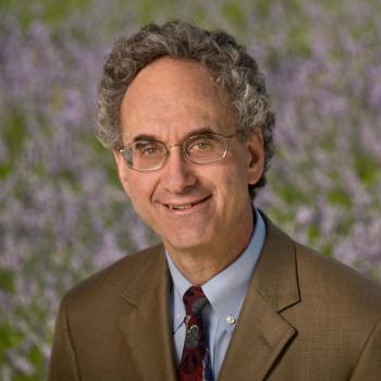 Douglas F. Levinson合作论文数Department of Psychiatry and Behavioral Sciences, School of Medicine, Stanford University10Meghan Curtin合作论文数9Shannon M. Lacy合作论文数9
 Mark Smithers合作论文数Mayne Academy of Surgery, Medical School, Faculty of Medicine, The University of Queensland9
 Jules Lin合作论文数Section of Thoracic Surgery, Department of Surgery, Medical School, University of Michigan-Ann Arbor9
 Matthias Truttmann合作论文数Truttmann Lab Site, Department of Molecular and Integrative Physiology, University of Michigan Medical School9Gotley David C合作论文数9
 David Watson合作论文数Department of Surgery, College of Medicine and Public Health, Flinders University9
 Sangeeta Jaiswal合作论文数9Graeme J. Walker合作论文数9Andrew C Chang合作论文数8
 Andrew Barbour合作论文数Diamantina Institute, Faculty of Medicine, The University of Queensland8
 Jeremy M Silverman合作论文数Department of Psychiatry, Icahn School of Medicine at Mount Sinai;Graduate School of Biomedical Sciences, Icahn School of Medicine at Mount Sinai;James J. Peters Veterans Affairs Medical Center8Jane M. Palmer合作论文数8
 Paul A Drew合作论文数8DA Nertney合作论文数7D P Lennon合作论文数7
 Dafydd Thomas合作论文数Department of Pathology, University of Michigan7
 Reddy Rishindra M合作论文数Department of Surgery, University of Michigan7
 Mitchell Stark合作论文数Dermatology Research Centre, Faculty of Health, Medicine and Behavioural Sciences, The University of Queensland;Frazer Institute, Faculty of Health, Medicine and Behavioural Sciences, The University of Queensland6
 Nancy C. Andreasen合作论文数Department of Psychiatry, College of Medicine, University of Iowa6Andrew Clouston合作论文数School of Biomedical Sciences, Faculty of Medicine, University of Queensland6
 Govindasamy Kumaramanickavel合作论文数Narayana Nethralaya/Aditya Jyot Eye Hospital6L Sharpe合作论文数5Richard C. Mohs合作论文数Eli Lilly and Company, Mount Sinai School of Medicine5Raymond R. Crowe合作论文数5
 Mark B Orringer合作论文数University of Michigan Medical School5
 Srinivasan Tirupati合作论文数5
 Sean Grimmond合作论文数Division of Medicine, Dentistry and Health Sciences, University of Melbourne;Centre for Cancer Research, University of Melbourne5R Kuick合作论文数4
 Lynn B. Jorde合作论文数Department of Human Genetics, School of Medicine, University of Utah4
 Donald W. Black合作论文数Roy J. and Lucille A. Carver College of Medicine, The University of Iowa;Iowa City Veterans Affairs Medical Center4Lampe Guy合作论文数4
 Chev Kidson合作论文数Mahidol University4




