Pulmonary neuroendocrine cells (PNECs) are rare chemosensory epithelial cells, facultative stem cells, and a cell-of-origin for neuroendocrine lung cancers, yet the mechanisms governing their differentiation and heterogeneity are poorly understood. Here we establish NEr-fAOs, a human fetal airway organoid platform that robustly enriches PNECs, and identify a synergistic requirement for dual GSK3 and NOTCH inhibition to drive directed PNEC differentiation. This strategy yields stable cultures with up to 60-fold expansion of PNECs whose transcriptomes closely match fetal and adult PNECs. In addition to PNEC-enrichment, NEr-fAOs retain diverse airway epithelial cell types, preserving epithelial complexity. Time-resolved single-cell transcriptomics maps PNEC trajectories in NEr-fAOs, resolving precursor and mature states. Comparative analyses further reveal a distal airway bias in NEr-fAOs and enrichment for lower-airway progenitors. NEr-fAOs thus provide a scalable, tractable platform to dissect human PNEC biology and distal airway progenitor hierarchies relevant to lung development, cancer, and disease. ### Competing Interest Statement The authors have declared no competing interest.
Supplementary Figure S4. Frag J/Jm mediated loss of misfolded Fluc-DM is independent of mutant p53 status.
The role of FGF is the least understood of the morphogens driving mammalian gastrulation. Here, we have investigated FGF function in a 2D gastruloid model for human gastrulation. We observed a ring of FGF-dependent ERK activity that closely follows the emergence of primitive streak (PS)-like cells but expands further inward. This ERK activity pattern depends on localized activation of basolateral FGF receptor 1 (FGFR1) by endogenous FGF gradients and is required for PS-like differentiation, with loss of PS-like cells upon FGF receptor inhibition rescued by direct ERK activation. Single cell transcriptome analysis confirmed that, among the ligands, FGF2 is broadly expressed, FGF8 is transiently expressed during PS-like differentiation and FGF4/17 are specifically expressed in PS-like cells - similar to the human and monkey embryo but different from the mouse. FGF4 knockdown greatly reduced PS-like differentiation, while FGF17 knockdown primarily affected subsequent mesoderm differentiation. FGF8 expression was spatially and temporally displaced from PS markers and FGF4 expression, while knockdown expanded PS-like cells, suggesting FGF8 may limit PS-like differentiation. Thus, we have identified a previously unreported role for FGF-dependent ERK signaling in 2D gastruloids and possibly the human embryo, where FGF4 and FGF17 signal through basolateral FGFR1 to induce PS-like cells and derivatives, potentially restricted by FGF8.
Supplementary Figure S7. Design and testing of scrambled peptides as negative controls.
Supplementary Figure S3. GRAIL1 is a glycosylated protein that undergoes processing.
Apicobasal polarization is crucial for tissue organization during in vivo development and in human organoid models. Extracellular matrix (ECM) signaling typically provides a basal cue, and intestinal and lung organoids reverse polarity from apical-in to apical-out after ECM removal. However, ECM-free brain organoids maintain apical-in polarity, suggesting that media components may influence polarity. Exposing brain organoids to serum induced apical-out orientation. Lysophosphatidic acid (LPA), present in the medium of prior apical-out techniques, was identified as the causative factor. LPA-induced apical-out orientation in brain organoids occurred within 1 day, lasted at least 1 month, and was optimal at human cerebrospinal fluid LPA concentrations. Sphingosine-1-phosphate (S1P) induced similar apical-out polarization. Pharmacological studies revealed that LPA/S1P act via a G-protein coupled receptor/RhoA pathway. Finally, LPA induced apical-out polarity in patient-derived human lung and intestinal organoids, iPSC spheres, and multilineage iPSC-derived intestinal organoids. These findings indicate that LPA signaling is a critical apical polarity cue in multiple tissues.
Background:Severe acute respiratory syndrome coronavirus 2 (SARS-CoV-2) infection remains a public health challenge worldwide. The gastrointestinal tract has emerged as an important site of infection and has been implicated as a reservoir for long-term infection, particularly for post-acute COVID-19 syndrome. However, whether vaccine-induced systemic antibodies can prevent intestinal infection with SARS-CoV-2 is unclear. Compared to Vero cells commonly used to assess SARS-CoV-2 neutralization, the intestinal epithelium has a functional interferon response and expresses higher levels of ACE2, enzymes, and antibody-binding Fc receptors that may impact SARS-CoV-2 immune elimination. Methods:We evaluated the potential of antibodies from both naturally infected and vaccinated human subjects to inhibit SARS-CoV-2 infection of the intestinal epithelium. Serum samples were collected from human volunteers who had undergone natural infection with SARS-CoV-2 in 2020 (n=5) or who had received the Pfizer BNT162b2 COVID-19 vaccine (n=13). Banked sera collected in 2016 served as negative controls (n=2). SARS-CoV-2 (WA01, Delta or Omicron) was pre-treated with sera and then used to infect iPSC-derived human intestinal organoids (HIO) or Caco-2 colonic epithelial cells, and SARS-CoV-2 infection was quantified by plaque assay, PCR, or immunofluorescence (IF) after 48-96 h. Results:Both HIOs and Caco-2 cells supported robust infection with SARS-CoV-2. In HIOs, pretreatment of SARS-CoV-2 with a high titer post-vaccine serum completely blocked replication of WA01. Similarly, sera from both naturally infected donors collected in 2020 and sera from individuals who had received a BNT162b2 vaccine significantly inhibited replication of the WA01 strain in Caco-2 cells. In contrast, none of the sera significantly inhibited infection with the Delta variant of SARS-CoV-2. For Omicron, only sera from individuals who had received an Omicron-based vaccine significantly inhibited infection with SARS-CoV-2 in the plaque assay. Across all virus types, sera from individuals who had received Omicron-based BNT162b2 boosters were the most effective at reducing infection in Caco-2 cells. Conclusion:Our results suggest that vaccine-induced antibody responses to SARS-CoV-2 are protective in the gut. Our study also supports previous reports indicating that SARS-CoV-2 vaccines need to be adapted to circulating virus strains to convey full protection from infection.
Using single-cell RNA sequencing (scRNA-seq) and histological approaches, we examine cross-species cellular identity, diversity and organization of mesenchymal (fibroblast) populations in the developing mouse and human intestines. In both species, we defined 7 fibroblast populations. Using cross-species integration and label transfer approaches we find that each mesenchymal cell subtype in the murine intestine is highly concordant to a cell type/state in the human intestine, and vice-versa, suggesting a strong conservation of mesenchymal cell types/states across species. Despite this conservation, we also observe that individual lineage-defining genes are not always shared and can be found in different mesenchymal populations. High resolution spatial analysis via fluorescent in situ hybridization (FISH) and immunofluorescence (IF) confirmed these findings and revealed that transcriptionally-defined sub-types of intestinal mesenchymal cells in mice and humans are organized within similar spatial domains.
 Yu-Hwai Tsai合作论文数42
 Michael K Dame合作论文数31
 Sha Huang (黄沙)合作论文数Chinese PLA General Hospital26Angeline Wu合作论文数25Hill David R合作论文数25
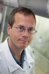 James M. Wells合作论文数Centre for Archaeology, English Heritage25
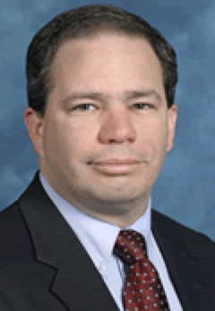 Peter D. R. Higgins合作论文数University of Michigan Health System;Higgins IBD Research Group, Medical School, University of Michigan20Emily M Holloway合作论文数Sch Med, Univ Michigan19
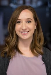 Alyssa J. Miller合作论文数19
 Tristan Frum合作论文数Dept Biochem & Mol Biol, Michigan State Univ17
 Vincent B. Young合作论文数Department of Microbiology & Immunology, Medical School, University of Michigan;Infectious Diseases Division, Department of Internal Medicine, Medical School, University of Michigan16Joshua H Wu合作论文数15
 Daysha Ferrer-Torres合作论文数15
 Ian A Glass合作论文数15Dye Briana R合作论文数14
 Priya H Dedhia合作论文数14Charlie J Childs合作论文数13
 Michael J. Czerwinski合作论文数Department of Cell and Developmental Biology, University of Michigan Medical School;Department of Cell and Developmental Biology, University of Michigan Medical School12Nagy Melinda S合作论文数12
 Sangeeta Jaiswal合作论文数12
 Michael A. Helmrath合作论文数Center for Stem Cell & Organoid Medicine, Cincinnati Children’s Hospital Medical Center;Center for Bariatric Research and Innovation, Cincinnati Children’s Hospital Medical Center;Department of Surgery, University of Cincinnati12Noah F. Shroyer合作论文数MLC, Cincinnati Children’s Hospital12
 Christiane E. Wobus合作论文数Department of Microbiology and Immunology, University of Michigan Medical School11
 Jarrett Camp合作论文数Department of Biosystems Science and Engineering, Eidgenössische Technische Hochschule Zürich11
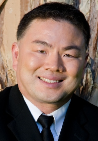 Thomas D. Wang合作论文数Department of Biomedical Engineering and Mechanical Engineering, University of Michigan11
 David G. Beer合作论文数Department of Surgery, Medical School, University of Michigan11May San Martinho合作论文数10Meghan Capeling合作论文数10Veda K. Yadagiri合作论文数10
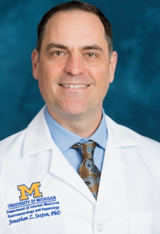 Jonathan Sexton合作论文数Department of Medicinal Chemistry, College of Pharmacy, University of Michigan;Division of Gastroenterology and Hepatology, Department of Internal Medicine, University of Michigan;University of Michigan Medical School;Curl Bio LLC10
 Derek Nancarrow合作论文数Thoracic Surgery Section, University of Michigan10
 Paramita Ray合作论文数Department of Radiation Oncology, Medical School, University of Michigan10
 Dipankar Ray合作论文数Dipankar Ray Laboratory, Department of Radiation Oncology, Medical School, University of Michigan10
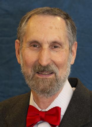 Theodore S Lawrence合作论文数Department of Radiation Oncology, Medical School, University of Michigan10
 Yatrik Shah合作论文数Department of Molecular & Integrative Physiology, Medical School, University of Michigan10
 Zhuwen Wang (王祝文)合作论文数10Meghan Curtin合作论文数9Srimathi Kasturirangan合作论文数9Shannon M. Lacy合作论文数9Alana M. Chin合作论文数9Caroline L. Mccarthy合作论文数Med Sch, Univ Michigan9Margaret S. Bohm合作论文数Med Sch, Univ Michigan9
 Kateryna Karpoff合作论文数9
 Charles Zhang合作论文数College of Pharmacy, University of Michigan9
 Kiran H Lagisetty合作论文数Department of Surgery, University of Michigan9
 Qianhui Yu合作论文数Institute of Molecular and Clinical Ophthalmology Basel (IOB), Basel, Switzerland9
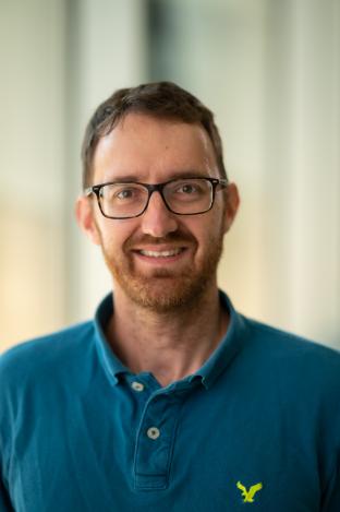 Matthias Truttmann合作论文数Truttmann Lab Site, Department of Molecular and Integrative Physiology, University of Michigan Medical School9
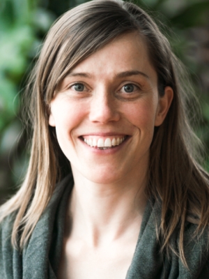 Barbara Treutlein合作论文数Department of Biosystems Science and Engineering, Eidgenossische Technische Hochschule Zurich9
 Durga Attili合作论文数Department of Pathology, University of Michigan Medical School9




