Objectives: To compare COVID-19-associated pulmonary mucormycosis (CAPM) with COVID-19associated rhino-orbital mucormycosis (CAROM), ascertain factors associated with CAPM among patients with COVID-19, and identify factors associated with 12-week mortality in CAPM. Methods: We performed a retrospective multicentre cohort study. All study participants had COVID-19. We enrolled CAPM, CAROM, and COVID-19 subjects without mucormycosis (controls; age-matched). We collected information on demography, predisposing factors, and details of COVID-19 illness. Univariable analysis was used to compare CAPM and CAROM. We used multivariable logistic regression to evaluate factors associated with CAPM (with hypoxemia during COVID-19 as the primary exposure) and at 12-week mortality. Results: We included 1724 cases (CAPM [n = 122], CAROM [n = 1602]) and 3911 controls. Male sex, renal transplantation, multimorbidity, neutrophil-lymphocyte ratio, intensive care admission, and cumulative glucocorticoid dose for COVID-19 were significantly higher in CAPM than in CAROM. On multivariable analysis, COVID-19-related hypoxemia (aOR, 2.384; 95% CI, 1.209-4.70 0), male sex, rural residence, diabetes mellitus, serum C-reactive protein, glucocorticoid, and zinc use during COVID-19 were independently associated with CAPM. CAPM reported a higher 12-week mortality than CAROM (56 of the 107 [52.3%] vs. 413 of the 1356 [30.5%]; p = 0.0001). Hypoxemia during COVID-19 (aOR [95% CI], 3.70 [1.34 -10.25]) and Aspergillus co-infection (aOR [95% CI], 5.40 [1.23-23.64]) were independently associated with mortality in CAPM, whereas surgery was associated with better survival. Discussion: CAPM is a distinct entity with a higher mortality than CAROM. Hypoxemia during COVID-19 illness is associated with CAPM. COVID-19 hypoxemia and Aspergillus co-infection were associated with higher mortality in CAPM. Valliappan Muthu, Clin Microbiol Infect 2024;30:368 (c) 2023 European Society of Clinical Microbiology and Infectious Diseases. Published by Elsevier Ltd. All rights reserved.
Pleural effusion is a common problem in our country, and most of these patients need invasive tests as they can't be evaluated by blood tests alone. The simplest of them is diagnostic pleural aspiration, and diagnostic techniques such as medical thoracoscopy are being performed more frequently than ever before. However, most physicians in India treat pleural effusion empirically, leading to delays in diagnosis, misdiagnosis and complications from wrong treatments. This situation must change, and the adoption of evidence-based protocols is urgently needed. Furthermore, the spectrum of pleural disease in India is different from that in the West, and yet Western guidelines and algorithms are used by Indian physicians. Therefore, India-specific consensus guidelines are needed. To fulfil this need, the Indian Chest Society and the National College of Chest Physicians; the premier societies for pulmonary physicians came together to create this National guideline. This document aims to provide evidence based recommendations on basic principles, initial assessment, diagnostic modalities and management of pleural effusions.
BACKGROUND:The International Society for Human and Animal Mycology (ISHAM) working group proposed recommendations for managing allergic bronchopulmonary aspergillosis (ABPA) a decade ago. There is a need to update these recommendations due to advances in diagnostics and therapeutics. METHODS:An international expert group was convened to develop guidelines for managing ABPA (caused by Aspergillus spp.) and allergic bronchopulmonary mycosis (ABPM; caused by fungi other than Aspergillus spp.) in adults and children using a modified Delphi method (two online rounds and one in-person meeting). We defined consensus as ≥70% agreement or disagreement. The terms "recommend" and "suggest" are used when the consensus was ≥70% and <70%, respectively. RESULTS:We recommend screening for A. fumigatus sensitisation using fungus-specific IgE in all newly diagnosed asthmatic adults at tertiary care but only difficult-to-treat asthmatic children. We recommend diagnosing ABPA in those with predisposing conditions or compatible clinico-radiological presentation, with a mandatory demonstration of fungal sensitisation and serum total IgE ≥500 IU·mL-1 and two of the following: fungal-specific IgG, peripheral blood eosinophilia or suggestive imaging. ABPM is considered in those with an ABPA-like presentation but normal A. fumigatus-IgE. Additionally, diagnosing ABPM requires repeated growth of the causative fungus from sputum. We do not routinely recommend treating asymptomatic ABPA patients. We recommend oral prednisolone or itraconazole monotherapy for treating acute ABPA (newly diagnosed or exacerbation), with prednisolone and itraconazole combination only for treating recurrent ABPA exacerbations. We have devised an objective multidimensional criterion to assess treatment response. CONCLUSION:We have framed consensus guidelines for diagnosing, classifying and treating ABPA/M for patient care and research.
Dear Editor, A large number of mucormycosis cases were reported in India following the second wave of COVID-19.[1] However, data on the imaging appearance of COVID-19-associated pulmonary mucormycosis (CAPM) are limited.[2] A few published case reports on CAPM and a systematic literature review suggested cavity and consolidation as the most common finding.[3-6] The primary objective of the current study was to describe the computed tomography (CT) thorax findings of CAPM at diagnosis. The secondary objectives were to compare the CT findings of (1) CAPM in patients with diabetes mellitus (DM) and those with no known traditional risk factors, and (2) survivors and non-survivors with CAPM. The MuCovi-2 study was a multicentre retrospective study conducted across 25 centres in India between 1 January 2021 and 30 June 2021. Data on COVID-19-associated mucormycosis (microbiologically or pathologically confirmed) cases and age-matched controls were collected in the original study.[7] COVID-19 was diagnosed by detecting SARS-CoV2 infection in nasopharyngeal or throat swabs by rapid antigen testing or reverse transcriptase polymerase chain reaction. Only subjects diagnosed with proven and probable CAPM were included in the present study, as defined previously.[2] The diagnosis and management of CAPM patients were per the prevalent guidelines.[2,8] The clinical presentation, risk factors, and outcome of the entire cohort of CAPM patients have been published recently.[9] For the current study, we excluded CAPM patients with inadequate CT data. We noted (as yes/no) the radiological findings on CT (cavity, consolidation, reversed halo sign (RHS), bird's nest sign, pneumothorax, mycotic aneurysm, pleural effusion, nodules, and others) at diagnosis of CAPM. Data are presented as numbers (percentage) or mean with standard deviation (SD). We used the Mann–Whitney U and Fisher's exact (or Chi-square) tests to analyse the difference between continuous and categorical variables. We assumed statistical significance as a P value <0.05. We included 84 subjects with CAPM [Table 1]. The participants' mean (SD) age was 53.8 (12.5) years. DM was the most common predisposing factor in 58/84 (69%) subjects. Glucocorticoid treatment for COVID-19 was prescribed in 82.3% (65/79) subjects, and nearly 20% of these subjects received inappropriate glucocorticoid therapy (i.e., in the absence of COVID-19-related hypoxemia). Cavity was the most common finding (56/84; 77.8%). RHS or bird's nest sign were observed in 14/84 (16.7%) subjects. Pleural effusion or >10 lung nodules were reported in 19/84 (22.6%) and 2/84 (2.4%) subjects, respectively. Tracheobronchial mucormycosis was noted in four subjects, one of whom had an isolated tracheal lesion with no other parenchymal or pleural abnormality. Of the 84 subjects, 43 (51.2%) and 41 (48.8%) had proven and probable CAPM, respectively. Bilateral disease was significantly more common in those with probable CAPM (20/41 [48.8%] versus 9/43 [20.9%] among proven cases), and pleural effusion was more frequent in proven CAPM (15/43 [34.9%] versus 4/41 [9.8%] in probable CAPM). None of the other findings were significantly different between proven or probable CAPM. The CT findings were similar in patients with COVID-19 as the only risk factor for CAPM versus those with DM, except for pleural effusion, which was significantly less common in DM [Table 2]. Survival at 6 weeks and 12 weeks was 43/79 (54.4%) and 37/73 (50.7%). We found no significant differences in CT findings among survivors versus non-survivors at 6-weeks [Table 3].Table 1: Baseline characteristics and imaging findings of CAPM (n=84) at diagnosisTable 2: Comparison of CT findings in CAPM patients with and without diabetes mellitusTable 3: Comparison of CT findings between survivors versus non-survivors at 6 weeks with CAPMTimely diagnosis improves survival in pulmonary mucormycosis (PM), a serious illness with a high mortality.[10,11] A CT scan provides one of the earliest clues for diagnosing PM [Figure 1].[12] However, most data on the CT findings of PM are from patients with risk factors such as haematological malignancies and organ transplant recipients.[13,14] Imaging appearances of CAPM are poorly studied. We found cavity (66.7%) the most common finding in CAPM, like in DM.[15,16] However, consolidation was less frequently reported in our study (13%) than in the previous studies (48%).[17] Interestingly, in the current study, consolidation was infrequent (12%), even in diabetic subjects with CAPM. The lesser frequency of consolidation and predominance of the cavities in our study could be related to the duration of illness before the CT scan.[15] The earliest imaging findings of PM include nodules, consolidation, and masses; cavitation occurs later when the lesions undergo central necrosis.[18] Neutropenic patients are evaluated early with chest CT and thus manifest nodules or masses. Contrarily, PM in DM or COVID-19 presents later and cavities are common. The current study participants (CAPM with and without DM) were possibly evaluated late during the illness due to severe COVID-19. Also, consolidation due to PM could have been attributed to COVID-19 or hospital-acquired pneumonia. Although insensitive, other notable findings in our study, like RHS and mycotic aneurysm, may suggest PM in those with predisposing factors (including COVID-19 and uncontrolled DM).Figure 1: Representative images showing a few typical signs of COVID-19-associated pulmonary mucormycosis: (a) consolidation in the right upper lobe along with diffuse ground glass opacities (GGOs; COVID-19 related) and pneumomediastinum, (b) reversed halo sign on the right side with patchy consolidation in bilateral lungs and a few GGOs, (c) cavity with air-fluid level in the right lower lobe and bilateral GGOs and (d) pulmonary artery aneurysm in a lesion in the right lower lobe; pneumothorax is also noted on the right sideOur study has a few limitations. The CT findings were retrospectively retrieved from patients' records, and the imaging was not centrally reviewed. Despite a multicentre study, the number of PM patients was relatively small. Further, the CT protocol, the use of contrast, and the differences in the reporting could have influenced our study results. The chest CT findings may vary depending on the duration of PM, and we do not have this information. We do not have a comparator group (e.g., invasive pulmonary aspergillosis); therefore, the specificity of the imaging findings is unknown. We included only individuals with a confirmed diagnosis of CAPM, and we might have missed cases where microbiological or pathological confirmation was unavailable. Isolated tracheobronchial mucormycosis (also reported in COVID-19) without any parenchymal abnormalities could have been missed in our series.[19] In summary, we found similar imaging findings in CAPM as previously reported in PM complicating DM. Cavity was the most common finding in CAPM patients in India. Prospective studies, including serial imaging performed on a larger number of patients with varying risk factors, are required. Financial support and sponsorship Nil. Conflicts of interest There are no conflicts of interest.
Background: Data on mixed mould infection with COVID-19-associated pulmonary aspergillosis (CAPA) and COVID-19-associated pulmonary mucormycosis (CAPM) are sparse. Objectives: To ascertain the prevalence of co-existent CAPA in CAPM (mixed mould infection) and whether mixed mould infection is associated with early mortality (<= 7 days of diagnosis). Methods: We retrospectively analysed the data collected from 25 centres across India on COVID-19-associated mucormycosis. We included only CAPM and excluded subjects with disseminated or rhino-orbital mucormycosis. We defined co-existent CAPA if a respiratory specimen showed septate hyphae on smear, histopathology or culture grew Aspergillus spp. We also compare the demography, predisposing factors, severity of COVID-19, and management of CAPM patients with and without CAPA. Using a case-control design, we assess whether mixed mould infection (primary exposure) were associated with early mortality in CAPM. Results: We included 105 patients with CAPM. The prevalence of mixed mould infection was 20% (21/105). Patients with mixed mould infection experienced early mortality (9/21 [42.9%] vs. 15/84 [17.9%]; p = 0.02) and poorer survival at 6 weeks (7/21 [33.3] vs. 46/77 [59.7%]; p = 0.03) than CAPM alone. On imaging, consolidation was more commonly encountered with mixed mould infections than CAPM. Co-existent CAPA (odds ratio [95% confidence interval], 19.1 [2.62-139.1]) was independently associated with early mortality in CAPM after adjusting for hypoxemia during COVID-19 and other factors. Conclusion: Coinfection of CAPA and CAPM was not uncommon in our CAPM patients and portends a worse prognosis. Prospective studies from different countries are required to know the impact of mixed mould infection.
Over the past decade, endobronchial ultrasound-guided transbronchial needle aspiration (EBUS-TBNA) has become an indispensable tool in the diagnostic armamentarium of the pulmonologist. As the expertise with EBUS-TBNA has evolved and several innovations have occurred, the indications for its use have expanded. However, several aspects of EBUS-TBNA are still not standardized. Hence, evidence-based guidelines are needed to optimize the diagnostic yield and safety of EBUS-TBNA. For this purpose, a working group of experts from India was constituted. A detailed and systematic search was performed to extract relevant literature pertaining to various aspects of EBUS-TBNA. The modified GRADE system was used for evaluating the level of evidence and assigning the strength of recommendations. The final recommendations were framed with the consensus of the working group after several rounds of online discussions and a two-day in-person meeting. These guidelines provide evidence-based recommendations encompassing indications of EBUS-TBNA, pre-procedure evaluation, sedation and anesthesia, technical and procedural aspects, sample processing, EBUS-TBNA in special situations, and training for EBUS-TBNA.
Case seriesthrombolyzed with 100 mg injection actilyse [recombinant tissue plasminogen activator (rt-PA)] over 2 hours.2D-echo was repeated again (Video 2) and showed reduction in the size of thrombus.After 10 days, oxygen requirement was reduced to 5-6 L/minute, and in another 10-11 days, patient was shifted out of ICU and was finally discharged after 24 days of admission.Following discharge, patient was advised to take dabigatran 150 mg 12 hourly. Case 2A 29-year-old obese female presented with severe COVID-19 pneumonia, with SpO 2 at 67% on room air and SpO 2 at 95% with 6 L/minute O 2 on mask in intensive care unit.Her oxygen requirement increased within 36-48 hours after admission.
We performed a case-control study across 25 hospitals in India for the period of January-June 2021 to evaluate the reasons for an COVID-19-associated mucormycosis (CAM) outbreak. We investigated whether COVID-19 treat-ment practices (glucocorticoids, zinc, tocilizumab, and oth-ers) were associated with CAM. We included 1,733 cases of CAM and 3,911 age-matched COVID-19 controls. We found cumulative glucocorticoid dose (odds ratio [OR] 1.006, 95% CI 1.004-1.007) and zinc supplementation (OR 2.76, 95% CI 2.24-3.40), along with elevated C-reactive protein (OR 1.004, 95% CI 1.002-1.006), host factors (re-nal transplantation [OR 7.58, 95% CI 3.31-17.40], diabetes mellitus [OR 6.72, 95% CI 5.45-8.28], diabetic ketoacidosis during COVID-19 [OR 4.41, 95% CI 2.03-9.60]), and rural residence (OR 2.88, 95% CI 2.12-3.79), significantly as-sociated with CAM. Mortality rate at 12 weeks was 32.2% (473/1,471). We emphasize the judicious use of COVID-19 therapies and optimal glycemic control to prevent CAM.
“We cannot change the outcome but we can affect the journey” – Ann Richardson Malignant pleural effusion (MPE) is a common and disabling complication of cancer and is associated with significant morbidity and mortality. Life expectancy in such patients is dismal and, depending on the primary tumour, the median survival ranges from 3 to 12 months.[1] With 1 in 8 men and 1 in 10 women developing cancer globally, overall cancer incidence has shown an ascending trend and hence it is only expected that the burden of MPE will rise in the future.[2] There is also a significant healthcare burden with MPE accounting for more than 125000 hospital admissions in the USA in 2012 and an estimated expense of more than 5 billion dollars per year.[3] There is a paucity of Indian data on MPE. MPE represents an advanced stage of cancer and in the large majority, treatment remains palliative based on symptom relief and preventing recurrence via early and successful pleural symphysis. Options include repeated thoracentesis, chest drain followed by chemical pleurodesis and indwelling pleural catheter (IPC). Pleurodesis and IPC offer ‘definitive’ management and are the widely favoured procedures. Both have their pros and cons and their adoption into clinical practice is based on numerous factors. While pleurodesis requires hospitalisation and has an approximately 50% failure rate at six months,[4] it remains the first procedure of choice in our country mainly due to the ease of administration, patient preference and its favourable economics. Talc remains the pleurodesis agent of choice with slurry being as effective as poudrage.[56] Cheaper and safe alternative like povidone iodine, with an efficacy rate of 89%, remains an attractive option in our part of the world.[7] IPC, introduced in late 1990s, is a 15.5 F fenestrated catheter with a proprietary one-way valve and polyester cuff which gets embedded in the tunneled subcutaneous tissue giving it the indwelling property for longer periods. Single-use pre-vacuum bottle is attached to drain fluid in a controlled manner. Inserted as a day-care procedure, IPC offers ambulatory advantage, effective symptom control, shorter hospital stays, reduced number of subsequent pleural interventions and minimal and tolerable complication rate.[8910] An added and unexplained benefit of IPC is the phenomenon of auto pleurodesis seen in 11-24% patients at the end of three months.[111213] In the last decade, a number of well conducted patient-centric randomised control trials (RCTs) on IPC have resulted in it being positioned as the first-line therapy for MPE in most parts of the western world.[10111213] Novel strategies like aggressive versus symptom-based drainage (ASAP and AMPLE 2) and IPC followed by talc pleurodesis (IPC-PLUS) have facilitated early pleural symphysis and catheter removal.[111213] Impregnating indwelling catheters with silver nitrate, however, failed to show superiority over standard IPC in pleurodesis efficacy (SWIFT).[14] MPE IN INDIA The projected incidence of cancer in India among males is 679,421 (94.1 per 100,000) and in females 712,758 (103.6 per 100,000) for the year 2020.[15] If one in six cancer patients go on to develop MPE, this would leave a substantial number of patients to be treated by our health personnel.[16] Managing this large group of patients will require judicious decision making based on patient selection, their preferences, available skills, resources and cost. IPC was introduced in India six years back but has yet to establish a strong foothold. Technical expertise, cost, erratic supplies, recurring expenditure, and skepticism on homecare failures are some deterrents to its widespread use. Increased risk of empyema remains a perceived rather than evident concern for the rates of infection, as experienced by the authors and other colleagues, is no worse than the ~ 5% as seen in other parts of the world (17, personal communication).[17] There is a pressing need for workers across the country to establish an IPC registry and share their experiences. In the current issue, Shrinath et al.[18] have made a small beginning in this direction by sharing their preliminary results in a research letter. Their observations on a very small number of patients conforms to available literature while excluding what would have been significant information on IPC in the Indian context, namely, the cost analysis and details of home care – two vital issues that need attention in our part of the world. IPC holds advantage over pleurodesis in the reduction of hospital visits and handing over the care and control to patients themselves. By bringing in all patients daily for the first week and thereafter three-weekly for drainage and inspection for complications defeats this purpose. Compulsions such as absence of healthcare inputs at home from trained nurse or family physician may have been a possible cause and herein lies the anticipated main contraindication of IPC in India which is the inability for the patient, family or healthcare services to manage home drainage. Unless efforts are made to institute and reinforce training for IPC home care, we may be in danger of causing more harm than benefit. Many centres in the country have larger experience of IPC insertions and follow-up of their patients and a wealth of information can be obtained if they publish their data. This will help to ideally position this procedure in the MPE management algorithm in our country which has a unique and complex heathcare system characterised by mixed ownership pattern, sociocultural differences, different types of providers and systems of medicine – all of which mandate that we may not always be successful in replicating the Western model of health care. We need to invent our indigenous pathways to manage MPE. This should include low-cost drainage systems, engaging our vast community and hospital-based health workers to provide home support to IPC patients and most importantly, keeping the overall cost affordable. It is incumbent on the manufacturers to consider an economical business model suited to the healthcare practices of our country if we want to expand the role of IPC beyond the trapped lung and failed pleurodesis scenario. Finally, we believe that an RCT is needed in India, comparing tube thoracostomy and pleurodesis with IPC, where the primary outcomes are infection rate and cost analysis.
COVID-19-associated pulmonary mucormycosis (CAPM) remains an underdiagnosed entity. Using a modified Delphi method, we have formulated a consensus statement for the diagnosis and management of CAPM. We selected 26 experts from various disciplines who are involved in managing CAPM. Three rounds of the Delphi process were held to reach consensus (>= 70% agreement or disagreement) or dissensus. A consensus was achieved for 84 of the 89 statements. Pulmonary mucormycosis occurring within 3 months of COVID-19 diagnosis was labelled CAPM and classified further as proven, probable, and possible. We recommend flexible bronchoscopy to enable early diagnosis. The experts proposed definitions to categorise dual infections with aspergillosis and mucormycosis in patients with COVID-19. We recommend liposomal amphotericin B (5 mg/kg per day) and early surgery as central to the management of mucormycosis in patients with COVID-19. We recommend response assessment at 4-6 weeks using clinical and imaging parameters. Posaconazole or isavuconazole was recommended as maintenance therapy following initial response, but no consensus was reached for the duration of treatment. In patients with stable or progressive disease, the experts recommended salvage therapy with posaconazole or isavuconazole. CAPM is a rare but under-reported complication of COVID-19. Although we have proposed recommendations for defining, diagnosing, and managing CAPM, more extensive research is required.
Background.Idiopathic pulmonary fibrosis (IPF) is a specific type of chronic fibrosing interstitial lung disease (ILD) that is progressive in course.Although evidence-based guidelines for IPF are available, these are difficult to interpret for the average physician and may not be suitable for use in resource-constrained settings.There was an unmet need to formulate guidelines that are pragmatic, easy to understand and suited for application in resource-limited settings.Methods.This statement was made by a group of expert pulmonologists.Twenty-five questions regarding diagnosis and management of IPF were framed.A literature search was conducted using the PubMed and EmBase databases.The expert group discussed available evidence relevant to each question and recommendations were arrived at by consensus. Results.A thorough clinical and laboratory evaluation should be performed in patients suspected to have ILDs and potential underlying causes should be ruled out.A high resolution computed tomography (HRCT) of the chest is essential to identify the pattern of ILD.The need for a lung biopsy should be decided based on the appearance on the HRCT.Once a diagnosis of IPF is made, anti-fibrotic drugs (pirfenidone or nintedanib) should be offered after discussing the expected benefits and potential adverse effects with the patient.Recommendations have been made on other issues in the management of IPF, such as management of cough and dyspnoea, role of supplemental oxygen, mechanical ventilation and lung transplantation. Conclusion.This consensus statement provides practical and easy-to-use recommendations for the diagnosis and management of IPF in resource-limited settings.
During the times of the ongoing COVID pandemic, aerosol-generating procedures such as bronchoscopy have the potential of transmission of severe acute respiratory syndrome coronavirus 2 to the healthcare workers. The decision to perform bronchoscopy during the COVID pandemic should be taken judiciously. Over the years, the indications for bronchoscopy in the clinical practice have expanded. Experts at the Indian Association for Bronchology perceived the need to develop a concise statement that would assist a bronchoscopist in performing bronchoscopy during the COVID pandemic safely. The current Indian Association for Bronchology Consensus Statement provides specific guidelines including triaging, indications, bronchoscopy area, use of personal protective equipment, patient preparation, sedation and anesthesia, patient monitoring, bronchoscopy technique, sample collection and handling, bronchoscope disinfection, and environmental disinfection concerning the coronavirus disease-2019 situation. The suggestions provided herewith should be adopted in addition to the national bronchoscopy guidelines that were published recently. This statement summarizes the essential aspects to be considered for the performance of bronchoscopy in COVID pandemic, to ensure safety for both for patients and healthcare personnel.
Pleura, while considered the outpost of the lung, has become the subject of intense interest among clinicians and researchers. The last two decades have seen a more in-depth and clear understanding of the pleural pathophysiology, new investigative tools, and ingenious devices for improved patient care. As we step into the third decade of the 21st century, we move forward confidently with the assurance that the work in progress will get consolidated as more researchers adopt pleura as their area of interest. The pharmaceutical and device manufactures have wholeheartedly supported these endevoures, and a wider section of the clinical community is getting better equipped to manage the increasing incidence of pleural diseases and its complexities.[1] The Pleural Juggernaut is truly rolling swiftly, smoothly, and safely. Medical thoracoscopy (MT) has acted as a major fuel in igniting the interest in pleural diseases. It has a lot to owe to the Swedish internist Hans Christian Jacobaeus for its origin, refinement, and extensive description (Jacobaeus operation). From the 1940s onward, with the discovery of streptomycin, the procedure witnessed a temporary decline in its popularity only to then begin its European renaissance in the systematic application for the diagnosis of many pleuro-pulmonary diseases. The excellent diagnostic utility of MT enveloped within a favorable safety profile has made it an attractive procedure for physicians to adapt.[2] The relatively easy learning curve[3] and the less demanding infrastructure has enabled it to enter the armamentarium of every modern intervention pulmonologist. MT has become a method of choice for investigating undiagnosed exudative pleural effusions, and its therapeutic application is seen, with a high success rate, in malignant pleural effusion and loculated empyema.[4] India has not remained aloof to this development. The urge to inspect the pleura saw several tuberculosis units using their existing fiberoptic bronchoscope as a thoracoscope.[5] In the early 1980s, thoracic oncosurgeons (personal communication Dr. Raman Deshpande) and pediatric surgical departments had already put the rigid thoracoscope to widespread use for a variety of video-assisted procedures.[6] The launch of semi-rigid thoracoscope in 2008 paved the way for pulmonary physicians to adapt and advance their skills in managing pleural diseases. Apex institutes in India published their successful experience with the procedure[78910] and then went on to push the boundaries by removing foreign objects,[11] carrying out pleural cryobiopsies[12] and even performing lung biopsies through this route.[13] Like any other medical procedure, MT too needs its checks and balance and its advancement in a systematic fashion. Uniformity helps in sharing a common language and improves practices through sharing experiences. A national guideline and its adherence is one way to achieve this objective. An important first step toward this goal is to gain insights into practices across a wide spectrum of health care existing in a vast country like ours. An effective way of doing so is to invite and involve as many exponents by the way of methods that are uniform and less cumbersome. Questionnaire-based survey is a useful tool that allows assessment of a wide cross-section of population with relative ease. Using electronic platforms further economizes the time, response rate, data compilation, and cost and has become the method of choice for large medical surveys. In the just published issue of Lung India,[14] Dr. Madan et al. have done well in adopting this method, having previously used it for the Indian Bronchoscopy survey.[15] Using the database of national respiratory organizations and sending out a carefully constructed questionnaire encompassing the key points of MT, they have meaningfully interpreted the results while acknowledging the limitations entailed in such methods and responses. It is reassuring to note that most respondents have adhered to the broad principles of the procedure albeit with varying degrees of benign transgressions. The increased need for incorporating a thoracoscopy program in our public health sector and encouraging participation of trainees should remain a prerogative. Although the concept of “see one, do one, teach one” is a time-tested traditional method of learning, only one-fifth of the respondents have undergone a formal training program, emphasizing the need for increased availability and participation in well-structured courses. Most of the exponents have addressed patient safety by ensuring the presence of adequate monitoring tools, resuscitative measures, and the availability of surgical backup. The necessity of documentation and accountability is well reflected in the findings that a substantial number (84%) have video-recording facilities. An interesting finding has been the use of rigid thoracoscope as the instrument of choice over the more popular flexi-rigid thoracoscope, which, by its similarity to the familiar flexible bronchoscope, would have been the more natural and preferable choice of most chest physicians. Undiagnosed pleural effusion remains the major indication of the procedure and is in line with worldwide practice. Managing adhesions in pleural space infections constitutes another important indication in our part of the world due to the nonavailability of more effective human recombinant DNase in combination with t-PA[1617] and the lack of widespread availability of enthusiastic thoracic surgeons. Talc pleurodesis is a close third indication though the TAPPS trial may well change this practice.[18] An important and concerning finding of the survey has been the complication rate reported by almost 70% of the respondents. The authors may have well analyzed this in their discussion, for it forms an important audit tool. With close to 30% of the respondents reporting postprocedural empyema and port-site infections, it leaves scope for more stringent infection control measures and to consider early strategies for trapped lung situation or referral to surgical colleagues. While the exact mortality figures are not available, almost 8% of the respondents have encountered this during the procedure, which is well beyond the accepted rate of 0.34%.[2] Thus, the complication rate needs serious self-introspection as to the cause and remedial measures that need to be taken. Formal training, careful patient selection, and a less cavalier approach may well be the first step in this direction. Primum non nocere must always remain the motto. The authors have done well to acknowledge their limitation, especially the dismal percentage of respondents from a national pool of more than 5000 chest physicians. The interpretations of such surveys, therefore, demand caution, which readers will undoubtedly exercise. The intention is to provide a nationwide snapshot of the current practice which can then be amalgamated into national consensus statements and guidelines. It is also hoped that the survey will feed into further clinical and epidemiological research, which is the need of the hour for a country like India to ensure both quality assurance and improvement in the care of its patients with pleural diseases. Tell me and I forget, teach me and I may remember, involve me and I learn – Benjamin Franklin
in TBLC has been reported not only with respect to complications but also with respect to the quality and size of the biopsies.In a previous study, Almeida and colleagues assessed 100 TBLCs performed in patients with suspected diffuse lung disease (2).When they compared the first 50 TBLCs with the next 50 TBLCs, they found that the length and area of the biopsies were smaller and the diagnostic yield was lower in the first group, and all parameters improved when the bronchoscopists gained more experience.In their study, Almeida and colleagues reported a median length of 5.0 mm in the first 50 biopsies and 6.0 mm in the next 50 biopsies.Romagnoli and colleagues reported a level of agreement between external blinded versus local pathology reports as fair to moderate, with k values of 0.22-0.51.The k values for individual pathologists are not presented, and as noted above for bronchoscopy, there may be a learning curve for pathologic evaluations of cryobiopsies.In support of this, previous studies (which included the same external pathologist as in the present study) reported k values between 0.59 and 0.61 (5,6).With regard to the agreement between the pathologic diagnosis based on the two types of specimens and the final diagnosis at the second multidisciplinary assessment or the final treatment (Table 2 andTable E3 in the online supplement of Reference 1), there is no statistically significant difference by conventional standards between the two types of specimens in terms of performance when evaluated by a chi-square test or Fisher's exact test on simple 2 3 2 tables, even though there is trend in favor of SLB.This emphasizes the need for further research into this important subject before any conclusions can be made.The TBLCs were compared with SLBs as the gold standard.However, the accuracy of SLB has never been proven, and previous studies (7) have clearly shown that SLBs can also provide discordant results when performed in different lobes; thus, the perception of SLB as the gold standard requires careful consideration.The study by Romagnoli and colleagues certainly indicates that more research into the accuracy of TBLC is warranted, but their results cannot stand alone and should not discourage the continued use of TBLC in interstitial lung disease.
Background: Bronchoscopic lung cryobiopsy (BLC) is a novel technique for obtaining lung tissue for the diagnosis of diffuse parenchymal lung diseases. The procedure is performed using several different variations of technique, resulting in an inconsistent diagnostic yield and a variable risk of complications. There is an unmet need for standardization of the technical aspects of BLC. Methodology: This is a position statement framed by a group comprising experts from the fields of pulmonary medicine, thoracic surgery, pathology, and radiology under the aegis of the Indian Association for Bronchology. Sixteen questions on various technical aspects of BLC were framed. A literature search was conducted using PubMed and EMBASE databases. The expert group discussed the available evidence relevant to each question through e-mail and a face-to-face meeting, and arrived at a consensus. Results: The experts agreed that patients should be carefully selected for BLC after weighing the risks and benefits of the procedure. Where appropriate, consideration should be given to perform alternate procedures such as conventional transbronchial biopsy or subject the patient directly to a surgical lung biopsy. The procedure is best performed after placement of an artificial airway under sedation/general anesthesia. Fluoroscopic guidance and occlusion balloon should be utilized for positioning the cryoprobe to reduce the risk of pneumothorax and bleeding, respectively. At least four tissue specimens (with at least two of adequate size, i.e., ≥5 mm) should be obtained during the procedure from different lobes or different segments of a lobe. The histopathological findings of BLC should be interpreted by an experienced pulmonary pathologist. The final diagnosis should be made after a multidisciplinary discussion. Finally, there is a need for structured training for performing BLC. Conclusion: This position statement is an attempt to provide practical recommendations for the performance of BLC in DPLDs.
Flexible bronchoscopy (FB) is commonly performed by respiratory physicians for diagnostic as well as therapeutic purposes. However, bronchoscopy practices vary widely across India and worldwide. The three major respiratory organizations of the country supported a national-level expert group that formulated a comprehensive guideline document for FB based on a detailed appraisal of available evidence. These guidelines are an attempt to provide the bronchoscopist with the most scientifically sound as well as practical approach of bronchoscopy. It involved framing appropriate questions, review and critical appraisal of the relevant literature and reaching a recommendation by the expert groups. The guidelines cover major areas in basic bronchoscopy including (but not limited to), indications for procedure, patient preparation, various sampling procedures, bronchoscopy in the ICU setting, equipment care, and training issues. The target audience is respiratory physicians working in India and well as other parts of the world. It is hoped that this document would serve as a complete reference guide for all pulmonary physicians performing or desiring to learn the technique of flexible bronchoscopy.
Asthma is a common, chronic inflammatory disease of the airways that affects people of all ages and imposes a substantial burden on patients, their families, and the community.[1] It causes respiratory symptoms that are interspersed with severe attacks, which can require urgent health care and may be fatal. The burden of asthma is immense, with more than 300 million individuals currently suffering from asthma worldwide, about a tenth of those living in India.[1,2] The prevalence of asthma has been estimated to range 3-38% in children and 2-12% in adults,[3] being the commonest chronic disorder among children. A recent Indian Study on Epidemiology of Asthma, Respiratory Symptoms and Chronic Bronchitis (INSEARCH) done with 85,105 men and 84,470 women from 12 urban and 11 rural sites in India estimated the prevalence of asthma in India to be 2.05% among those aged >15 years, with an estimated national burden of 18 million asthmatics.[4] Asthma causes limitations in daily activities, loss of school and work days, lung function impairment, reduced quality of life, and an adverse socioeconomic burden. About 15 million disability-adjusted life years are lost annually due to asthma, which represents 1% of the total global disease burden.[1] There are about 489,000 deaths attributable to asthma annually[5] and the majority of deaths occur in low- and middle-income countries, particularly Oceania, South and Southeast Asia, the Middle East, and Africa.[6] Patients from low- and middle-income countries have more severe symptoms than those in high-income countries, possibly due to incorrect diagnoses, poor access to health care, unaffordability of therapy, exposure to environmental irritants, and genetic susceptibility to more severe disease.[7] Achievement and maintenance of control through the assessment of clinical manifestations and future risk has become the aim of treatment over the years. In high-income areas, mortality due to asthma, which is predominantly an adult problem, has fallen substantially in recent decades with the spread of new guidelines for treatment that emphasize the use of inhaled steroids to control the disease.[6] While a number of guidelines exist regarding the management of asthma in general, substantial differences exist across countries regarding the insights, attitudes, and perceptions about asthma and its treatment that suggest unmet, country-specific cultural and educational needs. A large proportion of asthma patients overestimate their level of control. Indian asthmatics have a high frequency of reported exacerbations (67%), leading to substantial functional and emotional limitations.[8] This depicts poor control of asthma and reflects the inadequate treatment of such patients. The uptakes of bronchodilators, inhaled corticosteroids, and influenza vaccinations have been found to be low in lower-income countries low-income countries, including India, compared to those with higher per-capita income, suggesting the role of economics in determining the uptake of adequate therapy.[9] The use of inhaled steroids and bronchodilators is clearly a cost-effective strategy, because the amelioration of symptoms will not only improve individual symptoms but also shall be collectively be contributory to the economic productivity of this active workforce. The current supplement on asthma guidelines, a joint initiative by the Indian Chest Society (ICS) and the National College of Chest Physicians (NCCP), pulls together a locally relevant set of guidelines for general and pulmonary physicians of this country. While the guidelines aim to provide an evidence-based framework for physicians for the diagnosis and management of asthma in general, adherence to the recommendations may not ensure a successful outcome in every case. The ultimate judgment must be made by the appropriate health-care professional responsible for clinical decisions regarding a particular clinical procedure or treatment plan arrived at following discussion of the options with the patient, covering the diagnostic and treatment choices available. These guidelines were spearheaded by an astute pulmonologist of our times, Dr. (Prof.) Dheeraj Gupta, who, sadly, left for his heavenly abode on February 22. Dr. Gupta contributed immensely to the progress of the ICS. He was convener of the credentialing committee of ICS proving the prestigious fellowship. He was associate editor of “Lung India” from 2004 to 2010, and along with Prof. S. K. Jindal led to its refinement and continued surge in stature. Indexing in PubMed and the online version of journal started during his association with “Lung India.” Additionally, after January 2010, whenever the need arose, he always provided help and support to “Lung India” as a doyen. He was the key figure in in bringing the ICS and the NCCP together and formulate national evidence based guidelines for a number of important locally relevant respiratory diseases such as evidence-based guidelines for the management of some very important respiratory diseases, such as pneumonia, chronic obstructive pulmonary disease (COPD), and bronchial asthma.[10,11,12] Be it in the cyber world or during various conferences, odd hours of the day or the night, in the prime of his health or braving a damning glioblastoma, Dheeraj was prompt to discuss any issue with an unrelenting enthusiasm, leaving everyone astonished at his zeal and passion for science. A prolific researcher with more than 250 publications, and lead author of many landmark publications, he was ever a team man, never hesitating to attribute his achievements to all his colleagues. A perfect tribute to this noble soul would be to use the guidance in the current supplement to benefit countless children and adults through reduced suffering and hospitalizations.
Contents: Executive Summary Introduction Methodology Definition, Epidemiology and Risk Factors Diagnosis of Asthma Management of Stable Asthma Management of Acute Exacerbations of Asthma Miscellaneous Issues in Asthma Management EXECUTIVE SUMMARY Asthma is defined as a chronic inflammatory disorder of the airways which manifests itself as recurrent episodes of wheezing, breathlessness, chest tightness and cough. It is characterized by bronchial hyper-responsiveness and variable airflow obstruction, that is often reversible either spontaneously or with treatment. The prevalence of asthma in India is about 2%, and asthma is responsible for significant morbidity. In India, the estimated cost of asthma treatment per year for the year 2015 has been calculated at about 139.45 billion Indian rupees. 1. When should a diagnosis of asthma be considered? A clinical diagnosis of asthma should be suspected in the presence of recurrent/episodic wheezing, breathlessness, cough, and/or chest tightness with no alternative explanation for these symptoms. (1A) None of the symptoms and signs are specific for asthma. (UPP) Absence of signs and symptoms at the time of presentation does not rule out the presence of asthma. (1A) 2. What is the role of spirometry in the diagnosis of asthma? Wherever available, spirometry is recommended for all patients suspected to have asthma for confirming diagnosis (3A), assessing severity of airflow limitation (1A) and monitoring asthma control. (2A) A normal spirometry does not rule out asthma. (1A) The ratio of forced expiratory volume in the first second (FEV1) to forced vital capacity (FVC) below the lower limit of normal (lower 5th percentile of values from reference population) should be preferentially used as the criterion to diagnose airflow obstruction. (1A) When reference equations for lower limit of normal are not available a fixed cut off of FEV1/FVC <0.75 for older subjects and <0.8 for younger individuals may be used to diagnose airflow obstruction. (UPP) 3. What is the role of reversibility testing in asthma? Bronchodilator reversibility is a useful investigation in the diagnostic workup for asthma and is recommended if spirometry demonstrates presence of airflow limitation. (2A) If spirometry is not available, bronchodilator reversibility may be assessed with peak expiratory flow (PEF) meters. (3B) Presence of bronchodilator reversibility is neither diagnostic of asthma nor its absence rules out asthma. (1A) 4. What is the role of PEF monitoring in asthma? PEF measurements should not be used interchangeably with FEV1 measurements. (1A) Self-monitoring of PEF by patients is recommended for better asthma control. (1A) 5. Do bronchoprovocative tests help in the diagnosis and management of asthma? Bronchoprovocative testing is not recommended as a routine test in the diagnosis of asthma. (1A) Methacholine challenge can be used to exclude asthma as a differential especially when spirometry is normal. (2A) Tests for bronchial hyper-responsiveness are to be performed in specialized centers only. (UPP) 6. What is the role of chest radiography in asthma? Chest radiograph is not routinely recommended for patients suspected to have asthma. (2A) A chest radiograph in a stable asthmatic may be considered when alternate diagnosis or complication of asthma is suspected. (UPP) 7. What is the role of non-invasive markers of inflammation in asthma management? Quantification of eosinophil count in sputum (<2% normal, >2% suggestive of eosinophilic inflammation) can guide inhaled corticosteroid (ICS) therapy, thereby reducing the risk of exacerbations in adults with moderate to severe asthma. (2A) Measuring the exhaled breath fractional nitric oxide (FENO) is not recommended routinely in the management of asthma. (2A) 8. What is the role of testing the allergic status of an asthmatic patient? Tests for allergic status by measurement of total IgE, specific IgE to various environmental allergens, and skin prick tests are not recommended routinely for the diagnosis or management of asthma. (UPP) These tests may however be done in specialized centers when specific triggers are suspected. (UPP) 9. How to categorize the severity of stable asthma? We do not recommend classifying asthma based on severity of asthma. 10. How to assess asthma control during follow up? Asthma control should be classified as adequate or inadequate based on day time symptoms (or rescue medication use), night time symptoms/awakening, limitation of activities and pulmonary function (PEF, FEV1 %) as described in the Table below.Level of current asthma control (over the preceding 4 weeks)11. What is the role of inhaled corticosteroids (ICSs) in asthma? ICSs are the controller medication of choice for management of stable asthma. (1A) All the ICSs are equally efficacious when used in equipotent doses. (1A) Most of the clinical benefit from ICS is obtained at low to moderate doses. Only a minority of patients benefit from increasing the dose beyond this. (1A) ICS should be started at low to moderate dose (depending on the severity of symptoms at presentation) and used at lowest possible dose required. (1A) High-dose ICS use should preferably be avoided to decrease the risk of side effects, both local and systemic. (1A) We recommend the use of valved holding chambers/spacers whenever using moderate to high-dose ICS. (UPP) 12. What is the role of long-acting beta-2 agonists (LABA) in stable asthma? LABA monotherapy should not be used in the management of stable asthma. (1A) Addition of LABA to ICS is the preferred choice when symptoms are uncontrolled despite ICS monotherapy in moderate doses. (1A) 13. What is the role of leukotriene receptor antagonists (LTRAs) in stable asthma? Monotherapy with LTRA is inferior to monotherapy with ICS. (1A) Monotherapy with LTRA might be an alternative to ICS in patients with mild asthma if they are unwilling to use ICS or if they are not suitable for ICS therapy. (1B) As add-on to ICS, LTRAs are inferior to LABA. (1A) Addition of LTRA might be beneficial in patients whose asthma remain uncontrolled despite the ICS/LABA combination. (2B) 14. What is the role of long-acting anti-muscarinic agent tiotropium in the management of stable asthma? Tiotropium may be used as add-on therapy if asthma remains uncontrolled despite moderate-to-high-dose ICS and LABA combination therapy. (1A) 15. What is the role of long-acting methylxanthines in the management of stable asthma? Methylxanthine monotherapy is inferior to ICS monotherapy. (1A) When stepping up from ICS monotherapy, addition of methylxanthine to ICS is as effective as doubling the dose of ICS (1A) but inferior to the ICS/LABA combination. (2A) Methylxanthines may be used as an add-on therapy in patients who remain uncontrolled on a moderate to high ICS/LABA combination. (2B) Whenever used as an add-on to ICS, we recommend using low dose (200-400 mg/day) sustained release formulations of theophylline. (UPP) 16. What is the role of short-acting beta-2 agonists (SABAs) in stable asthma? SABA is the agent of choice for rescue medication in asthma. (UPP) Short-acting muscarinic antagonist (SAMA) is a less preferred alternative/add-on to SABA as reliever medication. (UPP) Formoterol monotherapy as a reliever should be avoided due to safety concerns with the use of LABA monotherapy. (1A) Oral beta-agonists should not be used as rescue medications.(UPP) 17. What is the role of using a single inhaler for maintenance and reliever therapy? We prefer the use of single inhaler therapy (SiT) using an ICS/LABA combination (formoterol-based) as both maintenance and reliever medication whenever feasible (steps 3-5, as described below). (1A)Proposed strategy for the management of asthma in the Indian setting18. What should be the strategy for management of stable asthma in the Indian context? We recommend a five-step approach for the management of stable asthma with an aim to achieve and maintain asthma control as shown in the Table below. 19. How is the severity of an asthma attack assessed? The classification of acute asthma exacerbation and the site of management of an acute attack is shown in the Table below.Assessment of the severity of acute asthma exacerbation20. How should patients with an acute exacerbation of asthma be evaluated? Oxygen saturation should be measured by pulse oximetry in all patients presenting with an acute attack of asthma. (UPP) Non-severe exacerbation does not require any investigation in most instances, except PEF and pulse oximetry. (UPP) Patient with a PEF less than 60% of predicted or personal best should be managed in the emergency department. (2A) Patients with a saturation of less than 92% should be managed in the emergency department or hospital ward and investigated further with an arterial blood gas analysis, if available. (2A) 21. What is the role of oxygen in the management of severe acute asthma? Oxygen should be used only in hypoxemic patients. (1A) Oxygen should be titrated to maintain a SpO2 between 93% and 95% (>95% in pregnancy). (1A) Lack of pulse oximetry/arterial blood analysis should not preclude administration of oxygen. (UPP) In patients in whom there is a need of oxygen >8 L/min, PaCO2 should be closely monitored. (2A) 22. What is the role of bronchodilators in severe acute asthma? Rapid-acting inhaled beta-2 agonists (salbutamol) are the bronchodilators of choice for managing acute exacerbation of asthma. (1A) Combination of ipratropium bromide with salbutamol produces better bronchodilation than either drug alone. Ipratropium (500 μg once then 250 μg q4-6 h) should be used in all patients with severe exacerbations of asthma. (1A) MDI with a spacer device is as effective as nebulizer in the management of acute asthma (1A). However, the dose required is higher with nebulizer with increased propensity for side-effects. In patients unable to use MDI with spacer, drugs can be delivered via a nebulizer. Once stabilized patient should be switched over to spacer from nebulizer. (UPP) Continuous (2.5 mg salbutamol every 15 min, or >4 nebulization per hour) nebulization is better than intermittent (2.5 mg salbutamol every 20 min, or ≤3 nebulization per hour) nebulization of rapid-acting SABA (1A). The subsequent dose of nebulized salbutamol should be 2.5 mg every 2-4 h depending on the clinical response. (UPP) Levosalbutamol has similar efficacy and safety as compared to salbutamol in acute asthma, and has no additional benefit in the management of severe acute asthma. (1A) Formoterol confers no added advantage over salbutamol, hence it is not recommended for routine use in acute asthma. (1A) Parenteral beta-2 agonists and theophylline should not be used routinely as they do not confer any advantage over inhaled beta-2 agonists but are associated with increased adverse reactions (1A). However, they may be used in exceptional circumstances where inhaled medications are ineffective. (UPP) 23. What is the role of corticosteroids in management of severe acute asthma? Systemic glucocorticoids should be used in all patients with severe acute asthma. (1A) Oral route is as effective as parenteral route except in very sick patients or those with contraindications to enteral feeding. (1A) Daily doses of glucocorticoids equivalent to 30-40 mg of prednisolone or equivalent (0.75 mg dexamethasone ~ 4 mg methylprednisolone ~ 5 mg prednisolone ~ 20 mg hydrocortisone) for 5-7 days are adequate in most patients. (1A) Systemic steroids can be stopped without tapering if given for less than 3 weeks. (1A) In non-severe exacerbations, patients should be initially managed with increase in dose of inhaled SABA (4-6 puffs of 100 μg salbutamol every 30 min). If there is no response in 1 h, oral prednisone 30-40 mg once a day for 5-7 days should be started. (UPP) ICSs do not provide any additional benefit when used along with systemic corticosteroids and ICSs are hence not recommended in acute asthma. (1A) The dose of inhaled steroids (in patients already on inhaled steroids) should be hiked up for 2-4 weeks at discharge from ED in addition to oral steroids. (2A) 24. What is the role of magnesium sulfate in the management of severe acute asthma? There is no role of intravenous or inhaled magnesium sulfate in routine management of acute exacerbation of asthma. (1A) Intravenous magnesium sulfate as a single dose of 2 gm over 20 min may be used in exceptional situations in those with severe asthma not responding to a combination of inhaled beta-2 agonist, anticholinergic and systemic glucocorticoids. (UPP) 25. What is the role of leukotriene inhibitors in severe acute asthma? Leukotriene modifiers have no role in the management of patients with acute asthma. (1A) 26. What is the role of antibiotics in management of severe acute asthma? Antibiotics should not be routinely used in acute asthma except in demonstrable bacterial infection. (1A) 27. What is the role of noninvasive ventilation (NIV) in severe acute asthma? There is paucity of data on the role of NIV in acute asthma and hence it should be judiciously used in asthma exacerbation. (2B) 28. What is the role of heliox in the management of severe acute asthma? Heliox should not be routinely used in treatment of acute asthma exacerbation. (1A) 29. What should be the strategy for management of acute exacerbation in the Indian context? The first step is to decide the severity of the exacerbation, which guides the site for management of the exacerbation. Once the site has been identified, further management should be done as outlined in Table below. 30. What is the management of difficult-to-treat asthma? Patients with difficult-to-treat asthma are defined as those whose symptoms are inadequately controlled despite optimal step 4 therapy for a period of 1-3 months. (UPP) Patient compliance to drug adherence and inhaler technique should be checked at each visit. (UPP) In patients with difficult-to-treat asthma, the possibility of asthma mimics (vocal cord dysfunction, tracheal tumors, and others) should be considered. (UPP) Patients with difficult-to-treat asthma should also be evaluated for the presence of ABPA. (UPP) Smoking cessation should be advised for all asthmatics who are smokers. (UPP) Patients with difficult-to-treat asthma with features of associated comorbidities (like rhinitis, obesity, obstructive sleep apnea, and gastro-esophageal reflux disease) should be evaluated and treated accordingly. (UPP) Addition of oral corticosteroids for difficult-to-treat asthma should be considered only if the patient's symptoms remain uncontrolled despite maximal step 4 therapy. (UPP) When considered, oral corticosteroids should be used at the lowest possible dose for the shortest possible duration and patients should be simultaneously monitored for drug-related adverse effects. (UPP) 31. What is the role of anti-IgE in asthma? Omalizumab may be considered as an adjunctive therapy to ICS in patients with moderate to severe asthma who have elevated serum IgE levels and a positive skin test to at least one perennial aero-allergen. (1B)Practical management of asthma exacerbations32. What is the role of bronchial thermoplasty in asthma? As of now, good quality evidence is lacking for recommending bronchial thermoplasty in the routine management of bronchial asthma. (2A) 33. What is the role of immunotherapy in asthma? Single allergen immunotherapy may provide a modest benefit to patients with mild-to-moderate asthma with demonstrable skin allergy to that antigen. (2B) Multiple allergen immunotherapy cannot be recommended at the moment based on currently available evidence. (2A) Immunotherapy carries the risk of severe reactions which can be life threatening. Therefore, it should be practiced only by well-trained personnel in centers experienced in performing the technique. (3A) Immunotherapy should not be used in patients with severe or poorly controlled asthma, and in patients with FEV1 <70% because of significantly higher risk of fatal reactions. (3A) 34. What is the role of patient education in asthma? Optimal self-management which involves a combination of patient education, self-monitoring, regular physician review, and self-management using a written asthma action plan is strongly recommended in the management of asthma. (1A) 35. What is the role of pulmonary rehabilitation in asthma? Pulmonary rehabilitation therapy in asthmatics produces significant improvement in exercise capacity. (2A) Pulmonary rehabilitation therapy in asthmatics improves asthma symptoms and quality of life. (3A) 36. What is the role of vaccination in the prevention of asthma exacerbations? Current evidence is insufficient to recommend influenza or pneumococcal vaccination routinely for patients with asthma. (3A) 37. What is the role of antibiotics in the prevention of asthma exacerbations? Available evidence does not suggest a role for antibiotics in the prevention of asthma exacerbations. (2A) 38. How should asthma be managed during pregnancy? Poorly controlled asthma and asthma exacerbations are associated with adverse pregnancy outcomes, while well-controlled asthma is associated with normal pregnancy outcomes. (2A) Most medications used for asthma have negligible effects on the fetus. (3A) Adequate asthma control in pregnancy should be attempted with routinely available asthma medications as in the non-pregnant state (including systemic steroids whenever indicated). (3A) Asthma during lactation should be managed similar to asthma during pregnancy. (3A) Caution should be exercised while using theophyllines during pregnancy and lactation. (3A) 39. How should exercise-induced asthma (EIA) be managed? Pretreatment with bronchodilator agents (SABA, SAMA, and LABA) as well as anti-inflammatory agents (LTRA but not ICS) is effective in attenuating the fall in FEV1 associated with EIA. (2A) Regular use of ICS or LTRAs is effective in prevention of exercise-induced bronchospasm. (2A) Regular use of LABA as prophylaxis for EIA should be avoided as long-term regular administration of LABA induces tolerance and may cause increase in adverse effects. (2A) 40. How should aspirin-induced asthma (AIA) be managed? Patients with AIA should avoid all NSAIDs which can inhibit the enzyme cyclo-oxygenase 1 (COX-1). (3A) COX-2 inhibitors can be safely used in patients with AIA. (3A) Patients with AIA can have cross-reactions to paracetamol (esp. in doses ≥1000 mg); however, these reactions tend to be mild. (3A) Aspirin desensitization may be useful in selected subjects with AIA. (3A) There is no sufficient evidence to suggest that the management of AIA should be different from that of allergic asthma apart from avoidance of NSAIDs. (UPP) 41. What are the recommendations for occupational asthma? Both removal of exposure and reduction of exposure improve symptoms of occupational asthma. Removal of exposure appears to be better than reduction of exposure. However, this should be considered against a background of increased risk of unemployment with the former. (2A) A. INTRODUCTION Bronchial asthma is a common respiratory disorder with prevalence ranging from 1-18% in different populations. It is an important public health problem in India with significant morbidity. The prevalence of asthma in India is about 2% with a burden of about 17 million asthmatic patients. Thus, asthma imposes a tremendous burden on the healthcare system and society of India due to loss of productivity, especially due to the fact that young individuals in the most efficient phase of their life, are affected. Several international guidelines for diagnosis and management of asthma are available, however there is a need for country-specific guidelines due to vast differences in availability and affordability of healthcare facilities across the globe. The two foremost societies of Respiratory Medicine in India namely the Indian Chest Society (ICS) and the National College of Chest Physicians (NCCP) of India have collaborated to develop evidence-based guidelines with an aim to assist physicians at all levels of healthcare in diagnosis and management of asthma in a scientific manner. Besides a systematic review of literature, the Indian studies were specifically analyzed to arrive at simple and practical recommendations. The evidence is presented under these five headings: (a) definitions, epidemiology and impact, (b) diagnosis, (c) pharmacologic management of stable disease, (d) management of acute exacerbations, and (e) nonpharmacologic management and special situations. B. METHODOLOGY The process of development of guidelines for diagnosis and management of patients of bronchial asthma in India was undertaken as a joint exercise of the two National Pulmonary Associations (Indian Chest Society and National College of Chest Physicians), by the Department of Pulmonary Medicine, Postgraduate Institute of Medical Education and Research, Chandigarh. The committee constituted for this purpose included representatives from the two associations, as well as experts from other institutes and medical colleges, including those from disciplines of Internal Medicine, Microbiology, and Pharmacology. For the development of guidelines, an extensive initial desk review was followed by a joint workshop. The review of literature was performed by searching the electronic databases (PubMed, EmBase, and Cochrane). The major international guidelines, including those available from the Global Initiative for Asthma (GINA), British Thoracic Society (BTS) and National Asthma Education and Prevention Program of the National Heart, Lung, and Blood Institute, were also reviewed. The search was conducted under five subgroups (a) definitions, epidemiology and impact, (b) diagnosis, (c) pharmacologic management of stable disease, (d) management of acute exacerbations, and (e) nonpharmacologic management and special situations. Important questions were framed on the basis of discussions on issues with reference to the Indian context. Literature review and discussions in each area were coordinated by Group Chairs and recorded by rapporteurs. The available evidence as well as the questions were circulated to all the group members before the joint workshop. Discussions for grading of evidence and recommendations were held independently in five parallel group sessions, and thereafter together in the joint meeting of all the groups. Final decisions in the joint group were based on a consensus approach. The modified GRADE system was used for classifying the quality of evidence as 1, 2, 3 or usual practice point (UPP) [Table 1].[1] The strength of recommendation was graded as A or B depending upon the level of evidence [Table 1]. Grade A recommendations in the guidelines should be interpreted as “recommended” and the grade B recommendations as “suggested.” While making a recommendation, the issues of practicality, costs, and feasibility in the country at different levels of healthcare were also taken into consideration.[2]Table 1: Classification of level of evidence and grading of recommendation based on the quality of evidence supporting the recommendationThe final document was reviewed by all the committee members, as well as by other external experts. C. DEFINITION, EPIDEMIOLOGY AND RISK FACTORS C1. What is the definition of asthma? Asthma was first defined in 1959 as “a disease characterized by wide variation over short periods of time in resistance to flow in the airways of the lung.”[3] Several definitions have been laid down in different guidelines,[456] but the most widely accepted definition is the one proposed by Global Initiative for asthma.[5] This definition involves several components, which are difficult to establish in routine clinical practice, especially in a resource-limited country like India. Therefore, we recommend the following clinical definition of asthma: “Asthma is defined as a chronic inflammatory disorder of the airways which manifests itself as recurrent episodes of wheezing, breathlessness, chest tightness and cough. It is characterized by bronchial hyper-responsiveness and variable airflow obstruction, that is often reversible either spontaneously or with treatment.”[7] C2. What is the prevalence of asthma? Asthma is one of the most common chronic diseases worldwide. The global prevalence of asthma, using a definition of clinical asthma or treated asthma, is estimated to be about 4.5% (95% confidence intervals [CI], 4.4-4.6).[8910] Using this prevalence figure, there are about 315 million people estimated to be suffering from asthma worldwide. Using a less rigorous definition for diagnosis of asthma, the global prevalence is approximately 8.6% (95% CI, 8.5-8.7) with a burden of 623 million asthmatic patients.[9] There has been an increase in prevalence of asthma over time, similar to other allergic disorders. Thus, an additional 100 million people worldwide are likely to develop asthma, by 2025.[10] In studies from several single centers, the prevalence of asthma in children in India ranged from 2.3% to 11.9% [Table 2],[11121314151617] while the prevalence of asthma in adults varied from 0.96% to 11.03% [Table 3].[1819202122] The major drawback of these studies is the small sample size; hence, these results cannot be used for the estimation of nationwide prevalence. Studies in special groups have reported prevalence ranging from 5.8% in petrol pump workers to 14.8% in industrial workers.[2324] One study using data from the third National Family Health Survey (NFHS 3) found the prevalence of self-reported asthma to be 1.9%.[25] In a recently conducted World Health Survey, the prevalence of wheezing, clinical asthma and doctor-diagnosed asthma was 9.63%, 3.3% and 3.16%, respectively in Indian adults.[9] The Indian Study on Epidemiology of Asthma, Respiratory Symptoms and Chronic Bronchitis (INSEARCH) in adults, which involved 16 centers across the country in two phases is the largest, prospective multicenter study on the prevalence of asthma in Indian adults.[2627] The prevalence of asthma in adults reported in this study, using a validated International Union against Tuberculosis and Lung Diseases questionnaire, was 2.05%, with an estimated burden of 17.23 million.[262728] Currently, it is reasonable to accept a prevalence of asthma in India of at least 2% till systematic studies on physician-diagnosed asthma are available.Table 2: Prevalence of asthma in Indian childrenTable 3: Single center studies from India reporting the population prevalence of asthmaC3. What are the implications of asthma on morbidity and mortality? Asthma is responsible for significant morbidity worldwide. It is the 25th leading cause of disability adjusted life years (DALYs) lost per year accounting for an estimated 15 million DALYs lost (about 1% of all lost DALYs).[1029] This is comparable to other common diseases like diabetes mellitus and schizophrenia. Asthma accounts for 1 of 250 deaths worldwide, however most of these deaths are preventable with appropriate management.[10] No data is available from India on mortality and morbidity. C4. What is the economic impact of asthma? In Europe, the estimated direct costs of asthma treatment are about 17.7 billion Euros every year while the indirect cost due to loss of productivity is about 9.8 billion Euros annually.[30] Similarly in the United States, the total additional cost of asthma to society was 56 billion dollars, with loss of productivity due to morbidity accounting for 3.8 billion dollars and productivity losses due to mortality amounting to 2.1 billion dollars.[31] In India, the estimated cost of asthma treatment per year for the year 2015 has been calculated at about 139.45 billion Indian rupees (approximately 2.3 billion US dollars). Interestingly, it has been deduced that this cost is likely to come down to about 48.5 billion Indian rupees if all asthmatics receive treatment according to evidence-based guidelines.[32] It is noteworthy that this estimate does not include the indirect costs of asthma.[32] C5. What are the risk factors for asthma? Several factors have been found to have a strong association with development of asthma and are considered as risk factors. However, no cause and effect relationship has been established for any of the etiological factors and development of asthma. I. Non-modifiable risk factors Age and gender: In two multicentre studies from India, the prevalence of asthma increased with advancing age. However, this association is likely the result of mathematic coupling of age rather than a true risk factor. Female gender has consistently been associated with higher prevalence of asthma in adults.[252627] In children, slight male predominance has been reported,[11] which is consistent with reports worldwide. Atopy: Atopy is production of abnormal amounts of IgE antibodies in response to common environmental allergens. A history of atopy is the strongest risk factor for development of asthma with an adjusted odds ratio of 12.3 (95% CI, 11.1-13.7).[26] Family history of asthma and/or atopy: A family history of atopy and/or asthma is strongly associated with development of asthma.[33] In INSEARCH I and II, the adjusted OR for asthma in those with family history of asthma was 6.1 (95% CI, 5.4-6.9) and 8.8 (95% CI, 8.1-9.6), respectively.[2627] Genetic risk factors: Several genetic factors have been implicated in different studies, however no cause and effect relation has been established.[34] Genome-wide association studies have identified a locus on chromosome 17q12-21 as a risk factor for childhood-onset asthma, but not for atopy or adult-onset asthma.[35] Studies from India have described polymorphism in different genes such as GSTM 1, GSTT 1, MBL2 and others,[3637383940414243] to be associated with asthma. However, there are no systematic genome-wide association studies on asthma from the In
Although a simple and useful pulmonary function test, spirometry remains underutilized in India. The Indian Chest Society and National College of Chest Physicians (India) jointly supported an expert group to provide recommendations for spirometry in India. Based on a scientific grading of available published evidence, as well as other international recommendations, we propose a consensus statement for planning, performing and interpreting spirometry in a systematic manner across all levels of healthcare in India. We stress the use of standard equipment, and the need for quality control, to optimize testing. Important technical requirements for patient selection, and proper conduct of the vital capacity maneuver, are outlined. A brief algorithm to interpret and report spirometric data using minimal and most important variables is presented. The use of statistically valid lower limits of normality during interpretation is emphasized, and a listing of Indian reference equations is provided for this purpose. Other important issues such as peak expiratory flow, bronchodilator reversibility testing, and technician training are also discussed. We hope that this document will improve use of spirometry in a standardized fashion across diverse settings in India.
 Ritesh Agarwal合作论文数Department of Pulmonary Medicine, Postgraduate Institute of Medical Education and Research, Chandigarh16
 Inderpaul Singh Sehgal合作论文数Department of Pulmonary Medicine, Postgraduate Institute of Medical Education and Research, Chandigarh11
 Sahajal Dhooria合作论文数Dept Pulm Med, Postgrad Inst Med Educ & Res10
 Ashutosh N. Aggarwal合作论文数Department of Pulmonary Medicine, Postgraduate Institute of Medical Education and Research10
 Arjun Srinivasan合作论文数9
 Valliappan Muthu合作论文数Department of Internal Medicine, Post Graduate Institute of Medical Education and Research9
 Ravindra L. Mehta合作论文数Department of Medicine, School of Medicine, University of California, San Diego9
 Prashant N. Chhajed合作论文数University Hospital Basel8
 Parvaiz A. Koul合作论文数Sher-i-Kashmir Institute of Medical Sciences7
 Madan Karan合作论文数Department of Pulmonary Medicine and Sleep Disorders, All India Institute of Medical Sciences (AIIMS), New Delhi, India. E-mail: drkaranmadan@gmail.com.7
 Dhar Raja合作论文数7Randeep Guleria合作论文数Department of Medicine, All India Institute of Medical Sciences7
 Balamugesh Thangakunam合作论文数Department of Pulmonary Medicine, Christian Medical College7Puneet Saxena合作论文数6
 Raj Kumar合作论文数6
 Nath Alok合作论文数6
 Deepak Talwar合作论文数Metro Centre for Respiratory Diseases, Metro Group of Hospitals6
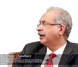 Chaudhry Dhruva合作论文数Postgraduate Institute of Medical Education and Research (PGIMER),, National College of Chest Physicians6
 Shivaprakash M Rudramurthy合作论文数Department of Medical Microbiology, Postgraduate Institute of Medical Education and Research6Maturu Venkata Nagarjuna合作论文数Department of Pulmonary Medicine, Postgraduate Institute of Medical Education and Research (PGIMER), Sector 12, Chandigarh, 160012, India.6
 Anand Mohan合作论文数Department of Internal Medicine, All India Institute of Medical Sciences6
 Mandeep Kumar Garg合作论文数Department of Radiodiagnosis and Imaging;graduate Institute of Medical Education and Research, Post;Department of Radiodiagnosis and Imaging|Postgraduate Institute of Medical Education and Research6Vallandaramam Pattabhiraman合作论文数5Surya Kant合作论文数5
 Jayanthi Savio合作论文数5
 Nandini Sethuraman合作论文数Department of Microbiology, Apollo Hospitals5
 Swarnakar Rajesh合作论文数Department of Respiratory Diseases, NKP Salve Institute of Medical Sciences5Goyal Rajiv合作论文数5
 Tanu Singhal合作论文数Department of Pediatrics, P.D. Hinduja National Hospital & Medical Research Center5
 Atul Patel合作论文数Infectious Diseases Clinic, “Vedanta” Institute of Medical, Sciences5Surinder K. Jindal合作论文数5Digambar Behera合作论文数4George Dsouza合作论文数4Piyush Gugale合作论文数4Pradeep Hosamani合作论文数4Anuradha Deshmukh合作论文数4Sunil Pawar合作论文数4Sanjiv Badhwar合作论文数4Gayathri Devi Rajagopal合作论文数4Hariprasad Kalpakkam合作论文数4Vishwanath Gella合作论文数4Manoj Radhakishan Shevkani合作论文数4Ashwini Tayade合作论文数4Srinivas Kishore Sistla合作论文数4Krishna Prabha Premachandran合作论文数4Pratik Savaj合作论文数4Vasant Nagvekar合作论文数4Raminder Pal Singh Sibia合作论文数4
 Bharat Gopal合作论文数4




