IKKα is a serine/threonine kinase that acts as a tumour suppressor in non-small cell lung cancer (NSCLC) in an NF-κΒ-independent manner, however, little is known about its downstream signalling pathways. To interrogate the IKKα signalling network in NSCLC, we investigated the impact of IKKα silencing on the miRNA expression profile of NSCLC cells. Nanostring miRNome analysis identified miR-9-5p, a known oncomir, as the top upregulated miRNA upon IKKα depletion. We show that overexpression of miR-9-5p in human lung cancer cells increases cell migration and invasion and promotes epithelial-to-mesenchymal cell transition (EMT) through loss of E-cadherin and activation of the Akt1/β-catenin pathway. In vivo tumour xenograft models show that overexpression of miR-9-5p promotes tumour growth and widespread transcriptomic reprogramming, altering the expression of genes related to EMT, apoptosis and NF-κB signalling. Overall, we show that miR-9-5p acts as an oncogenic effector upon IKKα loss, promoting tumour progression through EMT and Akt-1/GSK-3β/β-catenin pathway activation, leading to increased invasiveness and tumour growth.
AbstractNon-small cell lung cancer (NSCLC) constitutes one of the deadliest and most common malignancies. The LKB1/STK11 tumour suppressor is mutated in ∼ 30% of NSCLCs, typically lung adenocarcinomas (LUAD). We implemented zebrafish and human lung organoids as synergistic platforms to pre-clinically screen for metabolic compounds selectively targeting LKB1-deficient tumours. Interestingly, two kinase inhibitors, Piceatannol and Tyrphostin 23, appeared to exert synthetic lethality with LKB1 mutations. Although LKB1 loss alone accelerates energy expenditure, unexpectedly we find that it additionally alters regulation of the key energy homeostasis maintenance player leptin (LEP), further increasing the energetic burden and exposing a vulnerable point; acquired sensitivity to the identified compounds. We show that compound treatment stabilises Hypoxia-inducible factor 1-alpha (HIF1A) by antagonising Von Hippel-Lindau (VHL)-mediated HIF1A ubiquitination, driving LEP hyperactivation. Importantly, we demonstrate that sensitivity to piceatannol/tyrphostin 23 epistatically relies on a HIF1A-LEP-Uncoupling Protein 2 (UCP2) signaling axis lowering cellular energy beyond survival, in already challenged LKB1-deficient cells. Thus, we uncover a pivotal metabolic vulnerability of LKB1-deficient tumours, which may be therapeutically exploited using our identified compounds as mitochondrial uncouplers.
Severe respiratory infections such as COVID-19 are characterized by excessive inflammation leading to the development of pneumonia and acute respiratory distress syndrome. Bioactive lipid mediators (LMs) derived from ω6 and ω3 polyunsaturated fatty acids are central to the regulation of inflammation, controlling both its initiation and resolution. Still, their role in viral infections remains underexplored. By employing a holistic approach involving the analysis of white blood cell transcriptomes, targeted lipidomics, cytokine and immune cell profiling, we now show that LM patterns around hospital admission are profoundly altered in COVID-19, correlate with inflammatory responses, and stratify patients according to disease severity. Central to this are CYP450-derived LMs, such as 20-HETE, and lipoxygenase- or nonenzymatic-associated LMs such as 15-HETE, both exhibiting vasoactive function, along with lipid peroxidation metabolites such as 10-HDOHE. Among them, increased 20-HETE appears to be a promising prognostic biomarker for ICU admission and a potential therapeutic target for severe COVID-19 disease. Our study emphasizes the importance of LM patterns in COVID-19 pathophysiology and sheds light into the broader immune mechanisms beyond cytokines driving viral pneumonia in humans.
Background: Immunotherapy has significantly improved cancer treatment. However, it is not effective in all cancer patients, rendering the need to further delineate the differences among responders and non-responders at the molecular and cellular level. Unresponsiveness to immunotherapy has been attributed to dysfunctional immune cell states such as T-cell exhaustion and anergy, whereas the contribution of cellular senescence remains elusive. Herein, we have investigated the role of immune cell senescence in the response to checkpoint inhibitors in melanomas where these immunotherapies are applied as a first line treatment. Methods: Two senescence detecting complementary approaches were utilized in a case control study we conducted. First, we implemented a senescence molecular signature we developed, termed “SeneVick” retrospectively in a single cell RNA-seq dataset from melanoma patients who received immunotherapy. Prior to this analysis, the signature was extensively validated in a variety of cell/tissue contexts, senescence types and species. Second, cellular senescence was assessed via an established experimental algorithmic approach in circulating immune cells of an analogous melanoma clinical cohort. Results: Melanoma patients who did not respond to immunotherapy exhibited increased cellular senescence in their CD8+ T-cells, CD4+ T-cells, B-cells and NK cells compared to responders. This phenomenon was independent of patients’ age and not an outcome of immunotherapy, in contrast to conventional anti-cancer treatments. Interestingly, alterations of cell-cell interactions among the immune sub-populations in non-responders compared to responders were identified, suggesting the involvement of immune cell senescence in defective immune responses and treatment failure. Conclusion: Overall, our findings support cellular senescence of the immune cell compartment within the TME, as a potent determinant of the response to immunotherapy and pave the way for strategies targeting immune cell senescence, as promising approaches to improve the outcome of such interventions. ### Competing Interest Statement The authors have declared no competing interest. General Secretariat for Research and Technology, https://ror.org/04yeh8h63, 2020ΣЕ01300001, 2021ΝΑ11900006 Hellenic Foundation for Research and Innovation, https://ror.org/05v75r592, 3782, 2906, 20554 Nicholas and Sofula Kotopoulos Trust, donation
The emerging field of senolytics is centered on eliminating senescent cells to block their contribution to the progression of age-related diseases, including cancer, and to facilitate healthy aging. Enhancing the selectivity of senolytic treatments toward senescent cells stands to reduce the adverse effects associated with existing senolytic interventions. Taking advantage of lipofuscin accumulation in senescent cells, we describe here the development of a highly efficient senolytic platform consisting of a lipofuscin-binding domain scaffold, which can be conjugated with a senolytic drug via an ester bond. As a proof of concept, we present the generation of GL392, a senolytic compound that carries a dasatinib senolytic moiety. Encapsulation of the GL392 compound in a micelle nanocarrier (termed mGL392) allows for both in vitro and in vivo (in mice) selective elimination of senescent cells via targeted release of the senolytic agent with minimal systemic toxicity. Our findings suggest that this platform could be used to enhance targeting of senotherapeutics toward senescent cells. Exploiting the selective accumulation of lipofuscin in senescent cells, the authors developed a targeted senolytic platform. As proof of concept, they show that selective delivery of dasatinib elicits senolysis with minimal toxicity, in vitro, in organoids and in mice.
Post-Acute COVID-19 syndrome (PACS) is heterogeneous in phenotype and functional state. This prospective, observational study studied adults six months after acute COVID-19. We defined clinical phenotypes and profiled plasma mediators grouped into functional pathways (IL-1, IL-17, IFNγ/IFNγ-related cytokines, pro-/anti-inflammatory clusters). A subset underwent RNA-seq and ChIP-seq experiments. Three cohorts were analyzed (Exploratory n = 46; Discovery n = 591; Validation Cohort n = 289). PACS compatible symptoms were identified in 69.6 %; 59.2 % and 54.7 % respectively. Five phenotypes emerged. IL-1 cytokines (OR: 3.17, 95 % CIs: 1.94-5.19, p: 4.5 × 10⁻⁶), IL-17 cytokines (OR: 2.45, 95 % CIs: 1.47-4.07 p: 5.88 × 10⁻⁴) and the anti-inflammatory biomarkers (OR: 2.15, 95 % CIs: 1.34-3.45, p: 1.5 × 10⁻³) were upregulated in PACS patients. Respiratory phenotype was correlated with IL-1 upregulation (OR 4.23; 95 % CIs, 1.69-10.8, p = 0.0025). Transcriptomic and epigenomic changes were observed. Distinct phenotypes of PACS are driven by different immunological mechanisms at the DNA, transcriptomic, and protein levels.
Immunotherapy has significantly improved cancer treatment. However, it is not effective in all cancer patients, rendering the need to further delineate the differences among responders and non-responders at the molecular and cellular level. Unresponsiveness to immunotherapy has been attributed to dysfunctional immune cell states such as T-cell exhaustion and anergy, whereas the contribution of cellular senescence remains elusive. Herein, we have investigated the role of immune cell senescence in the response to checkpoint inhibitors in melanomas where these immunotherapies are applied as a first line treatment. Two senescence detecting complementary approaches were utilized in a case control study we conducted. First, we implemented a senescence molecular signature we developed, termed "SeneVick", retrospectively in a single cell RNA-seq dataset from melanoma patients who received immunotherapy. Prior to this analysis, the signature was extensively validated in a variety of cell/tissue contexts, senescence types and species. Second, cellular senescence was assessed via an established experimental algorithmic approach in circulating immune cells of an analogous melanoma clinical cohort. Melanoma patients who did not respond to immunotherapy exhibited increased cellular senescence in the CD8 + T-cell, CD4 + T-cell, B-cell (CD19 + /CD20 +) and NK cell compartments compared to responders. This phenomenon was independent of patients’ clinical features (age, sex, melanoma type, stage) and not an outcome of immunotherapy, in contrast to conventional anti-cancer treatments. Interestingly, alterations of cell-to-cell interactions among the immune sub-populations in non-responders compared to responders were identified, supporting, along with cytotoxicity assays, that senescent immune cells display immunosuppressive properties driving defective immune responses and treatment failure. Overall, our findings provide evidence that cellular senescence within the immune cell compartment of the tumor micro-environment is a potent determinant of the response to immunotherapy and pave the way for strategies targeting it as promising approaches to improve the outcome of such interventions.
Cellular reprogramming converts differentiated cells into a pluripotent state through extensive chromatin remodeling. The histone variant macroH2A has classically viewed as an epigenetic barrier stabilizing somatic identity and restricting pluripotency gene activation. Here, we show that during reprogramming, macroH2A1 nucleosomes undergo functional repurposing. Early in the process, mH2A1 nucleosomes act as a barrier to cellular plasticity, but later facilitate the establishment and maintenance of pluripotency by reshaping the epigenetic landscape. High-resolution chromatin profiling reveals that mH2A1.2 nucleosomes undergo rapid, large-scale repositioning, dissociating from promoters and reassembling ~30 bp away, frequently near NRF-1 binding sites. This repositioning occludes E2F4 binding, relieving cell-cycle arrest thereby enabling reprogramming. Likewise, mH2A1.1 nucleosomes display extensive mobility that culminates in deposition at pluripotency genes in ESCs, where they sustain their expression by assembling promoter transcriptional hubs. In this later role, mH2A1.1 functions as a chromatin bookmark stabilizing the Nanog, Sox2, and Oct4 network. These findings suggest that mH2A1 nucleosomal mobility underlines its context-dependent functional repurposing from reprogramming inhibitor to facilitator, illustrating how chromatin components evolve dynamic roles to coordinate cell-state transitions.
Identification and isolation of senescent cells is challenging, rendering their detailed analysis an unmet need. We describe a precise one-step protocol to fluorescently label senescent cells, for flow cytometry and fluorescence microscopy, implementing a fluorophore-conjugated Sudan Black -B analog, GLF16. Also, a micelle -based approach allows identification of senescent cells in vivo and in vitro, enabling live -cell sorting for downstream analyses and live in vivo tracking. Our protocols are applicable to cellular systems, tissues, or animal models where senescence is present. For complete details on the use and execution of this protocol, please refer to Magkouta et al.1
Objectives Age is the strongest risk factor of giant cell arteritis (GCA), implying a possible pathogenetic role of cellular senescence. To address this question, we applied an established senescence specific multimarker algorithm in temporal artery biopsies (TABs) of GCA patients. Methods 75(+) TABs from GCA patients, 22(−) TABs from polymyalgia rheumatica (PMR) patients and 10(−) TABs from non-GCA/non-PMR patients were retrospectively retrieved and analysed. Synovial tissue specimens from patients with inflammatory arthritis and aorta tissue were used as disease control samples. Senescent cells and their histological origin were identified with specific cellular markers; IL-6 and MMP-9 were investigated as components of the senescent associated secretory phenotype by triple costaining. GCA or PMR artery culture supernatants were applied to fibroblasts, HUVECs and monocytes with or without IL-6R blocking agent to explore the induction of IL-6-associated cellular senescence. Results Senescent cells were present in GCA arteries at higher proportion compared with PMR (9.50% vs 2.66%, respectively, p<0.0001) and were mainly originated from fibroblasts, macrophages and endothelial cells. IL-6 was expressed by senescent fibroblasts, and macrophages while MMP-9 by senescent fibroblasts only. IL-6(+) senescent cells were associated with the extension of vascular inflammation (transmural inflammation vs adventitia limited disease: 10.02% vs 4.37%, respectively, p<0.0001). GCA but not PMR artery culture supernatant could induce IL-6-associated senescence that was partially inhibited by IL-6R blockade. Conclusions Senescent cells with inflammatory phenotype are present in GCA arteries and are associated with the tissue inflammatory bulk, suggesting a potential implication in disease pathogenesis.
Severe COVID-19 is characterized by excessive inflammation leading to the development of pneumonia and acute respiratory distress syndrome. Bioactive lipid mediators (LMs) derived from ω6 and ω3 polyunsaturated fatty acids are central to the regulation of inflammation, controlling both its initiation and resolution. Still, their role in COVID-19 remains underexplored. By employing a holistic approach involving the analysis of white blood cell transcriptomes, targeted lipidomics, cytokine and immune cell profiling, across the spectrum of disease severity groups, we now show that LM networks are profoundly altered in COVID-19, correlate with inflammatory patterns, and stratify patients according to disease severity. Central to this are CYP450-derived LMs such as 20-HETE, lipid peroxidation metabolites such as iPF2a-VI, and lipoxygenase-derived LMs such as 12-HETE, all of which are major vasoactive mediators of inflammation. Among them, 20-HETE appears to be a promising prognostic biomarker for ICU admission and a potential therapeutic target for severe COVID-19 disease. Our study thus underscores the significance of LM networks in COVID-19 pathophysiology and sheds light into the broader mechanisms driving viral pneumonia in humans. ### Competing Interest Statement The authors have declared no competing interest.
Cellular reprogramming of somatic cells towards induced pluripotency is a multistep stochastic process mediated by the transcription factors Oct4, Sox2, Klf4 and c-Myc (OSKM), which orchestrate global epigenetic and transcriptional changes. We performed a large-scale analysis of integrated ChIP-seq, ATAC-seq and RNA-seq data and revealed the spatiotemporal highly dynamic pattern of OSKM DNA binding during reprogramming. We found that OSKM show distinct temporal patterns of binding to different classes of pluripotency-related enhancers. Genes involved in reprogramming are regulated by the coordinated activity of multiple enhancers, which are sequentially bound by OSKM for strict transcriptional control. Based on these findings, we developed an unbiased approach to identify Reprogramming-Inducible Enhancers (RIEs), constructed enhancer-traps and isolated cells undergoing reprogramming in real time. We used a representative RIE taken from the Upp1 gene fused to Gfp and isolated cells at different time-points during reprogramming and found that they have unique developmental capacities as they are reprogrammed with high efficiency due to their distinct molecular signatures. In conclusion, our experiments have led to the development of an unbiased method to identify and isolate reprogrammable cells in real time by exploiting the functional dynamics of OSKM, which can be used as efficient reprogramming biomarkers.
The currently available anti-cancer therapies, such as gamma-radiation and chemotherapeutic agents, induce cell death and cellular senescence not only in cancer cells but also in the adjacent normal tissue. New anti-tumor approaches focus on limiting the side effects on normal cells. In this frame, the potential anti-tumor properties of Pulsed Electromagnetic Fields (PEMFs) through the irradiation of breast cancer epithelial cells (MCF-7 and MDA-MB-231) and normal fibroblasts (FF95) were investigated. PEMFs had a frequency of 8 Hz, full-square wave type and magnetic flux density of 0.011 T and were applied twice daily for 5 days. The data collected showcase that PEMF application decreases the proliferation rate and viability of breast cancer cells while having the opposite effect on normal fibroblasts. Moreover, PEMF irradiation induces cell death and cellular senescence only in breast cancer cells without any effect in the non-cancerous cells. These findings suggest PEMF irradiation as a novel, non-invasive anti-cancer strategy that, when combined with senolytic drugs, may eliminate both cancer and the remaining senescent cells, while simultaneously avoiding the side effects of the current treatments.
CYLD is a tumor suppressor gene coding for a deubiquitinating enzyme that has a critical regulatory function in a variety of signaling pathways and biological processes involved in cancer development and progression, many of which are also key modulators of somatic cell reprogramming. Nevertheless, the potential role of CYLD in this process has not been studied. With the dual aim of investigating the involvement of CYLD in reprogramming and developing a better understanding of the intricate regulatory system governing this process, we reprogrammed control (CYLDWT/WT) and CYLD DUB-deficient (CYLDΔ9/Δ9) mouse embryonic fibroblasts (MEFs) into induced pluripotent stem cells (iPSCs) through ectopic overexpression of the Yamanaka factors (Oct3/4, Sox2, Klf4, c-myc). CYLD DUB deficiency led to significantly reduced reprogramming efficiency and slower early reprogramming kinetics. The introduction of WT CYLD to CYLDΔ9/Δ9 MEFs rescued the phenotype. Nevertheless, CYLD DUB-deficient cells were capable of establishing induced pluripotent colonies with full spontaneous differentiation potential of the three germ layers. Whole proteome analysis (Data are available via ProteomeXchange with identifier PXD044220) revealed that the mesenchymal-to-epithelial transition (MET) during the early reprogramming stages was disrupted in CYLDΔ9/Δ9 MEFs. Interestingly, differentially enriched pathways revealed that the primary processes affected by CYLD DUB deficiency were associated with the organization of the extracellular matrix and several metabolic pathways. Our findings not only establish for the first time CYLD's significance as a regulatory component of early reprogramming but also highlight its role as an extracellular matrix regulator, which has profound implications in cancer research.
The contemporary lifestyle of the last decade has undeniably caused a tremendous increase in oxidative-stress-inducing environmental sources. This phenomenon is not only connected with the rise of ROS levels in multiple tissues but is also associated with the induction of senescence in different cell types. Several signaling pathways that are associated with the reduction in ROS levels and the regulation of the cell cycle are being activated, so that the organism can battle deleterious effects. Within this context, autophagy plays a significant role. Through autophagy, cells can maintain their homeostasis, as if it were a self-degradation process, which removes the "wounded" molecules from the cells and uses their materials as a substrate for the creation of new useful cell particles. However, the role of autophagy in senescence has both a "dark" and a "bright" side. This review is an attempt to reveal the mechanistic aspects of this dual role. Nanomedicine can play a significant role, providing materials that are able to act by either preventing ROS generation or controllably inducing it, thus functioning as potential therapeutic agents regulating the activation or inhibition of autophagy.
Chromatin immunoprecipitation (ChIP) protocols have been used to reveal protein-DNA interactions of various cell types and tissues; however, optimization is required for each specific type of sample. Here, we present a ChIP protocol from murine inguinal white adipose tissue. We describe steps for tissue harvesting, crosslinking, chromatin extraction, shearing, immunoprecipitation, and puri-fication. We then detail procedures for analysis including library preparation, sequencing, and qRT-PCR validation.For complete details on the use and execution of this protocol, please refer to Antonia Katsouda et al. (2022).1
Background Prostate cancer is a major cause of cancer morbidity and mortality in men worldwide. Androgen deprivation therapy (ADT) has proven effective in early-stage androgen-sensitive disease, but prostate cancer gradually develops into an androgen-resistant metastatic state in the vast majority of patients. According to our oncogene-induced model for cancer development, senescence is a major tumor progression barrier. However, whether senescence is implicated in the progression of early-stage androgen-sensitive to highly aggressive castration-resistant prostate cancer (CRPC) remains poorly addressed. Methods Androgen-dependent (LNCaP) and –independent (C4-2B and PC-3) cells were treated or not with enzalutamide, an Androgen Receptor (AR) inhibitor. RNA sequencing and pathway analyses were carried out in LNCaP cells to identify potential senescence regulators upon treatment. Assessment of the invasive potential of cells and senescence status following enzalutamide treatment and/or RNAi-mediated silencing of selected targets was performed in all cell lines, complemented by bioinformatics analyses on a wide range of in vitro and in vivo datasets. Key observations were validated in LNCaP and C4-2B mouse xenografts. Senescence induction was assessed by state-of-the-art GL13 staining by immunocytochemistry and confocal microscopy. Results We demonstrate that enzalutamide treatment induces senescence in androgen-sensitive cells via reduction of the replication licensing factor CDC6. Mechanistically, we show that CDC6 downregulation is mediated through endogenous activation of the GATA2 transcription factor functioning as a CDC6 repressor. Intriguingly, GATA2 levels decrease in enzalutamide-resistant cells, leading to CDC6 stabilization accompanied by activation of Epithelial-To-Mesenchymal Transition (EMT) markers and absence of senescence. We show that CDC6 loss is sufficient to reverse oncogenic features and induce senescence regardless of treatment responsiveness, thereby identifying CDC6 as a critical determinant of prostate cancer progression. Conclusions We identify a key GATA2-CDC6 signaling axis which is reciprocally regulated in enzalutamide-sensitive and -resistant prostate cancer environments. Upon acquired resistance, GATA2 repression leads to CDC6 stabilization, with detrimental effects in disease progression through exacerbation of EMT and abrogation of senescence. However, bypassing the GATA2-CDC6 axis by direct inhibition of CDC6 reverses oncogenic features and establishes senescence, thereby offering a therapeutic window even after acquiring resistance to therapy.
Generation of induced pluripotent stem cells from specialized cell types provides an excellent model to study how cells maintain their stability, and how they can change identity, especially in the context of disease. Previous studies have shown that chromatin safeguards cell identity by acting as a barrier to reprogramming. We investigated mechanisms by which the histone macroH2A variants inhibit reprogramming and discovered that they work as gate keepers of the mesenchymal cell state by blocking epithelial transition, a step required for reprogramming of mouse fibroblasts. More specifically, we found that individual macroH2A variants regulate the expression of defined sets of genes, whose overall function is to stabilize the mesenchymal gene expression program, thus resisting reprogramming. We identified a novel gene network (MSCN, mesenchymal network) composed of 63 macroH2A-regulated genes related to extracellular matrix, cell membrane, signaling and the transcriptional regulators Id2 and Snai2, all of which function as guardians of the mesenchymal phenotype. ChIP-seq and KD experiments revealed a macroH2A variant-specific combinatorial targeting of the genes reconstructing the MSCN, thus generating robustness in gene expression programs to resist cellular reprogramming.
 Giannis Vatsellas合作论文数Ctr Basic Res, Acad Athens20
 Tom Maniatis合作论文数Department of Biochemistry and Molecular Biophysics, Columbia University19Vassilis G. Gorgoulis合作论文数18Alexander Polyzos合作论文数Joan and Sanford I. Weill Department of Medicine, Weill Cornell Medicine, Cornell University14Dimitrios Valakos合作论文数13
 Joseph Papamatheakis合作论文数13
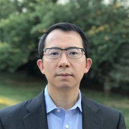 Junming Yie合作论文数Department of Biochemistry and Molecular, Columbia University11
 Polyzos Alexander合作论文数10
 Russell Petty合作论文数School of Medicine, University of Dundee;Division of Cancer Research, University of Dundee10M Merika合作论文数9
 Stavros Lomvardas合作论文数Vagelos College of Physicians & Surgeons, Columbia University Irving Medical Center9
 Konstantinos Evangelou合作论文数Medical School, University of Athens9Guoying Chen合作论文数9
 Nefeli Lagopati合作论文数Medical School, National and Kapodistrian University of Athens8
 Santa J. Ono (小野三太)合作论文数Department of Molecular, Cellular, and Developmental Biology, College of Literature, Science and the Arts, University of Michigan;Department of Ophthalmology and Visual Sciences, Faculty of Medicine, The University of British Columbia8
 Stravopodis Dimitrios J合作论文数Dept Cell Biol & Biophys, Univ Athens8
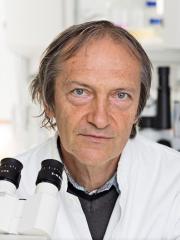 Jiri Bartek合作论文数Department of Medical Biochemistry and Biophysics, Karolinska Institutet8Dimitris Veroutis合作论文数7Marios Agelopoulos合作论文数7
 Aikaterini Polyzou合作论文数Department of Cellular and Molecular Immunology, Max Planck Institute of Immunobiology and Epigenetics, Freiburg, Germany7Munshi Nikhil V合作论文数Dept Internal Med, Div Cardiol, UT Southwestern Med Ctr7
 Antonis Kokkalis合作论文数7Foutadakis Spyros合作论文数Department of Biochemistry and Molecular Biology, University of Athens7
 Tucker Collins合作论文数The Childrens Hospital7
 Athanassios Kotsinas合作论文数Medical School, University of Athens7Theodora Agalioti合作论文数7
 Paul A. Townsend合作论文数Institute of Child Health, University College London7
 Angelos Papaspyropoulos合作论文数Medical School, National and Kapodistrian University of Athens6Ford Ethan合作论文数Australian Research Council Centre of Excellence in Plant Energy Biology, The University of Western Australia6Dimitris Kletsas合作论文数Institute of Biology, National Center for Scientific Research `Demokritos'6
 Konstantinos Vougas合作论文数Centre of Basic Research II, Foundation for Biomedical Research of the Academy of Athens6Sophia Havaki合作论文数Medical School, University of Athens6
 Sophia Magkouta合作论文数6angela maueroberthuer合作论文数5
 Effie Apostolou合作论文数Phyiology, Biophysics & Systems Biology, Weill Cornell Graduate School of Medical Sciences, Cornell University5
 Konstantinos Kambas合作论文数Lab Mol Hematol, Democritus Univ Thrace5
 Ema Anastasiadou合作论文数Brigham and Women's Hospital, Harvard Medical School5
 Sarantis Gagos合作论文数Academy of Athens, Academy of Athens5Pavlos Pantelis合作论文数4Eleftheria Klagkou合作论文数4sissy kolyva合作论文数4jutta steinkoetter合作论文数4MA READ合作论文数4Z Song合作论文数4James V Falvo合作论文数4Chrysa Nikopoulou合作论文数Max Planck Research Group ‘Chromatin and Ageing’, Max Planck Institute for Biology of Ageing4
 Periklis Makrythanasis合作论文数Department of Genetic Medicine and Development, University of Geneva;National and Kapodistrian University of Athens4
 Pippa Natassa合作论文数Faculty of Pharmacy, University of Athens4
 Andreas Papapetropoulos合作论文数Department of Pharmaceutical Chemistry, Faculty of Pharmacy, School of Health Sciences, National and Kapodistrian University of Athens4




