Pairs of cultured T and B lymphoid cells, derived from the same individual, were used as stimulating cells in MLC experiments. Blastogenic response to cultured B cells were much greater than those to the corresponding T cell line. These cells will provide a useful reagent for isolation and characterization of B cell specific antigens.
An HLA allele-specific cytotoxic T lymphocyte response is thought to influence the rate of disease progression in HIV-1-infected individuals. In a prior study of 139 HIV-1-infected homosexual men, we identified HLA class I alleles and observed an association of specific alleles with different relative hazards for progression to AIDS. Seeking an explanation for this association, we searched HIV-1 protein sequences to determine the number of peptides matching motifs defined by combinations of specific amino acids reported to bind 16 class I alleles. Analyzing complete sequences of 12 clade B HIV isolates, we determined the number of allele motifs that were conserved (occurring in all 12 isolates) and nonconserved (occurring in only one isolate), as well as the average number of allele motifs per isolate. We found significant correlations with an allele's association with disease progression for counts of conserved motifs in gag (R = 0.73; P = 0.002), pol (R = 0.58, P = 0.024), gp120 (R = 0.78, P = 0.00056), and total viral protein sequences (R = 0.67, P = 0.0058) and also for counts of nonconserved motifs in gag (R = 0.62, P = 0.013), pol (R = 0.74, P = 0.0017), gp41 (R = 0.52, P = 0.046), and total viral protein (R = 0.71, P = 0.0033). We also found significant correlations for the average number of motifs per isolate for gag, pol, gp120, and total viral protein. This study provides a plausible functional explanation for the observed association of different HLA alleles with variable rates of disease progression.
Major histocompatibility complex (MHC) genes (HLA in humans) regulate the immune response to foreign antigens. Molecular and serologic techniques were used to identify products of HLA class I, class II and transporter (TAP) genes (also part of the MHC) in homosexual seroconverters to human immunodeficiency virus type 1 (HIV-1). Comprehensive statistical analysis produced an HLA profile that predicted time from HIV-1 infection to the onset of AIDS. The profile was developed in a cohort of 139 men and evaluated in a second unrelated cohort of 102 men. In the evaluation cohort, the profile discriminated a sixfold difference between groups with the shortest and longest times to AIDS (P = 0.001). These findings support current theory about control of antigen processing by HLA genes and have implications for immunopathogenesis of HIV-1 and other infections.
The expression of the HLA class I molecules on the cell surface was investigated in hepatocellular carcinoma (HCC) cell lines using complement-mediated cytotoxicity (CMC) and flow cytometric analysis. Although HLA-A antigens were detected by CMC in all cell lines tested, HLA-B and -C antigens were not detectable in six of seven HCC cell lines. These results were also confirmed by flow cytometric analysis focusing on HLA-Bw4 and Bw6 public antigens. Furthermore, complementary DNA (cDNA) from each cell line was tested for the expression of HLA-A, -B, -C and the transporter associated with antigen processing genes (TAP1 and TAP2). Two cell lines showed a reduced level of one or both of the TAP messenger RNAs (mRNAs), and one of these showed a reduction of HLA-B and -C gene expression as well, but the others had detectable mRNA levels. These results demonstrate that hepatocellular carcinoma cell lines tested in the current study lose or decrease the expression of HLA-B and -C alleles on the cell surface, even though mRNA encoding these alleles is present, suggesting that the loss of the HLA molecules might be caused by posttranscriptional events or failure to transport and load peptides necessary for HLA expression. The selective loss of HLA-B and -C, but not -A, molecules (which also excludes a beta 2-microglobulin defect) is intriguing, and may be attributable to the ability of some of the HLA-A molecules to load signal peptides not requiring TAP transport, or to natural selection of HLA-B or -C locus-specific immune surveillance.
Background: Acquired immunodeficiency syndrome (AIDS) is associated with the occurrence of tumors such as Kaposi's sarcoma (KS) and B-cell lymphoma. However, no evidence exists yet that human immunodeficiency virus type 1, the causative agent of AIDS, is directly responsible for cell transformation. It is also not clear whether KS lesions, which are of complex cellularity, contain tumor cells derived from a true monoclonal malignancy (originating from a single malignant cell) or whether the lesions are just polyclonally hyperplastic in nature (containing increased numbers of normal cells). In fact, the presence of malignant KS cells has never been unequivocally shown in AIDS-associated KS, and previously isolated KS cell cultures were not immortal or malignant. Purpose: Our purpose was to (a) utilize technology that could facilitate isolation and enrichment of tumor cells from AIDS-associated KS lesions, (b) establish and characterize an immortalized KS cell line, and (c) test the malignant potential of such a cell line in animal models. Methods: Mononuclear cells were isolated from 2.5 L of pleural effusion from an AIDS-associated KS patient. T-lymphocytes, B-lymphocytes, monocytes/macrophages, and fibroblasts were removed by a cytotoxicity method, using monoclonal antibodies specific for cell surface markers and baby rabbit complement. KS cells were cultured in the absence of exogenous growth factors in an effort to select for transformed cells capable of self-sustained growth. The karyotype abnormalities were detected by G-banded marker studies, and phenotypic markers were determined by indirect immunofluorescence and immunocytochemical methods. Beige nude XID and severe combined immunodeficient mice mere used to evaluate the tumorigenic, angiogenic, and metastatic potentials of cells. Results: An immortalized cell line, named KS Y-1, was isolated. Its phenotype is similar to that of endothelial cells with positive CS34 and CD31 markers. Tetraploid chromosomal abnormalities were found in primary fresh KS tissue and in vitro passages of KS Y-1 cells. These cells promoted tumorigenesis, angiogenesis, and metastasis in immunodeficient mice. Tumors produced at the site of injection, as well as metastases in the lung, spleen, pancreas, gastrointestinal tract, and skin showed a human tetraploid karyotype. KS Y-1 cells show high plating efficiency. Conclusion: The KS Y-1 cell line could be the first evidence of AIDS-associated KS cells that may develop clones with an indisputable malignant cell phenotype. Implications: KS Y-1 cells in the in vivo mouse model can be used to study the effects of therapeutic compounds in advanced KS.
Macaques immunized with uninfected human cells have been shown to be protected from challenge with simian immunodeficiency virus (SIV) propagated in human cells. To identify the potential antigens involved in this protection, macaques were immunized with uninfected human cells, sucrose density gradient-purified culture fluid from uninfected human cells (mock virus), beta-2 microglobulin (beta 2M), immunoaffinity-purified HLA class I and class II proteins from these human cells, and adjuvant. Although all macaques immunized with beta 2M and HLA class I developed high antibody titers to beta 2M, these animals were not protected from a subsequent challenge with infectious SIV grown in human cells. In contrast, the macaques immunized with class II protein (HLA-DR) and mock virus developed antibodies to class II protein and were protected from the intravenous infectious virus challenge. The class II protein- and mock virus-immunized animals which were protected from challenge were given boosters of the appropriate antigen and challenged with the same SIV propagated in macaque cells. All animals became infected, indicating that the protection seen with human class II protein did not extend to protection from infection with SIV containing macaque class II proteins. Since the virus released from SIV-infected macaque cells would contain macaque class II proteins, our results suggest that the initial SIV infected was completely prevented. In addition, the lack of protection from challenge with SIV propagated in macaque cells provided strong evidence that the protection was due to an immune response to the cellular proteins and not to epitopes cross-reactive between class II proteins and the viral proteins, since the identical virus proteins were present in both challenge stocks. These results are the first demonstration that immunization with a purified cellular protein can protect from virus infection.
Objective: To assess the association between human leukocyte antigen (HLA) haplotypes and the incidence rates of CD4 decline to < 20% and to AIDS. Design: Retrospective cohort study of 95 HIV-1-infected hemophilic sibling pairs. Methods: HLA haplotype-sharing between siblings was assigned on the basis of serologic typing of HLA class I alleles and molecular typing of HLA class II alleles. Concordance of time to CD4 decline to < 20% and to AIDS within and between sibling pairs was assessed by analysis of variance models and calculations of intraclass correlation coefficients. The age-adjusted relative risks of these two endpoints for unique class II haplotypes were determined from proportional hazards models. Results: Sibling pairs sharing one or two haplotypes were significantly concordant in CD4 decline and AIDS status within 5 years of seroconversion. No concordance was found in pairs sharing zero haplotypes. At 6-10 years after seroconversion, significant concordance of these two endpoints was also observed in the pairs sharing one haplotype. The concordant results were not explained by the use of zidovudine within the pairs. Among the individuals in this cohort, the relative hazards for CD4 decline to < 20% and for AIDS were significantly elevated for one class II haplotype (DQB1*0501, DQA1*0101, DRB1*0101). In addition, the risk for AIDS was significantly increased for two other class II haplotypes (DQB1*0603, DQA1*0103, DRB1*1300, DRB3*0202 and DQB1*0301, DQA1*0501, DRB1*1400, DRB3*0202) and significantly decreased for one haplotype (DQB1*0302, DQA1*0301, DRB1*0401, DRB4*0101). Conclusions: These data demonstrate that HIV-1 disease progression is associated with the genes in the major histocompatibility complex that regulate the host's immune response.
Cultured peripheral blood lymphocytes (PBL) from HTLV-I-infected individuals proliferate in the absence of added mitogens and/or cytokines. In an attempt to answer questions regarding the activating signals for cells and virus, antibodies that react with cell surface components that are known to regulate cell activation and antibodies reacting with viral proteins were added to cultures of PBL from HTLV-I-infected, disease-free individuals. Spontaneous proliferation and virus production increased in the presence of antibodies reacting with CD3 andα/β T cell receptors (TCR) while antibodies to HLA class II and viral proteins had no effect. Addition of HLA class I antibodies shut down virus production and cell proliferation. These observations indicate that both virus and cell activation may occur through the α/β TCR on the infected cell. Cyclosporin A, however, markedly decreased cell proliferation but had only a modest suppressive effect on virus production. Thus, the uncoupling of cell proliferation from virus production by cyclosporin A suggests the possibility that the signal transduction pathways for these two events are different.
A flow cytometry-based assay for detection of immunoglobulin (Ig) class and subclass antibodies in human serum or plasma was developed. With use of this procedure, the presence and relative frequency of antibody activity in the Ig classes and subclasses (IgA1, IgA2, IgD, IgE, IgG1, IgG2, IgG3, IgG4, and IgM) to human immunodeficiency virus type 1 (HIV-1) proteins (gp160, gp120, p66, and p24) was determined in serum or plasma from a cohort of 47 HIV-1-infected, pregnant women. Antibody activity in each of the classes and subclasses was found with differences in frequency depending on the Ig class/subclass and the HIV-1 protein. IgG1 antibodies were the most frequently reactive Ig class/subclass to each protein. Intermediate frequencies of reactivity were found in IgA1, IgG2, IgG3, and IgM class and subclasses and antibodies of the IgA2, IgE, and IgG4 class/subclass the least frequently detected. An unexpected finding was the presence of IgD antibodies to HIV-1 proteins in approximately 50% of the individuals. The distributions of Ig class/subclass antibodies to the different HIV-1 proteins were compared in sera from 14 mothers giving birth to infants who were determined to be HIV-1 infected with sera from 25 individuals whose infants were not infected. Sera from transmitting mothers contained a broader distribution of class and subclass antibodies compared to sera from nontransmitting women. The single most frequent antibody-antigen combination that was found in the transmitting mother was IgG2-gp160.(ABSTRACT TRUNCATED AT 250 WORDS)
Journal Article The Role of the Major Histocompatibility Complex in Human Immunodeficiency Virus Infection—Ever More Complex? Get access Richard A. Kaslow, Richard A. Kaslow Reprints or correspondence: Dr. Richard A. Kaslow, Epidemiology and Biometry Branch, DMID, National Institute of Allergy and Infectious Diseases, National Institutes of Health, Solar Bldg. 3A24, Bethesda, MD 20892. Search for other works by this author on: Oxford Academic PubMed Google Scholar Dean L. Mann Dean L. Mann Search for other works by this author on: Oxford Academic PubMed Google Scholar The Journal of Infectious Diseases, Volume 169, Issue 6, June 1994, Pages 1332–1333, https://doi.org/10.1093/infdis/169.6.1332 Published: 01 June 1994 Article history Received: 28 January 1994 Published: 01 June 1994
HLA allele frequencies were examined for possible association(s) with the rate of disease progression and with the disease outcome (AIDS diagnosis) in a population of HIV-1-infected individuals. Certain alleles were associated with the relative rate of CD4+ T-cell decline. Association of particular alleles with several disease outcomes associated with infection was also observed. It is important to keep these two aspects (disease progression, AIDS diagnosis) separate when studying HLA in the HIV-1-infected population. Alleles that may play a role in the rate of virus speed by effecting the immune response may be different from those found to be associated with a particular disease. We feel that the only truly informative data, in this regard, can be generated from a relative precise determination of the time of infection (to study disease progression) and adequate numbers of individuals with specific diseases to study specific disease association. If such data can be generated we will have a much better understanding of the pathogenetic process(es) of HIV-1 infection.
The influence of cigarette smoking on T cell subsets has been studied in white subjects, but comparable data are not available for blacks. We analyzed peripheral blood mononuclear cell subsets in a population-based, stratified, random sample of healthy black adults using monoclonal antibodies and flow cytometry. The study population consisted of 94 men and 79 women, including 73 smokers (CS) and 100 nonsmokers (NS). Cigarette smoking was associated with a significant elevation in leukocyte (WBC) count (CS 7,270 +/- 230 cells/mm3 versus NS 6,260 +/- 160 cells/mm3; p = 0.001), although WBC counts for both groups were substantially lower than those reported for white smokers and nonsmokers. Smokers had a significantly lower proportion of CD4+ cells than nonsmokers (CS 55.4 +/- 0.9% versus NS 58.7 +/- 0.9; p = 0.01), adjusting for age and gender. No significant smoking-related changes were observed for CD8+ cells, the CD4/CD8 ratio, or total T cells (CD3+), monocytes (CD14+), or natural killer cells (CD16+). Among black smokers, a significant dose-related decrease in CD4+ cells was observed as the number of cigarettes smoked per day increased. Among black exsmokers, the level of WBC and CD4+ cells returned to the level observed in never smokers within 2 to 5 yr after smoking cessation. These results contrast sharply with the previously reported increase in CD4+ cells and decrease in natural killer cells associated with cigarette smoking in whites. The data suggest that the immunologic effects of cigarette smoking may be significantly modified by ethnic characteristics.
The effect of cryopreservation and longterm liquid nitrogen storage on peripheral blood mononuclear cell (PBMC) subsets was prospectively analyzed using monoclonal antibodies and flow cytometry. Brief cryopreservation did not significantly alter the proportion of positively stained cells for CD3+, CD4+, CD8+, CD14+, CD16+, and CD19+, cells. A small but statistically significant increase in the proportion of positive cells was observed for HLA‐DR+ and HLe‐1 + cells. Brief cryopreservation was associated with a decrease in the mean fluorescence intensity (MFl) values for CD3+, CD4+, and CD8+ cells; an increase in MFl values for CD14+ and HLA‐DR+ cells; and no change for CD16+, CD19+, and HLe‐1+ cells. There was no significant change in the proportion of CD3+, CD4+, or CD16+ cells during 20 months of storage in liquid nitrogen. Small but statistically significant decreases in the proportion of CD8+ and CD19+ cells were observed over the same interval, and the proportion of CD14+ cells (monocytes) was highly variable. Chronologic changes in fluorescence intensity during longterm storage were observed for all cell subsets except CD16+ and CD19+ cells. Cryopreservation is a valuable technique for longterm storage of viable cells. For many laboratory applications, the small changes noted in the present study will have no practical importance. However, for clinical and epidemiological investigations encompassing large numbers of samples, statistical techniques to adjust for small changes during storage should be considered.
 William Blattner合作论文数University of Maryland School of Medicine37
 JJ Goedert合作论文数17
 Jack Strominger合作论文数Department of Stem Cell and Regenerative Biology, Faculty of Arts and Sciences, Harvard University17
 Curtis C. Harris合作论文数Laboratory of Human Carcinogenesis, Center for Cancer Research, National Cancer Institute, National Institutes of Health16Mikulas Popovic合作论文数16
 Ainsley Weston合作论文数13R C Gallo合作论文数11CY NEULAND合作论文数9D. M. STRONG合作论文数9C Murray合作论文数9
 David J Tollerud合作论文数9MS REITZ合作论文数8M Carrington合作论文数Laboratory of Genomic Diversity, NCI-Frederick, Frederick, MD, USA8L M Brown合作论文数8
 Robert N. Hoover合作论文数Division of Cancer Epidemiology & Genetics, National Cancer Institute, National Institutes of Health8DK MANCHESTER合作论文数7J W Clark合作论文数7
 Georg Stingl合作论文数Department of Dermatology,Medical University of Vienna7As Fauci合作论文数6Ge Trivers合作论文数6K Wolff合作论文数6Haralampos M Moutsopoulos合作论文数6
 Peter Parham合作论文数Department of Structural Biology, School of Medicine, Stanford University;Department of Microbiology & Immunology, School of Medicine, Stanford University6
 Barton Ford Haynes合作论文数Department of Medicine, Duke Human Vaccine Institute6R.J Biggar合作论文数6Erwin Tschachler合作论文数6Ld Abelson合作论文数5Tm Chused合作论文数5m odonnell合作论文数5AH JOHNSON合作论文数5
 Dimitrios T. Boumpas合作论文数Fourth Department of Medicine, “Attikon” University Hospital5
 Timothy Alan Springer合作论文数Institute for Protein Innovation;Springer Lab, Harvard Medical School;Division of Hematology/Oncology, Department of Medicine, Boston Children’s Hospital5Israel Yust合作论文数5Klemens Rappersberger合作论文数5RJ GROSSMAN合作论文数4V Groh合作论文数4LO ARTHUR合作论文数4DM STRONG合作论文数4MJ NEWMAN合作论文数4Js Choi合作论文数4GS EISENBARTH合作论文数4
 Warren Strober合作论文数National Institute of Allergy and Infectious Diseases4Abelson L合作论文数4
 Joseph F. Fraumeni合作论文数Division of Cancer Epidemiology & Genetics, National Cancer Institute4Suzanne Gartner合作论文数4
 Cox Terhorst合作论文数Beth Israel Deaconess Medical Center4
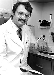 Samuel Broder合作论文数Celera Genomics4Richard A. Kaslow合作论文数4Nancy R. Mendell合作论文数Department of Applied Mathematics and Statistics, State University of New York at Stony Brook4




