BACKGROUND/AIM:There is concern that people who had COVID-19 will develop pulmonary fibrosis. Using mouse models, we compared pulmonary inflammation following injection of the spike protein of SARS-CoV-2 (COVID-19) to radiation-induced inflammation to demonstrate similarities between the two models. SARS-CoV-2 (COVID-19) induces inflammatory cytokines and stress responses, which are also common to ionizing irradiation-induced acute pulmonary damage. Cellular senescence, which is a late effect following exposure to SARS-CoV-2 as well as radiation, was investigated. MATERIALS AND METHODS:We evaluated the effect of SARS-CoV-2 spike protein compared to ionizing irradiation in K18-hACE2 mouse lung, human lung cell lines, and in freshly explanted human lung. We measured reactive oxygen species, DNA double-strand breaks, stimulation of transforming growth factor-beta pathways, and cellular senescence following exposure to SARS-CoV-2 spike protein, irradiation or SARS-COV-2 and irradiation. We also measured the effects of the antioxidant radiation mitigator MMS350 following irradiation or exposure to SARS-CoV-2. RESULTS:SARS-CoV-2 spike protein induced reactive oxygen species, DNA double-strand breaks, transforming growth factor-β signaling pathways, and senescence, which were exacerbated by prior or subsequent ionizing irradiation. The water-soluble radiation countermeasure, MMS350, reduced spike protein-induced changes. CONCLUSION:In both the SARS-Co-2 and the irradiation mouse models, similar responses were seen indicating that irradiation or exposure to SARS-CoV-2 virus may lead to similar lung diseases such as pulmonary fibrosis. Combination of irradiation and SARS-CoV-2 may result in a more severe case of pulmonary fibrosis. Cellular senescence may explain some of the late effects of exposure to SARS-CoV-2 spike protein and to ionizing irradiation.
BACKGROUND/AIM:Radiation oncologists are reluctant to treat cancer in Fanconi Anemia (FA) patients due to their lack of homologous recombination repair of DNA strand breaks in normal tissues. To determine the therapeutic effects of irradiation and combination chemotherapy on cancer in syngeneic, radiosensitive FA mice, we derived transplantable cancers of the same genotype in three FA mouse strains. MATERIALS AND METHODS:Fancd2-/- mice on a C57BL/6 or Sv/129 background and Fancg-/- mice (C57BL/6 background) that received 3-methylcholanthrene (3-MCA), were monitored for the development of subcutaneous tumors. RESULTS:Tumors were induced at the site of 3-MCA injection, and tumor cell lines were established and found to be transplantable. Explanted tumors were identified as pleomorphic/rhabdomyosarcomas using immunohistochemical biomarkers. CONCLUSION:These transplantable FA mouse tumor cell lines should be valuable for testing effects of new radiation therapy protocols including FLASH high dose rate radiation delivery, immunotherapies, and combined radiation and chemotherapy treatments for radiosensitive FA patients.
Purpose/Objective(s) Establish a novel therapeutic strategy for the management of ovarian cancer with LR-IL-22 gavage, an intestinal radioprotector, through its pivotal role in modifying the tumor microenvironment and subsequently improving the survival of female C57BL/6MUC-1 mice with widespread abdominal syngeneic 2F8cis ovarian cancer. Materials/Methods To assess differential gene expression following irradiation and LR-IL-22 gavage, we performed RNAseq analyses using Lgr5+GFP+ mice. Male and female mice were divided into four groups: control (0 Gy), 12 Gy total body irradiation (TBI), 12 Gy TBI + LR, and 12 Gy TBI + LR-IL-22. Twenty-four hours after TBI, mice received LR or LR-IL-22 gavage. In a separate experiment with female C57BL/6MUC-1 mice with widespread abdominal syngeneic 2F8cis ovarian cancer, flow analysis and immunohistochemistry were used to assess the tumor microenvironment following fractionated whole abdomen irradiation (WAI) using 6 Gy × 4 fractions with or without LR-IL-22 gavage. Results We observed that intestinal stem cell gene expression varied widely following exposure to TBI. Genes were either upregulated or downregulated following irradiation, and many were solely rescued by the genetically engineered probiotic LR-IL-22. In fact, 135 genes were uniquely rescued and thus downregulated by LR-IL-22, compared to 43 genes uniquely rescued and thus upregulated by LR-IL-22 following TBI. Moreover, the addition of fractionated WAI to LR-IL-22 not only induced PD-L1 protein expression in ovarian cancer cells and mobilized CD8+ T cells, but potentially reduced tumor growth rate through the migration of LR bacteria into the 2F8cis ovarian tumors. Conclusion Our data illustrates the effectiveness of LR-IL-22 as a radiation mitigator that manifests its role by modifying both, gene expression and the tumor microenvironment, following irradiation. Indeed, irradiation induced differential gene expression in intestinal stem cells, and significantly upregulated CD8+ T cells infiltrate in 2F8cis tumors, giving them an immunological ‘‘hot’’ phenotype which positively correlated with PD-L1 expression. This forms the basis for combining WAI with targeted therapies, including immune checkpoint inhibitors, in advanced or recurrent epithelial ovarian cancer.
Despite recent advances in cancer therapy, ovarian cancer remains the most lethal gynecological cancer worldwide, making it crucial and of the utmost importance to establish novel therapeutic strategies. Adjuvant radiotherapy has been assessed historically, but its use was limited by intestinal toxicity. We recently established the role of Limosilactobacillus reuteri in releasing IL-22 (LR-IL-22) as an effective radiation mitigator, and we have now assessed its effect in an ovarian cancer mouse model. We hypothesized that an LR-IL-22 gavage would enable intestinal radioprotection by modifying the tumor microenvironment and, subsequently, improving overall survival in female C57BL/6MUC-1 mice with widespread abdominal syngeneic 2F8cis ovarian cancer. Herein, we report that the LR-IL-22 gavage not only improved overall survival in mice when combined with a PD-L1 inhibitor by inducing differential gene expression in irradiated stem cells but also induced PD-L1 protein expression in ovarian cancer cells and mobilized CD8+ T cells in whole abdomen irradiated mice. The addition of LR-IL-22 to a combined treatment modality with fractionated whole abdomen radiation (WAI) and systemic chemotherapy and immunotherapy regimens can facilitate a safe and effective protocol to reduce tumor burden, increase survival, and improve the quality of life of a locally advanced ovarian cancer patient.
Background/Aim: Patients with radiation sensitive Fanconi anemia (FA) are presenting with cancers of the oral cavity, oropharynx, and other anatomic locations. Materials and Methods: Animal models for cancer in FA mice used orthotopic tumors from wild type mice. We derived a cancer cell line from Fanca-/- mice by topical application of the chemical carcinogen dimethyl benzanthracene (DMBA). Results: A Fanca-/- mouse rhabdomyosarcoma was derived from a Fanca-/- (129/Sv) mouse. The in vitro clonogenic survival of the Fanca-/- clone 6 cancer cell line was consistent with the FA genotype. Transplanted tumors demonstrated hypoxic centers surrounded by senescent cells. Conclusion: This Fanca-/- mouse syngeneic cancer should provide a valuable resource for discovery and development of new normal tissue radioprotectors for patients with FA and cancer.
Cellular senescence is involved in the development of pulmonary fibrosis as well as in lung tissue repair and regeneration. Therefore, a strategy of removal of senescent cells by senolytic drugs may not produce the desired therapeutic result. Previously we reported that tyrosine kinase Fgr is upregulated in ionizing irradiation-induced senescent cells. Inhibition of Fgr reduces the production of profibrotic proteins by radiation-induced senescent cells in vitro; however, a mechanistic relationship between senescent cells and radiation-induced pulmonary fibrosis (RIPF) has not been established. We now report that senescent cells from the lungs of mice with RIPF, release profibrotic proteins for target cells and secrete chemotactic proteins for marrow cells. The Fgr inhibitor TL02-59, reduces this release of profibrotic chemokines from the lungs of RIPF mice, without reducing numbers of senescent cells. In vitro studies demonstrated that TL02-59 abrogates the upregulation of profibrotic genes in target cells in transwell cultures. Also, protein arrays using lung fibroblasts demonstrated that TL02-59 inhibits the production of chemokines involved in the migration of macrophages to the lung. In thoracic-irradiated mice, TL02-59 prevents RIPF, significantly reduces levels of expression of fibrotic gene products, and significantly reduces the recruitment of CD11b+ macrophages to the lungs. Bronchoalveolar lavage (BAL) cells from RIPF mice show increased Fgr and other senescent cell markers including p16. In human idiopathic pulmonary fibrosis (IPF) and in RIPF, Fgr, and other senescent cell biomarkers are increased. In both mouse and human RIPF, there is an accumulation of Fgr-positive proinflammatory CD11b+ macrophages in the lungs. Thus, elevated levels of Fgr in lung senescent cells upregulate profibrotic gene products, and chemokines that might be responsible for macrophage infiltration into lungs. The detection of Fgr in senescent cells that are obtained from BAL during the development of RIPF may help predict the onset and facilitate the delivery of medical countermeasures.
LR-IFN-β is both a feasible and effective radiation mitigator that could potentially improve the management of ovarian cancer patients. Furthermore, the subsequent addition of platinum/taxane-based chemotherapy to the combination of WAI and LR-IFN-β should reduce tumor volume while protecting the intestine and thus improve overall survival in ovarian cancer patients.
BACKGROUND/AIM:The earliest cellular and molecular biologic changes in the esophagus that lead to esophageal cancer were evaluated in a mouse model. We correlated numbers of senescent cells with the levels of expression of potentially carcinogenic genes in sorted side population (SP) cells containing esophageal stem cells and non-stem cells in the non-side population cells in the 4-nitroquinolone oxide (NQO)-treated esophagus. MATERIALS AND METHODS:We compared stem cells with non-stem cells from the esophagus of mice treated with the chemical carcinogen 4-NQO (100 μg/ml) in drinking water. We also compared gene expression in human esophagus samples treated with 4-NQO (100 μg/ml media) to non-treated samples. We separated and quantitated the relative levels of expression of RNA using RNAseq analysis. We identified senescent cells by luciferase imaging of p16+/LUC mice and senescent cells in excised esophagus from tdTOMp16+ mice. RESULTS:A significant increase in the levels of RNA for oncostatin-M was found in senescent cells of the esophagus from 4-NQO-treated mice and human esophagus in vitro. CONCLUSION:Induction of OSM in chemically-induced esophageal cancer in mice correlates with the appearance of senescent cells.
Irradiation can be an effective treatment for ovarian cancer, but its use is limited by intestinal toxicity. Thus, strategies to mitigate toxicity are important and can revitalize the current standard of care. We previously established that LR-IL-22 protects the intestine from WAI. We now hypothesize that LR-IFN-β is an effective radiation protector and mitigator and is rapidly cleared from the digestive tract, making it an option for intestinal radioprotection. We report that the gavage of LR-IFN-β during WAI provides improved intestinal barrier integrity and significantly preserves the numbers of Lgr5+GFP+ intestinal stem cells, improving survival. The rapid clearance of the genetically engineered probiotic from the digestive tract renders it a safe and feasible radiation mitigator. Therefore, the above genetically engineered probiotic is both a feasible and effective radiation mitigator that could potentially revolutionize the management of OC patients. Furthermore, the subsequent addition of platinum/taxane-based chemotherapy to the combination of WAI and LR-IFN-β should reduce tumor volume while protecting the intestine and should improve the overall survival in OC patients.
(1) Background: The systemic administration of therapeutic agents to the intestine including cytokines, such as Interleukin-22 (IL-22), is compromised by damage to the microvasculature 24 hrs after total body irradiation (TBI). At that time, there is significant death of intestinal microvascular endothelial cells and destruction of the lamina propria, which limits drug delivery through the circulation, thus reducing the capacity of therapeutics to stabilize the numbers of Lgr5+ intestinal crypt stem cells and their progeny, and improve survival. By its direct action on intestinal stem cells and their villus regeneration capacity, IL-22 is both an ionizing irradiation protector and mitigator. (2) Methods: To improve delivery of IL-22 to the irradiated intestine, we gavaged Lactobacillus-reuteri as a platform for the second-generation probiotic Lactobacillus-reuteri-Interleukin-22 (LR-IL-22). (3) Results: There was effective radiation mitigation by gavage of LR-IL-22 at 24 h after intestinal irradiation. Multiple biomarkers of radiation damage to the intestine, immune system and bone marrow were improved by LR-IL-22 compared to the gavage of control LR or intraperitoneal injection of IL-22 protein. (4) Conclusions: Oral administration of LR-IL-22 is an effective protector and mitigator of intestinal irradiation damage.
The role of cellular senescence in radiation-induced pulmonary fibrosis (RIPF) and the underlying mechanisms are unknown. We isolated radiation-induced senescent tdTOMp16 positive mesenchymal stem cells, established their absence of cell division, then measured levels of irradiation-induced expression of biomarkers of senescence by RNA-seq analysis. We identified a Log2 6.17-fold upregulation of tyrosine kinase Fgr, which was a potent inducer of biomarkers of fibrosis in target cells in non-contact co-cultures. Inhibition of Fgr by shRNA knockdown did not block radiation-induced senescence in vitro; however, both shRNA knockdown, or addition of a specific small-molecule inhibitor of Fgr, TL02-59, abrogated senescent cell induction of profibrotic genes in transwell-separated target cells. Single-cell RNA-seq (scRNAseq) analysis of mouse lungs at day 150 after 20 Gy thoracic irradiation revealed upregulation of Fgr in senescent neutrophils, and macrophages before detection of lung fibrosis. Thus, upregulated Fgr in radiation-induced senescent cells mediates RIPF and is a potential therapeutic target for the prevention of this radiation late effect.
We defined the time course of ionizing radiation-induced senescence in lung compared to bone marrow of p16+/LUC mice in which the senescence-induced biomarker (p16) is linked to a luciferase reporter gene. Periodic in situ imaging revealed increased luciferase activity in the lungs of 20 Gy thoracic irradiated, but not 8 Gy total-body irradiated (TBI) mice beginning at day 75 and increasing to day 170. In serial sections of explanted lungs, senescent cells appeared in the same areas as did fibrosis in the 20 Gy thoracic irradiated, but not the 8 Gy TBI group. Lungs from 8 Gy TBI mice at one year did show increased RNA levels for p16, p21, p19 and TGF-β. Individual senescent cells in 20 Gy irradiated mouse lung included those with epithelial, endothelial, fibroblast and hematopoietic cell biomarkers. Rare senescent cells in the lungs of 8 Gy TBI mice at one year were of endothelial phenotype. Long-term bone marrow cultures (LTBMCs) were established at either day 60 or one year after 8 Gy TBI. In freshly removed marrow at both times after irradiation, there were increased senescent cells. In LTBMCs, there were increased senescent cells in both weekly harvested single cells and in colonies of multilineage hematopoietic progenitor cells producing CFU-GEMM (colony forming unit-granulocyte, erythrocyte, monocyte/macrophage, mega-karyocyte) that were formed in secondary cultures when these single cells were plated in semisolid media. LTBMCs from TBI mice produced fewer CFU-GEMM; however, the relative percentage of senescent cell-containing colonies was increased as measured by both p16-luciferase and β-galactosidase. Therefore, 20 Gy thoracic radiation, as well as 8 Gy TBI, induces senescent cells in the lungs. With bone marrow, 8 Gy TBI induced senescence in both hematopoietic cells and in colony-forming progenitors. The p16+/LUC mouse strain provides a valuable system in which to compare the kinetics of radiation-induced senescence between organs in vivo, and to evaluate the potential role of senescent cells in irradiation pulmonary fibrosis.
Abstract Ovarian cancer is the most lethal gynecological cancer worldwide with an estimated 152,000 deaths per year. Despite optimal management with radical cytoreductive surgery and subsequent platinum/taxane-based chemotherapy, most patients, will suffer recurrence within 18 months. Our laboratory has recently discovered a new therapeutic agent for intestinal radiation protection, namely the novel second-generation probiotic Lactobacillus reuteri (LR) genetically engineered to produce the radioprotective cytokine Interleukin-22 (IL-22) (Zhang et al. In Vivo, 34(1):39-50, 2020 Jan-Feb). To demonstrate that LR-IL-22 could protect the intestines from irradiation, we used three mouse models (total body irradiation (TBI), whole abdomen irradiation (WAI) and partial body irradiation (PBI)). For TBI, C57BL/6 mice were irradiated to 9.25 Gy to the entire body. For WAI, C57BL/6 mice were irradiated using a linear accelerator so that only the abdomen was irradiated to 19.75 Gy with the remainder of the body shielded from the irradiation. PBI was performed with the right rear leg shielded with the rest of the body irradiated to 15 Gy. In all three models the mice were gavaged 24 hours after irradiation with 1 × 10⁹ LR-IL-22 cells. The mice were followed for development of either the hematopoietic syndrome (TBI) or gastrointestinal syndrome (WAI or PBI). In separate experiments we determined if mice irradiated as above had decreased irradiation induced inflammation using a Luminex assay on the intestine and blood plasma from mice sacrificed on days 0, 1, 2, 3, 5 and 7 following irradiation. We also determined whether intraoral gavage of LR-IL-22 24 hours prior to irradiation might protect the tumor in a mouse ovarian tumor model. Murine ovarian tumor cells 2F8-cis were injected intraperitoneally into Muc1 transgenic mice. Seventy-two hours later the mice were irradiated to 16 Gy WAI and followed for tumor growth. In the TBI model, mice treated with LR-IL-22 24 hours prior to irradiation had an increased survival of 80% compared to 0% in control irradiated mice (p = 0.0001). In the WAI model, mice treated with LR-IL-22 had a 40% survival following 19.75 Gy compared to 0% in the control irradiated group (p = 0. 0100). Following the PBI dose of 15 Gy, mice treated with LR-IL-22 had a 70% survival compared to 0% for the control irradiation only mice (p = 0.0006). Decreased expression of several inflammatory proteins such as TNF-α, IL-6 and IFN-γ (p = 0.0423, 0.0473 and 0.0024, respectively) was also detected in mice treated with LR-IL-22 24 hours prior to WAI compared to control irradiated mice. Furthermore, two weeks after injecting Muc1 transgenic mice with tumors, the nonirradiated mice had more than 200 small tumor nodules disseminated throughout the peritoneum while control WAI mice or mice treated with intraoral LR-IL-22 prior to WAI had no more than 10 tumor nodules. Hence, intraoral LR-IL-22 prior to chemoradiation may protect the intestines and result in increased survival of ovarian cancer patients. Citation Format: Diala Fatima Hamade, Renee Fisher, Wen Hou, Donna Shields, Michael W. Epperly, Joel S. Greenberger. LR-IL-22 protects the intestine to facilitate whole abdomen irradiation in ovarian cancer [abstract]. In: Proceedings of the AACR Virtual Special Conference on Radiation Science and Medicine; 2021 Mar 2-3. Philadelphia (PA): AACR; Clin Cancer Res 2021;27(8_Suppl):Abstract nr PO-081.
BACKGROUND/AIM:The role of senescence and bone marrow-derived cells in silica-induced pulmonary fibrosis is unknown.MATERIALS AND METHODS:C57BL/6HNsd, p16+/LUC, and tdTOMp16+ mice were intratracheally injected with 200 mg/kg crystalline silica or irradiated (20 Gy) to the thoracic cavity and followed for the development of lung fibrosis.RESULTS:The p16+/LUC mice demonstrated senescence by day 7 after silica exposure. C57BL/6 mice exposed to silica demonstrated upregulation of p16, p21, and tyrosine kinase Fgr by day 7, whereas thoracic irradiation induced p21 and Fgr by day 50 and p16 by day 110. Silica exposed GFP+ bone marrow chimeric C57BL/6 mice demonstrated senescent cells and gfp+/Fgr+ monocyte/macrophages in the lungs on day 21. The Fgr inhibitor TL02-59 abrogated monocyte/macrophages recruitment in in vitro transwell experiments.CONCLUSION:Both silica and radiation exposure induce senescence and upregulate tyrosine kinase Fgr for the recruitment of bone marrow-derived monocyte/macrophages and the development of pulmonary fibrosis.
Abstract Acute Radiation Syndrome and the multiorgan failure from delayed effects of acute radiation exposure present challenging consequences of radiation terrorism or a radiological accident. Recent research suggests that radiation-induced cellular senescence plays an important role in radiation-induced pulmonary fibrosis (RIPF), and that clearance of senescent cells (SCs) could be an effective therapeutic strategy. However, the identification and targeted removal of only senescent cells using drugs have been difficult. Here we have established a bone-marrow stromal cell line from a tdTOMp16+ mouse. We show that 5 Gy irradiation induces ~9% cellular senescence in tdTOMp16+ stromal line after 10 days and can be isolated as a pure population of red tdTOMp16+ cells by FACS sorting. To confirm that these irradiated and then sorted red cells are senescent, we cultured them as single cells in 96-well plates and followed for four weeks. None of the 960 irradiated red cells we plated divided after 2 weeks, 137 of these cells remained intact and assumed large and flat cellular morphology. In contrast, 50 of 800 cells that were irradiated but didn’t turn red, divided, as did 104 of 560 nonirradiated control cells (0 Gy) divided. There was significant upregulation of senescent cell markers including SA-ß-gal, p16, and p21 in the irradiated and sorted red cells when compared to irradiated nonred cells. Sorted irradiated red, non-irradiated, or irradiated non-red cells were placed on the top well of transwells. There was a significant induction of fibrotic genes Ctgf, Tgf- ß, collagen 1a, and collagen 3 in C57/B6 stromal target cells in the bottom layer by irradiated red cells, but not the other cell populations irradiated non-red. Thus, radiation-induced biomarkers of fibrosis are induced by senescent cells. Citation Format: Amitava Mukherjee, Michael Epperly, Donna Shields, Wen Hou, Renee Fisher, Diala Hamade, Joel S. Greenberger. Radiation-induced and FACS-sorted senescent tdTOMp16+ cells upregulate profibrotic genes in C57BL/6 stromal target cells [abstract]. In: Proceedings of the AACR Virtual Special Conference on Radiation Science and Medicine; 2021 Mar 2-3. Philadelphia (PA): AACR; Clin Cancer Res 2021;27(8_Suppl):Abstract nr PO-025.
The data indicate that sorted irradiated red senescent tdTOMp16 stromal cells can be isolated as a pure non-dividing population and induce fibrotic markers in target cells in non-contact transwell cultures. This unique system can now be used to define the mechanism of senescent cell-induced radiation fibrosis, and the effects of senolytic drugs.
We sought to determine whether specific taxa in the communities of the C57BL/6J mouse intestinal microbiome were associated with survival after the LD50/30 dose of total body irradiation (TBI), and if administration of a second-generation probiotic producing anti-inflammatory Interleukin-22 (IL-22) mitigated Gastrointestinal Syndrome inducing doses of total body irradiation (TBI). Female C57BL/6J mice were irradiated to LD50/30 9.25 Gy TBI, or 19.75 Gy whole abdominal irradiation, and evaluated for primary endpoint of survival and secondary endpoint of expression of inflammatory proteins and bone marrow CFU-GEMMs per 104 cells. Daily collected fecal samples were analyzed for 16sRNA associated with 15 major taxa in the intestinal microbiome. Second-generation probiotic Lactobacillus reuteri or Escherichia-coli producing IL-22 (LR-IL-22 or E. coli-IL-22, respectively) was administered to subgroups by gavage at 24 hours after irradiation. The results were compared to subcutaneous administration of IL-22 (n = 12). Thirty-day TBI survivors of 9.25 Gy had at day 14 a predictive increased relative abundance of Lactobacillus, Roseburia, and Akkermansia, compared to other taxa, as did radiation mitigator treated (G-CSF or JP4-039) mice. Administration of LR-IL-22 at 24 hours after irradiation increased survival (p = 0.0144), as did E. coli-IL-22. GFP-IL-22 fusion protein producing probiotics showed uptake in intestinal villi at 2 h after gavage and clearance by day 5. At 24 hrs. after 9.25 Gy TBI, mice with 4 antibiotic-cleared intestinal microbiomes (5 weeks administration in drinking water of vancomycin, neomycin trisulfate, metronidazole, and ampicillin) had increased survival when treated with E. coli-IL-22, (p < 0.0001). On day 5 after 9.25 Gy TBI, the intestine and bone marrow were isolated. Luminex assay showed significantly decreased inflammatory proteins in mice gavaged with LR-IL-22, and bone marrow had significantly increased CFU-GEMMs per 104 cells compared to 9.25 Gy only (15.1 ± 1.1 and 9.1 ± 1.5, respectively, p = 0.0351). Whole abdomen irradiation to 19.75 Gy followed by gavage at 24 hrs. of LR-IL-22, or E. coli-IL-22 significantly increased survival (p = 0.0138, 0.0473). These data indicate that the relative abundance of specific taxa in the intestinal microbiome correlates with survival after total body irradiation. Furthermore, gavage of LR-IL-22 improves survival after GI Syndrome inducing doses of irradiation. Elucidation of the molecular mechanism of interaction of pro-survival microbiome communities with intestinal stem cells and regenerating crypts should identify new targets for intestinal radiation protection and mitigation.
Mitigation of total-body irradiation (TBI) in C57BL/6 mice by two drugs, which target apoptosis and necroptosis respectively, increases survival compared to one drug alone. Here we investigated whether the biomarker (signature)directed addition of a third anti-ferroptosis drug further mitigated TBI effects. C57BL/6NTac female mice (30–33 g) received 9.25 Gy TBI, and 24 h or later received JP4-039 (20 mg/kg), necrostatin-1 (1.65 mg/kg) and/or lipoxygenase-15 inhibitor (baicalein) (50 mg/kg) in single-, dual- or three-drug regimens. Some animals were sacrificed at days 0, 1, 2, 3, 4 or 7 postirradiation, while the majority in each group were maintained beyond 30 days. For those mice sacrificed at the early time points, femur bone marrow, intestine (ileum), lung and blood plasma were collected and analyzed for radiation-induced and mitigator-modified levels of 33 pro-inflammatory and stress response proteins. Each single mitigator administered [JP4-039 (24 h), necrostatin-1 (48 h) or baicalein (24 h)] improved survival at day 30 after TBI to 25% (P = 0.0432, 0.2816 or 0.1120, respectively) compared to 5% survival of 9.25 Gy TBI controls. Mice were administered the drug individually based on weight (mg/kg). Drug vehicles comprised 30% cyclodextrin for JP4-039 and baicalein, and 10% Cremphor-EL/10% ethanol/80% water for necrostatin-1; thus, dual-vehicle controls were also tested. The dual-drug combinations further enhanced survival: necrostatin-1 (delayed to 72 h) with baicalein 40% (P = 0.0359); JP4-039 with necrostatin-1 50% (P = 0.0062); and JP4-039 with baicalein 60% (P = 0.0064). The three-drug regimen, timed to signature directed evidence of onset after TBI of each death pathway in marrow and intestine, further increased the 30-day survival to 75% (P = 0.0002), and there was optimal normalization to preirradiation levels of inflammatory cytokine and stress response protein levels in plasma, intestine and marrow. In contrast, lung protein levels were minimally altered by 9.25 Gy TBI or mitigators over 7 days. Significantly, elevated intestinal proteins at day 7 after TBI were reduced by necrostatin-1-containing regimens; however, normalization of plasma protein levels at day 7 required the addition of JP4-039 and baicalein. These findings indicate that mitigator targeting to three distinct cell death pathways increases survival after TBI.
 Joel S. Greenberger合作论文数UPMC Shadyside Radiation Oncology,UPMC Cancer Pavilion22
 Michael W. Epperly合作论文数Departments of Radiation Oncology, University of Pittsburgh Cancer Institute21
 Julie P. Goff合作论文数14Tracy Dixon合作论文数10Darcy Franicola合作论文数10
 Hong Wang合作论文数9
 Peter Wipf合作论文数Department of Chemistry, Dietrich School of Arts and Sciences, University of Pittsburgh;Department of Bioengineering, Swanson School of Engineering, University of Pittsburgh7Shaonan Cao合作论文数6
 Valerian Kagan合作论文数Department of Environmental and Occupational Health, Graduate School of Public Health, University of Pittsburgh;McGowan Institute for Regenerative Medicine, University of Pittsburgh3
 Christopher James Bakkenist合作论文数Department of Radiation Oncology, University of Pittsburgh;UPMC Hillman Cancer Center3
 John C. Flickinger合作论文数Department of Radiation Oncology, School of Medicine, University of Pittsburgh2Julie Glowacki合作论文数2Melissa M. Sprachman合作论文数2Maury Rosenstein合作论文数2
 Shuanhu Zhou合作论文数Department of Orthopedic Surgery, Brigham and Women's Hospital, Boston, Mass., USA2Eva C. Guinan合作论文数2
 John S. Lazo合作论文数Department of Chemistry, University of Virginia;Department of Pharmacology, University of Virginia1
 John C. Morrison合作论文数Jackson1
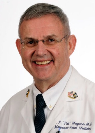 Everett F. "Pat" Magann合作论文数Department of Obstetrics And Gynecology, University of Arkansas for Medical Sciences1
 Yulia Y Tyurina合作论文数Department of Environmental and Occupational Health, School of Public Health, University of Pittsburgh1
 Leaf Huang合作论文数Division of Molecular Pharmaceutics, Eshelman School of Pharmacy, University of North Carolina at Chapel Hill;Department of Biomedical Engineering, University of North Carolina at Chapel Hill1Xianmin Song (宋献民)合作论文数1Lei Qian合作论文数1
 Sanjiang Li合作论文数Faculty of Engineering and Information Technology, University of Technology, Sydney;Centre for Quantum Software and Information, University of Technology, Sydney1
 Richard D. Wood合作论文数Department of Epigenetics and Molecular Carcinogenesis, Division of VP, Research, The University of Texas MD Anderson Cancer Center1
 Suneet P. Chauhan合作论文数Department of Obstetrics, Gynecology and Reproductive Sciences, McGovern Medical School, University of Texas Health Science Center at Houston1
 Dorota A Doherty合作论文数1Stephen D. Dertinger合作论文数1
 Jianmin Wang (王健民)合作论文数Changhai Hospital1Nu Wang合作论文数1Joe Fred合作论文数1
 Barbara Beck合作论文数1Emily E. Greenberger合作论文数1Mineaki Seki合作论文数1Charalambos Athanassiou合作论文数1Tongying Shun合作论文数1Alexander Doemling合作论文数1Shane M. Gjesdal合作论文数1




