BACKGROUND CONTEXT: Prolonged bedrest induces accumulation of intramuscular lipid concentration (ILC) in the lumbar musculature; however, spatial distribution of ILC has not been determined. Artificial gravity (AG) mitigates some adaptations induced by 60 day bedrest by creating a head-to-feet force while participants are in a supine position. PURPOSE: To quantify the spatial distribution of accumulation of ILC in the lumbar musculature after 60 day bedrest, and whether this can be mitigated by AG exposure. STUDY DESIGN: Prospective longitudinal study. PATIENT SAMPLE: Twenty-four healthy individuals (8 females) participated in the study: Eight received 30 min continuous AG (cAG); Eight received 6 x 5 min AG (iAG), interspersed with rests; Eight were not exposed to AG (CRTL). OUTCOME MEASURES: From 3T magnetic resonance imaging (MRI), axial images were selected to assess lumbar multifidus (LM), lumbar erector spinae (LES), quadratus lumborum (QL), and psoas major (PM) muscles from L1/L2 to L5/S1 intervertebral disc levels. Chemical shift-based 2-echo lipid and/or water Dixon sequence was used to measure tissue composition. Each lumbar muscle was segmented into four equal quartiles (from medial to lateral). METHODS: Participants arrived at the facility for the baseline data collection before undergoing a 60 day strict 6 degrees head-down tilt (HDT) bedrest period. MRI of the lumbopelvic region was conducted at baseline and Day-59 of bedrest. Participants performed all activities, including hygiene, in 6 degrees HDT and were discouraged from moving excessively or unnecessarily. RESULTS: At the L4/L5 and L5/S1 intervertebral disc levels, 60-day bedrest induced a greater increase in ILC in medial and lateral regions (similar to+4%) of the LM than central regions (similar to+2%; p<.05). A smaller increase in ILC was induced in the lateral region of LES (similar to+1%) at L1/L2 and L2/L3 than at the centro-medial region (similar to+2%; p<.05). There was no difference between CRTL and intervention groups. CONCLUSIONS: Inhomogeneous spatial distribution of accumulation of ILC was found in the lumbar musculature after 60 day bedrest. These findings might reflect pathophysiological mechanisms related to muscle disuse and contribute to localized lumbar spine dysfunction. Altered spatial distribution of ILC may impair lumbar spine function after prolonged body unloading, which could increase injury risk to vulnerable soft tissues, such as the lumbar intervertebral discs. These novel results may represent a new biomarker of lumbar deconditioning for astronauts, bedridden, sedentary individuals, or those with chronic back pain. Changes are potentially modifiable but not by the AG protocols tested here. (C) 2021 The Authors. Published by Elsevier Inc.
Reduced muscle size and accumulation of paraspinal muscle fat content (PFC) have been reported in lumbopelvic muscles after spaceflights and head-down tilt (HDT) bed rest. While some information is available regarding reconditioning programs on muscle atrophy recovery, the effects on the accumulation of PFC are unknown. Recently, a device (the Functional Re-adaptive Exercise Device—FRED) has been developed which aims to specifically recruit lumbopelvic muscles. This study aimed to investigate the effects of a standard reconditioning (SR) program and SR program supplemented by FRED (SR + FRED) on the recovery of the lumbopelvic muscles following 60-day HDT bed rest. Twenty-four healthy participants arrived at the facility for baseline data collection (BDC) before the bed rest period. They remained in the facility for 13-day post-HDT bed rest and were randomly allocated to one of two reconditioning programs: SR or SR + FRED. Muscle volumes of the lumbar multifidus (LM), lumbar erector spinae (LES), quadratus lumborum (QL), and psoas major (PM) muscles were measured from axial T1-weighted magnetic resonance imaging (MRI) at all lumbar intervertebral disc levels. PFC was determined using a chemical shift-based lipid/water Dixon sequence. Each lumbopelvic muscle was segmented into four equal quartiles (from medial to lateral). MRI of the lumbopelvic region was conducted at BDC, Day-59 of bed rest (HDT59), and Day-13 after reconditioning (R13). Comparing R13 with BDC, the volumes of the LM muscle at L4/L5 and L5/S1, LES at L1/L2, and QL at L3/L4 had not recovered (all—p < 0.05), and the PM muscle remained larger at L1/L2 (p = 0.001). Accumulation of PFC in the LM muscle at the L4/L5 and L5/S1 levels remained higher in the centro-medial regions at R13 than BDC (all—p < 0.05). There was no difference between the two reconditioning programs. A 2-week reconditioning program was insufficient to fully restore all volumes of lumbopelvic muscles and reverse the accumulation of PFC in the muscles measured to BDC values, particularly in the LM muscle at the lower lumbar levels. These findings suggest that more extended reconditioning programs or alternative exercises may be necessary to fully restore the size and properties of the lumbopelvic muscles after prolonged bed rest.
Exposure to spaceflight and head-down tilt (HDT) bed rest leads to decreases in the mass of the gluteal muscle. Preliminary results have suggested that interventions, such as artificial gravity (AG), can partially mitigate some of the physiological adaptations induced by HDT bed rest. However, its effect on the gluteal muscles is currently unknown. This study investigated the effects of daily AG on the gluteal muscles during 60-day HDT bed rest. Twenty-four healthy individuals participated in the study: eight received 30 min of continuous AG; eight received 6 × 5 min of AG, interspersed with rest periods; eight belonged to a control group. T1-weighted Dixon magnetic resonance imaging of the hip region was conducted at baseline and day 59 of HDT bed rest to establish changes in volumes and intramuscular lipid concentration (ILC). Results showed that, across groups, muscle volumes decreased by 9.2% for gluteus maximus (GMAX), 8.0% for gluteus medius (GMED), and 10.5% for gluteus minimus after 59-day HDT bed rest (all p < 0.005). The ILC increased by 1.3% for GMAX and 0.5% for GMED (both p < 0.05). Neither of the AG protocols mitigated deconditioning of the gluteal muscles. Whereas all gluteal muscles atrophied, the ratio of lipids to intramuscular water increased only in GMAX and GMED muscles. These changes could impair the function of the hip joint and increased the risk of falls. The deconditioning of the gluteal muscles in space may negatively impact the hip joint stability of astronauts when reexpose to terrestrial gravity.
This study investigated whether artificial gravity (AG), induced by short-radius centrifugation, mitigated deterioration in standing balance and anticipatory postural adjustments (APAs) of trunk muscles following 60-day head-down tilt bed rest. Twenty-four participants were allocated to one of three groups: control group (n = 8); 30-min continuous AG daily (n = 8); and intermittent 6 × 5 min AG daily (n = 8). Before and immediately after bed rest, standing balance was assessed in four conditions: eyes open and closed on both stable and foam surfaces. Measures including sway path, root mean square, and peak sway velocity, sway area, sway frequency power, and sway density curve were extracted from the center of pressure displacement. APAs were assessed during rapid arm movements using intramuscular or surface electromyography electrodes of the rectus abdominis; obliquus externus and internus abdominis; transversus abdominis; erector spinae at L1, L2, L3, and L4 vertebral levels; and deep lumbar multifidus muscles. The relative latency between the EMG onset of the deltoid and each of the trunk muscles was calculated. All three groups had poorer balance performance in most of the parameters (all P < 0.05) and delayed APAs of the trunk muscles following bed rest (all P < 0.05). Sway path and sway velocity were deteriorated, and sway frequency power was less in those who received intermittent AG than in the control group (all P < 0.05), particularly in conditions with reduced proprioceptive feedback. These data highlight the potential of intermittent AG to mitigate deterioration of some aspects of postural control induced by gravitational unloading, but no protective effects on trunk muscle responses were observed.NEW & NOTEWORTHY This study presents novel insights into the effect of artificial gravity (AG) on the deterioration of standing balance and anticipatory postural adjustments (APAs) of trunk muscles induced by 60-day strict head-down bed rest. The results indicated severe balance dysfunction and delayed APAs during rapid arm movement. AG partially mitigated the deterioration in standing balance and may thus be considered as a potential countermeasure for future planetary surface explorations. Optimization of AG protocols might enhance effects.
This study presents novel insights into the morphological adaptations occurring in the lumbar spine after 60-day head-down bed rest and the potential role of artificial gravity (AG) to mitigate them. Results demonstrated no protective effect of AG protocols used in this study. In atrophied paraspinal muscles, the ratio of lipids versus intramuscular water increased in the postural lumbar muscles, which could impair muscle function during upright standing. These findings have relevance for future space explorations.
Reduced paraspinal muscle size and flattening of spinal curvatures have been documented after spaceflight. Assessment of trunk adaptations to hypogravity can contribute to development of specific countermeasures. In this study, parabolic flights were used to investigate spinal curvature and muscle responses to hypogravity. Data from five trials at 0.25 g, 0.50 g, and 0.75 g were recorded from six participants positioned in a kneeling-seated position. During the first two trials, participants maintained a normal, upright posture. In the last three trials, small-amplitude perturbations were delivered in the anterior direction at the T10 level. Spinal curvature was estimated with motion capture cameras. Trunk displacement and contact force between the actuator and participant were recorded. Muscle activity responses were collected by intramuscular electromyography (iEMG) of the deep and superficial lumbar multifidus, iliocostalis lumborum, longissimus thoracis, quadratus lumborum, transversus abdominis, obliquus internus, and obliquus externus muscles. The root mean square iEMG and the average spinal angles were calculated. Trunk admittance and muscle responses to perturbations were calculated as closed-loop frequency-response functions. Compared with 0.75 g, 0.25 g resulted in lower activation of the longissimus thoracis (P = 0.002); lower responses of the superficial multifidus at low frequencies (P = 0.043); lower responses of the superficial multifidus (P = 0.029) and iliocostalis lumborum (P = 0.043); lower trunk admittance (P = 0.037) at intermediate frequencies; and stronger responses of the transversus abdominis at higher frequencies (P = 0.032). These findings indicate that exposure to hypogravity reduces trunk admittance, partially compensated by weaker stabilizing contributions of the paraspinal muscles and coinciding with an apparent increase of deep abdominal muscle activity.NEW & NOTEWORTHY This study presents for the first time novel insights into the adaptations to hypogravity of spinal curvatures, trunk stiffness, and paraspinal muscle activity. We showed that exposure to hypogravity reduces the displacement of the trunk by an applied perturbation, partially compensated by weaker stabilizing contributions of the paraspinal muscles and concomitant increase in abdominal muscle responses. These findings may have relevance for future recommendations for planetary surface explorations.
Mechanistic studies of the Functional Re-adaptive Exercise Device (FRED) have shown it automatically recruits Lumbar Multifidus (LM) and Transversus Abdominis (TrA) - two deep-spinal muscles that are atrophied and show altered motor control in low back pain (LBP). No studies have investigated the time required to familiarise to FRED exercise, which is required to inform future FRED based clinical trial protocols. This study therefore determined the effect of time, during FRED exercise, on biomechanical outcome measures, to establish the familiarisation period, and assess for loss of technique throughout a ten minute trial. A cohort comparison study of 148 participants, 70 experiencing low back pain, had lumbopelvic kinematics, exercise frequency and movement variability measured during a 10 minute trial. Magnitude-based inference was used to assess for familiarisation, using plots of variation over time with familiarised reference ranges. The no pain group took 170 seconds, and the back pain group took 150 seconds, to familiarise. A familiarisation period of at least 170 seconds (2.8 minutes) is recommended. This justifies, and provides a familiarisation time for use of the FRED as a motor control intervention.
Physical inactivity and lumbopelvic deconditioning have been linked to increased incidence of non-specific low back pain (LBP) and spinal injury in those who are exposed to microgravity (e.g. astronauts and individuals on long-duration bed rest) and in the general population. Astronauts have an increased risk of experiencing moderate to severe LBP during microgravity exposure and herniated intervertebral discs within 1 year following spaceflight. Atrophy and reduced motor control of the lumbar multifidus (LM) and transversus abdominis (TrA) muscles resulting from periods of deconditioning are linked to non-specific LBP and spinal injury risk in both post-flight astronauts and general populations. However, voluntary recruitment of these two key muscles is difficult and presents a rehabilitation challenge. This chapter reviews the concept of spinal stability as it relates to microgravity, discusses how existing exercise countermeasures used in space do not successfully maintain lumbopelvic muscle size, and introduces the functional readaptive exercise device (FRED) that shows potential to activate the LM and TrA muscles automatically and in a tonic fashion, which has relevance to rehabilitation of both astronaut and terrestrial populations.
This study investigated the effects of a single exercise session using a device developed for postural muscle training on the function of postural muscles in healthy, pain free individuals. During standardised rapid arm movements, timing of onset of electromyography (EMG) was measured using intramuscular and surface recordings of the transversus abdominis (TrA), obliquus internus abdominis (OI), obliquus externus abdominis (OE), lumbar multifidus (LM) and lumbar erector spinae (LES) muscles. A single exercise session with the device led to significantly (main effect of time: P = 0.03) earlier LES EMG onset in advance of the postural perturbation induced by rapid forward arm movements from -1 ms (SD: 32 ms) at baseline to -11 ms (SD: 27 ms) post-exercise and -16 ms (SD: 22 ms) at 10-min Wash-Out after the FRED exercise bout. The timing of EMG onset of the other trunk muscles was not affected by the single bout of exercise. A significant correlation was found between background activity and the EMG onset times of of TrA (r = 0.6; P < 0.001), OI (r = 0.59; P < 0.001), LES (r = 0.32; P = 0.046) and LMs (r = 0.77; P < 0.001). Higher levels of trunk muscle background activity were associated with later onset times. The present findings suggest that a single exposure to the postural training device can induce small changes in spinal muscle function in healthy pain free individuals.
ObjectivesTo determine the intrarater reliability and precision of lumbar multifidus and transversus abdominis thickness measurements using freehand sonography in a range of static and dynamic conditions.MethodsFifteen asymptomatic participants performed a range of exercises while sonography was used to measure absolute muscle thickness and changes in muscle thickness from rest. Exercise conditions included the abdominal drawing‐in maneuver, active straight leg raise, contralateral arm lift, both unloaded and loaded, treadmill walking, and using the Functional Readaptive Exercise Device. Intraday and interday reliability was assessed by intraclass correlation coefficients, and the standard error of measurement was used to assess measurement precision.ResultsGood to excellent reliability was achieved for absolute transversus abdominis and lumbar multifidus thickness in all conditions. Measurement precision for absolute lumbar multifidus thickness was ≤2.8 mm for the unloaded contralateral arm lift, ≤1.8 mm for the loaded contralateral arm lift, ≤3.1 mm for treadmill walking, and ≤3.8 mm for the Functional Readaptive Exercise Device; for absolute transversus abdominis thickness, precision was ≤0.6 mm for the abdominal drawing‐in maneuver, ≤0.5 mm for the active straight leg raise, ≤0.7 mm for treadmill walking, and ≤0.5 mm for the Functional Readaptive Exercise Device. Good to excellent reliability was achieved for relative transversus abdominis and lumbar multifidus thickness in all conditions. Measurement precision for relative lumbar multifidus thickness was ≤3.7% for the unloaded contralateral arm lift, ≤3.8% for the loaded contralateral arm lift, ≤6.3% for treadmill walking, and ≤7.6% for the Functional Readaptive Exercise Device; for relative transversus abdominis thickness, precision was ≤13.6% for the abdominal drawing‐in maneuver, ≤6.9% for the active straight leg raise, ≤11.1% for treadmill walking, and ≤7.2% for the Functional Readaptive Exercise Device.ConclusionsAcceptable reliability and precision of measurement is achieved for absolute and relative measures of deep spinal muscle thickness using freehand sonography in relatively static and dynamic exercises.
Gravitational unloading leads to adaptations of the human body, including the spine and its adjacent structures, making it more vulnerable to injury and pain. The Functional Re‐adaptive Exercise Device (FRED) has been developed to activate the deep spinal muscles, lumbar multifidus (LM) and transversus abdominis (TrA), that provide inter‐segmental control and spinal protection. The FRED provides an unstable base of support and combines weight bearing in up‐right posture with side alternating, elliptical leg movements, without any resistance to movement. The present study investigated the activation of LM, TrA, obliquus externus (OE), obliquus internus (OI), abdominis, and erector spinae (ES) during FRED exercise using intramuscular fine‐wire and surface EMG. Nine healthy male volunteers (27 ± 5 years) have been recruited for the study. FRED exercise was compared with treadmill walking. It was confirmed that LM and TrA were continually active during FRED exercise. Compared with walking, FRED exercise resulted in similar mean activation of LM and TrA, less activation of OE, OI, ES, and greater variability of lumbo‐pelvic muscle activation patterns between individual FRED/gait cycles. These data suggest that FRED continuously engages LM and TrA, and therefore, has the potential as a stationary exercise device to train these muscles.
Background: No studies have been published on an astronaut population to assess the effectiveness of countermeasures for limiting physiological changes in the lumbopelvic region caused by microgravity exposure during spaceflight. However, several studies in this area have been done using spaceflight simulation via bed-rest. The purpose of this systematic review was to evaluate the effectiveness of countermeasures designed to limit physiological changes to the lumbopelvic region caused by spaceflight simulation by means of bed-rest.Methods: Electronic databases were searched from the start of their records to November 2014. Studies were assessed with PEDro, Cochrane Risk of Bias and a bed-rest study quality tool. Magnitude based inferences were used to assess countermeasure effectiveness.Results: Seven studies were included. There was a lack of consistency across studies in reporting of outcome measures. Some countermeasures were found to be successful in preventing some lumbopelvic musculoskeletal changes, but not others. For example, resistive vibration exercise prevented muscle changes, but showed the potential to worsen loss of lumbar lordosis and intervertebral disc height.Conclusion: Future studies investigating countermeasures should report consistent outcomes, and also use an actual microgravity environment. Additional research with patient reported quality of life and functional outcome measures is advocated. (C) 2016 Elsevier Ltd. All rights reserved.
The full-text may be used and/or reproduced, and given to third parties in any format or medium, without prior permission or charge, for personal research or study, educational, or not-for-pro t purposes provided that: • a full bibliographic reference is made to the original source • a link is made to the metadata record in DRO • the full-text is not changed in any way The full-text must not be sold in any format or medium without the formal permission of the copyright holders. Please consult the full DRO policy for further details.
Introduction: Exposure to microgravity has various effects on the human musculoskeletal system. During spaceflight many astronauts experience low back pain and the risk of spine injuries is significantly greater post-flight. Nonetheless, the increased lumbo-pelvic injury risk is not specifically addressed by current countermeasures. Considering this, a novel exercise device has been developed to specifically counteract atrophy of deep spinal and postural muscles. The aim of the present study was to test the possibility of transferring this exercise concept from earth to space using a biomechanical simulation.Methods: A biomechanical model of the exercise device was developed and validated using intramuscular electromyographic (EMG) data as previously acquired on a terrestrial prototype of the exercise device. The model was then modified to the needs of a 0-g environment, creating gravity-like conditions using shoulder straps.Results: Modelled activation patterns of the investigated muscles were in line with the experimental data, showing a constant activation during exercise. The microgravity modifications of the model lead to increased muscle activation of deep spinal muscles and to decreased activation of superficial moment creating trunk muscles.Discussion: The results of the biomechanical model suggest that the exercise concept can be transferred from 1-g to space conditions. The present study is a first step in the investigation process of a novel exercise concept and human studies should be conducted to confirm the present theoretical investigation. (C) 2015 IAA. Published by Elsevier Ltd. All rights reserved.
The present study examined the acute effects of active gaming on energy intake (EI) and appetite responses in 8-11-year-old boys in a school-based setting. Using a randomised cross-over design, twenty-one boys completed four individual 90-min gaming bouts, each separated by 1 week. The gaming bouts were (1) seated gaming, no food or drink; (2) active gaming, no food or drink; (3) seated gaming with food and drink offered ad libitum; and (4) active gaming with food and drink offered ad libitum. In the two gaming bouts during which foods and drinks were offered, EI was measured. Appetite sensations - hunger, prospective food consumption and fullness - were recorded using visual analogue scales during all gaming bouts at 30-min intervals and at two 15-min intervals post gaming. In the two bouts with food and drink, no significant differences were found in acute EI (MJ) (P=0·238). Significant differences were detected in appetite sensations for hunger, prospective food consumption and fullness between the four gaming bouts at various time points. The relative EI calculated for the two gaming bouts with food and drink (active gaming 1·42 (sem 0·28) MJ; seated gaming 2·12 (sem 0·25) MJ) was not statistically different. Acute EI in response to active gaming was no different from seated gaming, and appetite sensations were influenced by whether food was made available during the 90-min gaming bouts.
The aim of this study was to investigate the influence of an exercise device, designed to improve the function of lumbopelvic muscles via low-impact weight-bearing exercise, on electromyographic (EMG) activity of lumbopelvic, including abdominal muscles. Surface EMG activity was collected from lumbar multifidus (LM), erector spinae (ES), internal oblique (IO), external oblique (EO) and rectus abdominis (RA) during overground walking (OW) and exercise device (EX) conditions. During walking, most muscles showed peaks in activity which were not seen during EX. Spinal extensors (LM, ES) were more active in EX. Internal oblique and RA were less active in EX. In EX, LM and ES were active for longer than during OW. Conversely, EO and RA were active for a shorter duration in EX than OW. The exercise device showed a phasic-to-tonic shift in activation of both local and global lumbopelvic muscles and promoted increased activation of spinal extensors in relation to walking. These features could make the exercise device a useful rehabilitative tool for populations with lumbopelvic muscle atrophy and dysfunction, including those recovering from deconditioning due to long-term bed rest and microgravity in astronauts.
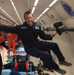 Nicholas Caplan合作论文数1 School of Psychology;and Sport Sciences;Northumbria University;School of Psychology|and Sport Sciences, Northumbria University20
 Andrew Winnard合作论文数13
 Julie A. Hides合作论文数10
 Jonathan Matthew Scott合作论文数9
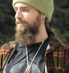 Salomoni Sauro E合作论文数Center for Sensory-Motor Interaction (SMI),, Aalborg University9
 Tobias Weber合作论文数National Center for Tumor Diseases, Heidelberg University Hospital9
 Enrico De Martino合作论文数Faculty of Health and Life Sciences, Northumbria University8
 Paul Hodges合作论文数School of Health and Rehabilitation Sciences, Faculty of Health and Behavioural Sciences, The University of Queensland8
 Kirsty Lindsay合作论文数6
 Jonathan Cook合作论文数Centre for Statistics in Medicine, Nuffield Department of Orthopaedics, Rheumatology and Musculoskeletal Sciences, Medical Sciences Division, University of Oxford;St Hugh’s College, University of Oxford6
 David Beard合作论文数Nuffield Department of Orthopaedics, Rheumatology and Musculoskeletal Sciences, Medical Sciences Division, University of Oxford6
 Simon N Evetts合作论文数5Mick Wilkinson合作论文数5
 Mark a Hoggarth合作论文数Loyola University Medical Center5Jochen Zange合作论文数4
 James A. Elliott合作论文数Department of Materials Science & Metallurgy, University of Cambridge4Caroline Dodd-Reynolds合作论文数3
 Penny L S Rumbold合作论文数3
 Susan Allsop合作论文数3Benjamin P. Green合作论文数2Angela Hibbs合作论文数2Petersen Nora合作论文数2
 Karl Gibbon合作论文数Faculty of Health &Life Sciences, Northumbria University;Faculty of Health & Life Sciences, Northumbria University2
 Maria Stokes合作论文数Southampton Marine and Maritime Institute, Faculty of Engineering and Physical Sciences, University of Southampton2Gunda Lambrecht合作论文数2
 Volker Damann合作论文数European Astronaut Center, European Space Agency2
 Vienna Tran合作论文数1Robert Ekman合作论文数1Gordon Cable合作论文数1Arran Parmar合作论文数1François Hug合作论文数1
 Edith Elgueta-Cancino合作论文数School of Sport, Exercise and Rehabilitation Sciences, University of Birmingham1
 Sherveen Riazati合作论文数1Anja Niehoff合作论文数1
 Jörn Rittweger合作论文数Departement of Pediatrics and Adolescent Medicine, University of Cologne;Department of Muscle and Bone Metabolism, Institute of Aerospace Medicine, German Aerospace Center1
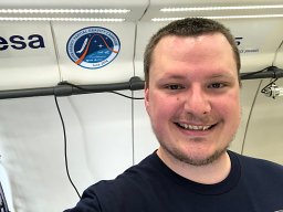 McCarty Kristofor合作论文数Faculty of Health and Life Sciences, Northumbria University1
 Ramdharry Gita合作论文数Institute of Neurology, University College London1
 Jaap Van Dieen合作论文数Faculty of Behavioural and Movement Sciences, Vrije Universiteit Amsterdam1Brace Hal合作论文数1
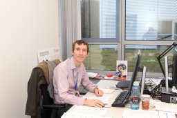 Green David A合作论文数1
 Carly McKay合作论文数Department of Community Health Sciences, Cumming School of Medicine, University of Calgary1
 Jacob Bloomberg合作论文数1Catherine Gibb合作论文数1Filippo Castrucci合作论文数1Mirko Pinotti合作论文数1Rebecca Cusack合作论文数Specialist Registrar in Intensive Care, St George's Hospital1Colin Chandler合作论文数1
 Alan St Clair Gibson合作论文数MRC/UCT Bioenergetics of Exercise Research Unit, Sport Science Institute of South Africa1Marcos Pinotti合作论文数Departamento de Engenharia Hidráulica e Recursos Hídricos;Faculdade de Medicina de São José do Rio Preto - SP;Departamento de Engenharia Hidráulica e Recursos Hídricos, Faculdade de Medicina de São José do Rio Preto - SP1




