Pulse oximetry devices systematically overestimate blood oxygen saturation (SaO₂) in hypoxemic patients, potentially masking critical conditions. We hypothesize that this bias stems from wavelength-dependent differences in optical pathlength (OPL), which vary with SaO₂ levels. This letter investigates the change in photon pathlength in tissue between different wavelengths and proposes the iso-pathlength (IPL) point, an angular position identifiable by constant intensity between media of varying scattering coefficients, as a solution. Using Monte Carlo (MC) simulations on a finger model (5% blood volume, 60-100% SaO₂), we analyzed the OPL ratio at various exit angles for the standard 660/940nm wavelength pair and a closer pair with 760/840nm. Analysis revealed that at the standard pulse oximetry transmission position, the OPL ratio for the 660/940nm pair is highly variable, ranging from 1.25 to 1.45 across SaO₂ levels, while the OPL ratio for the alternative 760/840nm pair changed from 1.05 to 1.12. In contrast, the OPL ratio at the IPL point remains stable, not exceeding 1.05 across all SaO₂ levels. These MC results provide mechanistic insight into pulse oximetry bias and demonstrate that IPL technique could help in the detection of critical hypoxemia, where current devices are most prone to dangerous overestimation.
Separating absorption and scattering in turbid media remains a major challenge in optical characterization. This work takes a step toward addressing it by applying non-contact estimation of the absorption coefficient (µa) within a phase-analysis framework originally developed for extracting the reduced scattering coefficient (μs'). The iterative multi-plane optical properties extraction (IMOPE) technique reconstructs diffuse-reflectance (DR) phase from multi-plane intensity measurements, enabling phase-based analysis for μs' extraction. Here, the DR model is extended to incorporate sensitivity to variations in µa, establishing a route for phase-driven absorption assessment, demonstrating opposing trends for µa and μs' increments. Monte Carlo simulations and TiO2-based phantoms with independently controlled μs' and µa validate the theoretical predictions. This study experimentally validates a phase-based µa extraction scheme at λ1 = 473 nm, with cross-wavelength verification at λ2 = 632.8 nm, demonstrating strong agreement, 91%, 95% for λ1, λ2, respectively, between extracted and designed absorption values. A detailed TiO2 phantom preparation protocol is provided to support experimental reproducibility. Finally, we present an initial framework for estimating µa from the reconstructed phase, laying the groundwork for future quantitative separation of absorption and scattering in turbid media.
Optical polarization techniques provide valuable tools for non-invasive sensing of turbid biological media, yet multiple scattering induces depolarization and complicates signal interpretation. In this work, we investigate the effect of polarization on the iso-pathlength (IPL) point - a geometrical feature where detected intensity becomes independent of the scattering coefficient. Using linearly polarized illumination and analyzing the co-polarized and cross-polarized detected light, we observe a distinct polarization-dependent IPL shift. For a 10 mm tube, the IPL occurs at an azimuthal angle of 100.3∘,104.9∘, and 111.7∘ for cross-polarized, unpolarized, and co-polarized detection, respectively. The IPL angle increases linearly with tube diameter, a trend confirmed by polarization-sensitive Monte Carlo simulations based on Stokes vector formalism.
Significance:Medical examination of human tissue is preferably performed by imaging the tissue surface. Optical imaging techniques are limited by low penetration depth due to high tissue scattering, whereas sensing techniques can detect changes deeper inside the tissue. Near-infrared sensing methods such as oximetry and fNIRS are already used clinically but have not yet been applied in endoscopy. Aim:We investigate the existence of iso-pathlength (IPL) points in endoscopic geometry, with the goal of extending the concept of IPL points from cylindrical and half-infinite geometries into hollow cylindrical tissue relevant to endoscopy. In addition, we demonstrate the ability to extract the absorption properties of a tissue at this structure by the IPL and demonstrate it by ex vivo experiment. Approach:The IPL point is a unique position in the full scattering profile, independent of tissue scattering and dependent only on the tissue absorption and geometry. We studied two directions in cylindrical endoscopic geometry: azimuthal and longitudinal. First, diffusion theory with extrapolated zero-boundary conditions was applied to predict IPL positions. These predictions were then tested using Monte Carlo simulations of photon distribution and validated experimentally using phantoms with cylindrical air holes measured by endoscopy. Finally, using the experimentally identified IPL point and applying the same procedure to a standard phantom, a hemoglobin-agar phantom, and chicken breast tissue, we were able to estimate the absorption coefficient of the chicken tissue. Results:Both azimuthal and longitudinal IPL points were identified. The experimental azimuthal IPL point was found at an angle of 144 deg ± 3 deg , whereas the longitudinal IPL point appeared at a distance of 0.33 ± 0.05 cm from the laser spot center. These findings confirm the theoretical and simulation predictions. Moreover, from the ex vivo experiment of a chicken breast, the IPL point enables us to calculate the absorption coefficient and get μ a = 0.94 cm - 1 , within the range of 0.2 cm - 1 ≤ μ a ≤ 2 cm - 1 . Conclusions:The demonstration of IPL points in endoscopic geometry provides a new framework for depth-resolved optical sensing in hollow cylindrical tissues. This approach may enable self-calibrated absorption measurements and open the way for improved diagnostic tools in the digestive system, esophagus, and other hollow organs where conventional endoscopy lacks depth information.
The editorial provides an overview of the JBO Special Section Nanoscale Imaging, Sensing, and Actuation and reflects on the future of nanoscale biophotonics.
Cutting-edge optical neural networks are often still trained by backpropagation,which is computationally intensive and originally for conventional artificial neural networks.Inspired by Pavlov's experiment,we drew upon the principles of biological memory to establish an associative learning framework for training optical neural networks that mimics the mechanisms of associative learning and synaptic plasticity using dual-wavelength stimuli(i.e.ultraviolet and visible light)on a dual-color photoinitiator resin.Sequential light irradiation was shown to induce fluorescence switching and encode associative memory in the resin,which can serve as a physical substrate for an optical neural network.In optical experiments,the established framework was applied to pattern recognition of the letters'N,''V,'and'Z.'Simulations were conducted that extended its application to the recognition of handwritten digits.Compared to the current mainstream'bottom-up'optical neural network fabrication approach that requires'weight calculation followed by hardware implementation',this work presents a novel'top-down'in-situ training methodology that eliminates the need for weight computation.The proposed method holds significant implications for large-scale,low-cost,and rapid fabrication of optical neural networks intended for edge computing applications.This study bridges biological learning principles with optical neural networks to provide a foundation for next-generation adaptive and scalable artificial intelligence systems.
Organic and inorganic contaminants threaten water quality and require monitoring methods that are rapid, simple, and selective in mixed turbid samples. Traditional laboratory techniques require extensive sample preparation and typically do not provide real-time measurements, while faster optical methods often struggle with analysis of complex turbid media. Therefore, this study aims to monitor and analyze mixed turbid water samples containing both organic and inorganic contaminants using a unique optical framework. Our approach combines full scattering profile (FSP) measurements with the iso-pathlength (IPL) phenomenon, enabling intrinsic calibration by separating scattering from absorption. In this proof-of-concept study, we extend this approach by constructing optical metrics for the independent isolation and quantification of cyanobacteria and heavy metals in mixed samples. Angularly resolved spectra were collected and normalized using the IPL phenomenon. Our results showed detection of each contaminant using specific spectral features: Microcystis aeruginosa a green cyanobacteria (CBG), detected through a second-derivative metric at 680 nm (1 ppb, R2 = 0.98), copper chloride via deviations in normalized intensity at the IPL point (20 ppm, R2 = 0.99), and ferric oxide through a shape-based spectral metric (300 ppb, R2 = 0.90) with condition-dependent calibration behavior. Concentration-level validation further supported the classification capability of the selected metrics, with the strongest performance obtained for CBG and CuCl2, whereas Fe2O3 showed lower but still effective performance due to composition dependency. Spectrophotometric measurements supported the physical basis of the extracted metrics. These results demonstrate a low-complexity, self-calibrated optical framework for real-time screening of multiple contaminants with potential for integration into automated monitoring systems.
Achieving precise tumor ablation without damaging surrounding healthy tissue remains a significant challenge in cancer therapy, particularly for deep-seated or irregularly shaped tumors. Traditional laser-based approaches, although minimally invasive, are often limited by insufficient tissue penetration, uncontrolled thermal damage, and narrow therapeutic windows. We introduce GHz high-repetition-rate pulsed lasers as a transformative modality for tumor ablation. This approach capitalizes on the thermal accumulation effect of GHz pulse trains, in which the pulse interval is significantly shorter than the thermal relaxation time of biological tissue. Such a regime enables efficient and localized heat deposition in tumor regions. By precisely tuning the repetition frequency, pulse duration, and energy density, we establish a dynamic "ablation-cooling" cycle: rapid energy delivery followed by transient inter-pulse cooling. This thermal modulation ensures sharply confined ablation zones with reduced collateral damage. Our systematic investigation of laser-tissue interaction parameters demonstrates that GHz lasers offer superior spatial selectivity, minimized off-target injury, and enhanced treatment safety, presenting a compelling rationale for clinical translation of this paradigm in precision photothermal oncology.
Significance:Integrating multiple biosensors improves the sensitivity and precision of physiological measurements in healthcare monitoring. By combining sensors that target different physiological parameters, a more comprehensive assessment of a subject's health can be achieved. Aim:We evaluate the performance of two biosensors for extracting cardiac parameters: a textile-based strain sensor for measuring respiratory rate and an optical sensor for measuring heart rate, SpO 2 , and respiratory rate. The objective is to determine optimal placement conditions for each sensor and assess their feasibility for integration into a single wearable system. Approach:Two experimental setups were tested. In the first, the strain sensor was placed on the subject's shirt, while the optical sensor was positioned on the external wrist. In the second, both sensors were placed on the chest, under the shirt. The accuracy and performance of each sensor were analyzed in both configurations. Results:The optical sensor demonstrated improved accuracy when placed on the chest compared to the wrist, whereas the strain sensor provided similar results for both configurations. Conclusions:We demonstrate that sensor placement significantly affects measurement quality, emphasizing the importance of optimizing placement when integrating multiple biosensors. Future work will focus on developing a unified wearable system that leverages the strengths of both sensors for comprehensive physiological monitoring.
Developing noninvasive techniques that can probe how solvents modulate the nucleation pathways of bioorganic molecules in solution remains an active and open area of research. Herein, we investigate the crystallization of the amino acid L-Cysteine and show that both the structure of the crystal and its intrinsic fluorescence can be drastically altered by the solvent. Crystals formed in heavy water exhibit markedly different intermolecular packing as well as strikingly different monomer conformations compared to those in light water. Remarkably, these differences in the supramolecular packing result in significantly elevated intrinsic fluorescence in the crystal that is formed in heavy water. Using a combination of experimental techniques and advanced electronic structure approaches, we elucidate the molecular interactions within the crystals that govern both the electronic origins and the intensity of their emission. These findings demonstrate how tuning the solvent by changing its isotope leads to the emergence of design principles for new intrinsic fluorophores that could serve as novel sensing probes for biomedical applications.
Objectives: This study aimed to assess the effectiveness of gold nanoparticles conjugated with anti-EGFR monoclonal antibodies (GNPs-EGFR) in distinguishing between benign and malignant salivary gland tumors. Methods: A total of 49 oral salivary gland tissue samples were analyzed, including 22 malignant salivary gland tumors (MSGTs), 15 benign salivary gland tumors (BSGTs), and 12 control samples. For each sample, three 5 μm consecutive tissue sections were prepared. The first section was stained with hematoxylin and eosin (H&E) to confirm the diagnosis, the second was immunohistochemically stained for anti-EGFR, and the third was treated with GNPs-EGFR followed by hyperspectral microscopy to analyze the reflectance spectrum. Results: Reflectance intensity was significantly higher (p < 0.001) in MSGTs compared to BSGTs and controls, with intensity levels increasing alongside tumor grade. The average hyperspectral reflectance values were strongly correlated with the GNPs-EGFR immunohistochemical score and varied significantly between subgroups (p < 0.001). Conclusions: GNPs-EGFR reflection measurements effectively differentiate MSGTs from BSGTs with high sensitivity. This diffusion–reflection technique holds potential as a valuable tool for tumor detection, surgical margin assessment, and intraoperative identification of residual disease in salivary gland tumors.
ABSTRACT Nowadays, many biodegradable materials are offered for biomedical applications, but there are only a few in vivo methods for their detection and monitoring. In this work, implants based on biodegradable polyester copolymers were labeled with indocyanine green (ICG) for fluorescence imaging in combination with tissue optical clearing (TOC) and magnetic resonance imaging (MRI). The results include in vitro degradation modeling followed by in vivo imaging of copolymer samples that were subcutaneously implanted in BALB/c mice. TOC with 70% glycerol has been demonstrated to significantly improve sample visualization. The TOC efficiency parameter Q demonstrated the variability of effects correlating with the timing of follow‐up in the postimplantation period. It has been shown that nonhealing wounds, peri‐implantation inflammation, or fibrosis, confirmed by MRI, affect the effectiveness of TOC in the range from Q = −30% to 70%.
 Hamootal Duadi合作论文数Engineering School, Bar Ilan University87
 Ankri Rinat合作论文数Faculty of Engineering and the Institute of Nanotechnology and Advanced Materials, Bar Ilan University34
 Zeev Zalevsky合作论文数Faculty of Engineering, Bar-Ilan University;Nano-Photonics Center, Institute of Nanotechnology and Advanced Materials, Bar-Ilan University;Z-square Ltd;ContinUse Biometrics Ltd;LensFree Ltd;Cogni-fiber Ltd;Nano-drops Ltd;SuperSpectra Ltd25
 Inbar Yariv合作论文数Faculty of Engineering and the Institute of Nanotechnology and Advanced Materials, Bar Ilan University22
 Popovtzer Rachela合作论文数Fac Engn, Bar Ilan Univ18Motiei Menachem合作论文数18
 Gilad Yahav合作论文数Fac Engn, Bar Ilan Univ17
 Shweta Vinod Pawar合作论文数Bar Ilan Inst Nanotechnol & Adv Mat BINA, Bar Ilan Univ14
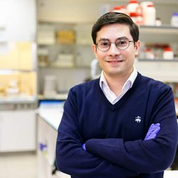 Ariel Ashkenazy合作论文数Inst Nanotechnol & Adv Mat, Bar Ilan Univ14Feder Idit合作论文数14
 Javier García Monreal合作论文数Opto-Electronic Processing Image Group, University of Valencia13Barnoy Eran合作论文数11Ruchira Chakraborty合作论文数10
 Vicente Micó合作论文数10Channa Shapira合作论文数10
 Asher Shainberg合作论文数BAU10
 Nitza Goldenberg-Cohen合作论文数Department of Pediatric Ophthalmology, Schneider Children’s Medical Center of Israel10Mordechai Deutsch合作论文数9Anat Lipovsky合作论文数9Leshem-Lev Dorit合作论文数9
 Lior Turgeman合作论文数Department of Physics and Advanced Materials and Nanotechnology Institute, Bar-Ilan University9
 Rachel Lubart合作论文数8
 Pavitra S R合作论文数7
 Eliahu Cohen合作论文数Faculty of Engineering, Bar Ilan University;Institute of Nanotechnology and Advanced Materials, Bar Ilan University7
 Moshe Deutsch合作论文数Department of Physics, Bar Ilan University7Reuven Tirosh合作论文数7
 David Iluz合作论文数Beit Berl College, Bar-Ilan University7
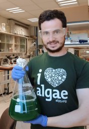 Said Abu-Ghosh合作论文数Mina & Everard Goodman Fac Life Sci, Bar Ilan Univ6Nayhoz Tsviya合作论文数6
 Aharon Gedanken合作论文数Department of Chemistry, Bar-Ilan University6
 Eli I. Lev合作论文数Cardiology Department, Rabin Medical Center6
 Sivan Gershanov合作论文数6Alon Tzroya合作论文数5Yitzchak Weber合作论文数5Yafit Fleger合作论文数5
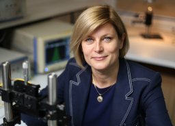 Malgorzata Szczerska合作论文数Dept Metrol & Optoelect, Gdansk Univ Technol5
 Mali Salmon-Divon合作论文数Dept Mol Biol, Ariel Univ5Aviram Gur合作论文数5Avraham Hirshberg合作论文数5
 Zvy Dubinsky合作论文数The Mina & Everard Goodman Faculty of Life Sciences, Bar-Ilan University5
 Edith Hochhauser合作论文数FMRC, TAU5Marianna Beiderman合作论文数4Tamar Sadan合作论文数4
 Ran Kornowski合作论文数Herzliya Medical Center;Department of Cardiology, School of Medicine, Sackler Faculty of Medicine, Tel Aviv University4Krishanu Ray合作论文数Tata Institute of Fundamental Research4
 Daqing (Daching) Piao合作论文数School of Electrical and Computer Engineering, College of Engineering, Architecture and Technology, Oklahoma State University4Naomi Zurgil合作论文数4
 Ilya Novikov合作论文数The Institute of Hematology, Tel Aviv University4
 Yevgeny Beiderman合作论文数4




