Introduction:Glioblastoma is characterized by heterogeneous tumor characteristics and infiltrative tumor boundaries, making accurate delineation difficult with extensive manual annotations. Chemical exchange saturation transfer (CEST) is a non-invasive MRI technique used for in vivo assessment of metabolic and macromolecular information through a Z-spectrum. CEST may provide insight into metabolic changes present in early-stage disease that are not visible in routine clinical imaging, thereby improving tumor delineation. In this work, we use an unsupervised anomaly detection (UAD) strategy to learn the distribution of features present in Z-spectra of healthy tissue and capture their deviations in pathology, foregoing the need for extensive labels. The approach leverages the metabolic information provided by CEST to improve the detection and delineation of glioblastoma and inform further treatment planning. Methods:A 1D convolutional autoencoder (CAE) was implemented to reconstruct Z-spectra from individual tissue voxels. The network was trained on Z-spectra acquired at 9.4T from healthy Sprague-Dawley rats and tested on data acquired from F98 glioma-bearing rats post Gd-administration. For baseline comparisons, Isolation Forest and Local Outlier Factor, which have shown success in anomaly detection, were implemented. For the CAE, our anomaly score was determined to be the mean squared reconstruction error. To facilitate clinical translation and evaluate the robustness of our model for under sampled Z-spectra, acceleration factors of 2x and 7x were performed with two sampling schemes: uniformly skipping frequency offsets and selecting offsets based on feature importance identified by Shapley value analysis and Integrated Gradients (IG). Binarization was performed by determining an optimal anomaly threshold, followed by comparison to ground truth tumor masks. Metrics related to model performance were assessed for baseline anomaly detectors on the fully sampled dataset and for the CAE on fully and under sampled datasets. Results:The best baseline anomaly detector was Isolation Forest, with an ROC-AUC of 0.967 and an F1-score of 0.584. Our method, the CAE, accurately reconstructed Z-spectral features, achieving Dice scores of up to 0.72 and outperforming the baseline model with an ROC-AUC of 0.968 and F1-score of 0.642. This model performance remained robust across sampling schemes and acceleration factors, with ROC-AUCs of ~0.96 and similar Dice scores (up to 0.7). Feature importance analysis indicated that offsets in the range of ±3.0 to 5.0ppm contributed most to the anomaly score. Discussion:This study successfully demonstrated a UAD pipeline utilizing the Z-spectrum from CEST MRI for metabolically informed tumor delineation. The framework captures biochemical deviations that may precede or extend beyond morphologic abnormalities, enabling sensitive detection of tumor regions and intra-tumoral heterogeneity that previous methods may fail to capture. The offsets from the feature analysis indicated a strong contribution from the magnetization transfer (MT) pool to the spectral deviations captured by the model, with additional contributions from relayed nuclear Overhauser effect (rNOE) and amide proton transfer (APT). Model robustness with under sampling further highlights the pipeline's potential in accelerated acquisitions, thus improving clinical practicality. While there is a need for validation on larger cohorts and clinical datasets, the current results demonstrate that this label-free, Z-spectral anomaly mapping can serve as an interpretable and scalable tool for monitoring tumor heterogeneity and progression, with potential applicability to other diffuse or metabolically subtle pathologies.
Cerebral glucose hypometabolism in Alzheimer’s disease (AD) leads to enhanced metabolism of fatty acids (FAs) and branched-chain amino acids (BCAAs) as a compensatory mechanism. While there have been some 13C labeled studies investigating the metabolism of FAs and BCCAs, their clinical translation is challenging. In this study, we investigated the potential of measuring neurometabolic perturbations through macromolecular signal at 0.9 ppm (MM09) in proton magnetic resonance (1H MR) spectrum. This signal represents a composite macromolecular signal with contributions from lipids and BCAA associated methyl resonances and may be sensitive to metabolic alterations occurring during glucose hypometabolism in AD. MM09 levels were measured from localized 1H MR spectra in the hippocampus and thalamus/hypothalamus of male and female APPNL−F/NL−F (AD) mice. In addition, the levels of glutamate in these regions were also recorded as it is known to be reduced under glucose hypometabolism in AD. We further studied the metabolic association of MM09 with glutamate in Pearson correlation plots. To find the statistical significance of difference two-way ANOVA analysis with post-hoc Tukey HSD tests were used. Male AD mice exhibited significantly reduced MM09 (15.42 ± 1.32 vs. 16.93 ± 1.15 mM; p = 0.008) and glutamate levels (15.27 ± 1.65 vs. 17.24 ± 1.21 mM; p = 0.004) in the hippocampus. Female AD mice did not show any changes in glutamate or MM09 levels. MM09 also showed a strong positive correlation with glutamate (R = 0.74; p < 0.0001). The observed reductions in MM09 and glutamate in male AD mice are consistent with neurometabolic alterations associated with impaired glucose metabolism, whereas the absence of such changes in female AD mice may reflect sex-specific metabolic resilience. The strong association between MM09 and glutamate suggests that MM09 may capture neurochemical changes linked to metabolic adaptations in AD. Because the MM09 resonance occurs in a relatively uncrowded region of the 1H MR spectrum, it may represent a promising spectroscopic marker for investigating metabolic shifts in AD and warrants further evaluation in clinical studies.
Tumor pseudo-progression (PsP) refers to an initial increase in tumor size or the appearance of new lesions. These pseudo-progressive lesions are predominantly composed of infiltrative inflammatory cells, such as macrophages. This phenomenon commonly occurs in patients undergoing radiation therapy or immunotherapy and typically indicates a positive treatment response. However, it often leads to premature treatment cessation due to misinterpretation as disease progression. Non-invasive imaging biomarkers capable of distinguishing pseudo-progression from true progression would greatly aid in treatment decision-making. In our preliminary study, we explored the potential of gadoterate meglumine (Gd-DOTA, a macrocyclic Gd-contrast) in combination with amine chemical-exchange saturation transfer (amine-CEST) imaging to differentiate tumor from radiation necrosis by assessing Gd-DOTA uptake by infiltrating immune cells, such as macrophages. To evaluate whether amine-CEST, in combination with Gd-DOTA, can differentiate macrophages from cancer cells, we incubated them with Gd-DOTA for 30 minutes. Subsequently, the cells were processed, and amine-CEST imaging was performed on a 9.4 Tesla preclinical scanner. Upon treatment with Gd-DOTA, we did not observe a significant change in amine-CEST contrast in F98 cells compared with untreated cells, whereas treated macrophages exhibited a marked decrease (~40%) in amine-CEST signal compared with untreated macrophages. This reduction in signal was attributed to the uptake of Gd-DOTA by macrophages, which notably shortened water T1 relaxation, thereby quenching the amine-CEST signal. Conversely, cancer cells showed no appreciable change in the amine-CEST signal, indicating no Gd-DOTA uptake. Furthermore, to validate that T1 shortening influences amine-CEST signal, cancer cells were also treated with manganese chloride (MnCl2) for 30 minutes. The uptake of MnCl2 by cancer cells similarly induced T1 shortening, as observed in macrophages, resulting in a decrease in the amine-CEST signal from these cells. Next, we performed the amin-CEST imaging on F98 tumor-bearing rats and radiation necrotic rats. Post-injection with Gd-DOTA showed no appreciable change in the amine-CEST contrast in the tumor-bearing rat, whereas a significant decrease in contrast was observed in the radiation necrotic rat. This further demonstrates that no change in the amine-CEST contrast in tumor-bearing rats is due to cancer cells failing to take up Gd-DOTA. The decrease in amine-CEST contrast in radiation-treated rats reflects the uptake of Gd-DOTA by macrophages infiltrating the radiation-necrotic regions. This straightforward imaging approach holds promise for clinical translation. It offers a novel method for characterizing pseudo-progressive lesions and monitoring diverse treatment responses in cancer patients using standard clinical scanners.
Background:Lipid dysregulation and myelin breakdown are hallmark features of multiple sclerosis (MS), yet current noninvasive biomarkers have limited sensitivity for detecting both focal demyelination and diffuse microstructural injury. We evaluated transient nuclear Overhauser effect (tNOE) MRI, a lipid-sensitive imaging technique, for mapping myelin-related alterations in MS. Methods:tNOE-MRI was performed in four MS patients and four healthy controls using a 7T scanner equipped with a 32-channel receive phased-array coil. Scan reproducibility was assessed across repeated sessions, and tNOE signal patterns were analyzed in major white-matter regions, including corpuscallosum and internal-capsule. Results:tNOE maps demonstrated strong contrast in highly myelinated structures, like splenium of corpuscallosum and internal-capsule. Inter-day and intra-day coefficients of variation were < 5%, indicating high measurement consistency. Quantitative analysis revealed that MS patients had significantly lower tNOE contrast in white-matter (12.6%vs16.1%) and grey-matter (5.9%vs7.2%) compared to healthy controls. The splenium exhibited the greatest tNOE reduction (~ 36%) in MS. Discussion:Reduced tNOE contrast corresponded with known regional differences in myelin density and suggests sensitivity to myelin-integrity. Lower tNOE signals in both white and gray matter indicate diffuse tissue alterations beyond focal lesions. With an acquisition time of ~ 3 minutes, tNOE-MRI shows promise as a rapid biomarker for MS diagnosis.
Purpose. Chemical Exchange Saturation Transfer (CEST) MRI relies on multiple saturation offsets to correct B0 inhomogeneity-induced quantification errors at the cost of a prolonged scan time. We previously developed a deep learning-based method for B0 inhomogeneity correction in Glutamate-weighted CEST (GluCEST) using a parsimonious number of Z-spectrum offset images, which can significantly reduce scan time and provide better correction quality. In this study, we propose a Transformer-based model that achieves B0 correction using only downfield Z-spectrum offset images, further reducing scan time by about 50%. Methods. B0 correction was performed separately for the positive and negative sides of the Z-spectrum using reduced saturation offset acquisitions. We constructed distinct Swin transformer networks for each side, training them to learn the nonlinear mapping from a limited number of GluCEST images at various frequencies on the positive side to the specific 3 ppm points where GluCEST peaks. A similar methodology was applied to NOE CEST imaging, optimizing each network to effectively handle the unique characteristics of each spectrum side. Results. The Transformer-based models significantly outperformed the previous deep learning methods both visually and quantitatively. By limiting inputs to only the positive Z-spectrum offsets, we achieved a 50% reduction in data acquisition time compared with previous deep learning approaches while maintaining B0 inhomogeneity correction accuracy. Conclusion. Efficient B0 inhomogeneity correction in GluCEST and NOE MRI can be achieved by using a select number of offset images from the downfield Z-spectrum, reducing the acquisition time by over 80%. The proposed transformer-based model demonstrates superior performance over traditional convolutional neural networks, offering a robust and efficient solution for performing CEST MRI in clinical practice.
Advanced MR imaging techniques like steady state Nuclear Overhauser enhancement (ssNOE), transient NOE (tNOE), and myelin water fraction (MWF) provide a non-invasive way to assess the biochemical and structural integrity of brain tissue. Their sensitivity to endogenous lipids and macromolecules allows for the early detection of neuropathological changes, making them valuable tools in studying brain health and disease progression. In this study, we systematically evaluate the repeatability and sensitivity of NOEMTR, tNOE, and MWF for quantifying lipid and myelin content in the brains of wild-type (WT) mice, correlating the results with immunohistochemistry (IHC). Five 6-month-old C57BL6/J mice were imaged using 3D-NOE, and four mice underwent imaging with 2D tNOE and MWF across four repeated sessions using a 9.4T Scanner. For ssNOE imaging, CEST-weighted images at 56 frequency offsets were acquired using B1rms of 1.0 µT and 3s saturation duration. For tNOE, 52 offsets were acquired with a hyperbolic secant inversion pulse (bandwidth = 400 Hz, duration = 44ms) and a mixing time of 200ms. For MWF, a multi-echo spin-echo (MESE) sequence was acquired with 40 evenly spaced echoes from 5.5ms to 200ms. For both ssNOE and tNOE, B0 correction was performed using WASSR. Repeatability was quantified using intra- and inter-subject coefficients of variation (COV
RATIONALE AND OBJECTIVES:Accurate determination of energy metabolism is crucial for disease indication and measurement of tissue health in disease conditions. Creatine recovery kinetics following exercise correlates to oxidative phosphorylation (OXPHOS) in tissues and can be used as a marker for understanding energy metabolism. In this study, we demonstrate that creatine chemical exchange saturation transfer (CrCEST), when combined with proton magnetic resonance spectroscopic imaging (1H-MRSI), provides a more accurate assessment of creatine recovery kinetics in a muscle-specific manner when compared to the traditional approach, 31P spectroscopy. MATERIAL AND METHODS:Three male subjects were scanned on a 7T MRI scanner using either a dual-tuned 1H-31P surface coil or a 1H 28-channel knee coil. Each scan session included baseline, in-magnet plantar flexion, and post-exercise acquisitions, with protocols tailored for mild or intense exercise. RESULTS:Using 31P-MRS, signal from different muscle components cannot be separated in phosphocreatine dynamics (Fig 1) and the Pi peak displays broad signal changes between 4-4.8 ppm (Fig 4). In contrast, 1H-based CrCEST and MRSI techniques provide spatially localized, muscle-specific information to detect creatine dynamics (Figs. 2, 3) and pH variations after exercise (Fig 5). Importantly, 1H-MRSI allows confirmation that mild exercise was performed by revealing minimal pH changes (e.g., <0.1 shift), ensuring CrCEST signal detection without acidosis for assessment of mitochondrial function (Fig 6). CONCLUSION:This combined approach enables precise, muscle-specific detection of creatine recovery kinetics. CrCEST provides accurate measurement of creatine recovery time and thus OXPHOS, while rapid 1H-MRSI acquisition captures muscle-specific pH variations to track acidosis. CATEGORY:Metabolic Imaging and Spectroscopy, Chemical Exchange Saturation Transfer, Creatine, pH.
Precise demarcation of brain tumor boundaries is critical for optimizing treatment strategies and improving patient outcomes. In vivo characterization of tumor using PET/CT and MRI is clinical standard. PET/CT highlights the metabolic aspects of the tumor, while MRI provides information on functional, metabolic and structural changes. Even with technological advancements in both PET/CT and MRI, a method that can precisely delineate infiltrative tumor boundaries from normal-appearing brain regions (NABR) in vivo is still lacking. To address this limitation, we explored a relatively new MR imaging method, the Nuclear Overhauser Effect Magnetization Transfer Ratio (NOE MTR ), in conjunction with a gadolinium-based contrast agent (Gd-DOTA), to precisely delineate the tumor boundaries in a rat model of infiltrative gliosarcoma. NOE MTR imaging was performed in the rat model (n=5) before and after Gd-DOTA administration. The post-Gd-DOTA NOE MTR map was subtracted from the pre-Gd-DOTA map and compared with contrast-enhanced T 1 -weighted images and immuno-histological findings. The resulting NOE MTR difference map clearly highlighted both the tumor core and infiltrative boundaries, which was not discernible on the post-contrast T 1 -weighted images. The extended tumor boundaries observed on the NOE MTR difference map corroborated with the IHC image, which confirmed the presence of infiltrative tumor cells and macrophages in these regions. Guided by the NOE MTR difference map, regions of interest (ROI) were drawn to quantify NOE MTR signal changes in the tumor core, tumor boundaries, and NABR post-Gd-DOTA. Tumor core showed a significant ∼43% reduction in NOE MTR signal (plJ=lJ0.003), while the tumor periphery exhibited a moderate reduction of ∼10%, (plJ=lJ0.045). No appreciable change in was observed in the NABR (plJ=lJ0.371). In contrast, the post contrast T 1 -weighted signal changes in tumor core, tumor periphery and NABR were, 33.32% (p = 0.092), 3.8% (p = 0.478), and 8.7% (p = 0.464) respectively. These findings suggest that NOE MTR imaging provides enhanced tumor contrast, particularly at the infiltrative tumor margins, where conventional contrast enhanced T 1 -weighted MRI may underestimate tumor extent. Histological validation confirmed the presence of infiltrative tumor cells and macrophages in the tumor periphery, as highlighted by the NOE MTR difference map. Overall, NOE MTR imaging, in combination with Gd-DOTA administration, demonstrates superior delineation of brain tumor boundaries compared to conventional MRI. As NOE MTR imaging is a fast acquisition scan (under 10 minutes) and performed on standard 3 Tesla, it can be easily integrated into clinical protocols. By improving visualization of tumor infiltration and distinguishing tumor regions from NABR, NOE MTR imaging holds promise for advancing neuro-oncological diagnostics and treatment planning.
BACKGROUND:The etiology of exercise intolerance in heart failure with preserved ejection fraction (HFpEF) is multifactorial. Several contributing pathways may be improved by ketone ester (KE). OBJECTIVES:This study aims to determine whether KE improves exercise tolerance in HFpEF. METHODS:KETO-HFpEF (Ketogenic Exogenous Therapies in HFpEF) is a randomized, crossover, placebo-controlled trial of acute KE dosing in 20 symptomatic HFpEF participants. Coprimary endpoints include peak oxygen consumption (VO2) during incremental cardiopulmonary exercise testing and time to exhaustion during an additional constant-intensity exercise (75% peak workload) bout. RESULTS:The average age was 71 ± 8 years, 60% were women, and 65% were White. KE did not improve peak VO2 (KE: 10.4 ± 3.6 vs placebo: 10.5 ± 4.0 mL/kg/min; P = 0.75). At rest, heart rate, biventricular systolic function, and cardiac output (0.6 L/min [95% CI: 0.3-1.0 L/min]) were greater with KE vs placebo, whereas total peripheral resistance (-3.2 WU [95% CI: -5.2 to -1.2 WU]) and the arteriovenous oxygen content difference (-0.7 mL of O2/dL blood [95% CI: -1.2 to -0.2 mL]) were lower. These differences mostly disappeared during incremental exercise. KE did not improve exercise endurance during the constant-intensity protocol (9.7 ± 7.3 minutes vs 8.7 ± 4.4 minutes; P = 0.51). In 6 participants receiving 6,6-2H2-glucose infusions during constant-intensity exercise, plasma glucose appearance rate before and during exercise was lower with KE (-0.24 mg/kg/min; P < 0.001). During both exercise protocols, KE lowered: 1) respiratory exchange ratios, demonstrating decreased systemic carbohydrate use; 2) nonesterified fatty acids and glucose; and 3) estimated left ventricular filling pressures (E/e'). CONCLUSIONS:Despite robust ketosis, shifting substrate use away from carbohydrates, and decreasing estimated left ventricular filling pressures, acute KE supplementation did not improve peak VO2 or constant-intensity exercise in HFpEF. (Ketogenic Exogenous Therapies in HFpEF [KETO-HFpEF]; NCT04633460).
Creatine chemical exchange saturation transfer (CrCEST) MRI is an emerging high resolution and noninvasive method for measuring muscle specific oxidative phosphorylation (OXPHOS). However, CrCEST measurements are sensitive to changes in muscle pH, which might confound the measurement and interpretation of creatine recovery time (τCr). Even with the same prescribed exercise stimulus, the extent of acidification and hence its impact on τCr is expected to vary between individuals. To address this issue, a method to measure pH pre- and post-exercise and its impact on CrCEST MRI with high temporal resolution is needed. In this work, we integrate carnosine 1H- magnetic resonance spectroscopy (MRS) and 3D CrCEST to establish "mild" and "moderate/intense" exercise stimuli. We then test the dependence of CrCEST recovery time on pH using different exercise stimuli. This comprehensive metabolic imaging protocol will enable personalized, muscle specific OXPHOS measurements in both healthy aging and myriad other disease states impacting muscle mitochondria.
Purpose The nuclear Overhauser effect (NOE) quantification from the steady-state NOE imaging suffers from multiple confounding non-NOE-specific sources, including direct saturation, magnetization transfer, and relevant chemical exchange species, and is affected by B-0 and B-1(+) inhomogeneities. The B-0-dependent and B-1(+)-dependent data needed for deconvolving these confounding effects would increase the scan time substantially, leading to other issues such as patient tolerability. Here, we demonstrate the feasibility of brain lipid mapping using an easily implementable transient NOE (tNOE) approach. Methods This 7T study used a frequency-selective inversion pulse at a range of frequency offsets between 1.0 and 5.0 parts per million (ppm) and -5.0 and -1.0 ppm relative to bulk water peak. This was followed by a fixed/variable mixing time and then a single-shot 2D turbo FLASH readout. The feasibility of tNOE measurements is demonstrated on bovine serum albumin phantoms and healthy human brains. Results The tNOE measurements from bovine serum albumin phantoms were found to be independent of physiological pH variations. Both bovine serum albumin phantoms and human brains showed broad tNOE contributions centered at approximately -3.5 ppm relative to water peak, with presumably aliphatic moieties in lipids and proteins being the dominant contributors. Less prominent tNOE contributions of approximately +2.5 ppm relative to water, presumably from aromatic moieties, were also detected. These aromatic signals were free from any CEST signals. Conclusion In this study, we have demonstrated the feasibility of tNOE in human brain at 7 T. This method is more scan-time efficient than steady-state NOE and provides NOE measurement with minimal confounders.
Purpose: Nuclear Overhauser effect (NOE) is based on dipolar cross-relaxation mechanism that enables the indirect detection of aliphatic protons via the water proton signal. This work focuses on determining the reproducibility of NOE magnetization transfer ratio (NOEMTR) and isolated or relayed NOE (rNOE) contributions to the NOE MRI of the healthy human brain at 7 Tesla (T).Methods: We optimized the B-1(+) amplitude and length of the saturation pulse by acquiring NOE images with different B-1(+) values with multiple saturation lengths. Repeated NOE MRI measurements were made on five healthy volun-teers by using optimized saturation pulse parameters including correction of B-0 and B-1(+) inhomogeneities. To isolate the individual contributions from z-spectra, we have fit the NOE z-spectra using multiple Lorentzians and calculated the total contribution from each pool contributing to the overall NOEMTR contrast.Results: We found that a saturation amplitude of 0.72 mu T and a length of 3 s pro-vided the highest contrast. We found that the mean NOEMTR value in gray matter (GM) was 26%, and in white matter (WM) was 33.3% across the 3D slab of the brain. The mean rNOE contributions from GM and WM values were 8.9% and 9.6%, which were similar to 10% of the corresponding total NOEMTR signal. The inter-subject coefficient of variations (CoVs) of NOEMTR from GM and WM were 4.5% and 6.5%, respectively, whereas those -rNOE were 4.8% and 5.6%, respectively. The intrasubject CoVs of the NOE(MTR )range was 2.1%-4.2%, and rNOE range was 2.9%-10.5%.Conclusion: This work has demonstrated an excellent reproducibility of both inter-and intrasubject NOEMTR and rNOE metrics in healthy human brains at 7 T.
Non-invasive methods of detecting early-stage Alzheimer's disease (AD) can provide valuable insight into disease pathology, improving the diagnosis and treatment of AD. Nuclear Overhauser enhancement (NOE) MRI is a technique that provides image contrast sensitive to lipid and protein content in the brain. These macromolecules have been shown to be altered in Alzheimer's pathology, with early disruptions in cell membrane integrity and signaling pathways leading to the buildup of amyloid-beta plaques and neurofibrillary tangles. We used template-based analyzes of NOE MRI data and the characteristic Z-spectrum, with parameters optimized for increase specificity to NOE, to detect changes in lipids and proteins in an AD mouse model that recapitulates features of human AD. We find changes in NOE contrast in the hippocampus, hypothalamus, entorhinal cortex, and fimbria, with these changes likely attributed to disruptions in the phospholipid bilayer of cell membranes in both gray and white matter regions. This study suggests that NOE MRI may be a useful tool for monitoring early-stage changes in lipid-mediated metabolism in AD and other disorders with high spatial resolution.
Quantitative magnetic resonance imaging (MRI) techniques are powerful tools for the study of human tissue, but, in practice, their utility has been limited by lengthy acquisition times. Here, we introduce the Constrained, Adaptive, Low-dimensional, Intrinsically Precise Reconstruction (CALIPR) framework in the context of myelin water imaging (MWI); a quantitative MRI technique generally regarded as the most rigorous approach for noninvasive, in vivo measurement of myelin content. The CALIPR framework exploits data redundancy to recover high-quality images from a small fraction of an imaging dataset, which allowed MWI to be acquired with a previously unattainable sequence (fully sampled acquisition 2 hours:57 min:20 s) in 7 min:26 s (4.2% of the dataset, acceleration factor 23.9). CALIPR quantitative metrics had excellent precision (myelin water fraction mean coefficient of variation 3.2% for the brain and 3.0% for the spinal cord) and markedly increased sensitivity to demyelinating disease pathology compared to a current, widely used technique. The CALIPR framework facilitates drastically improved MWI and could be similarly transformative for other quantitative MRI applications.
AbstractAimsSkeletal muscle (SkM) abnormalities may impact exercise capacity in patients with heart failure with preserved ejection fraction (HFpEF). We sought to quantify differences in SkM oxidative phosphorylation capacity (OxPhos), fibre composition, and the SkM proteome between HFpEF, hypertensive (HTN), and healthy participants.Methods and resultsFifty‐nine subjects (20 healthy, 19 HTN, and 20 HFpEF) performed a maximal‐effort cardiopulmonary exercise test to define peak oxygen consumption (VO2, peak), ventilatory threshold (VT), and VO2 efficiency (ratio of total work performed to O2 consumed). SkM OxPhos was assessed using Creatine Chemical‐Exchange Saturation Transfer (CrCEST, n = 51), which quantifies unphosphorylated Cr, before and after plantar flexion exercise. The half‐time of Cr recovery (t1/2, Cr) was taken as a metric of in vivo SkM OxPhos. In a subset of subjects (healthy = 13, HTN = 9, and HFpEF = 12), percutaneous biopsy of the vastus lateralis was performed for myofibre typing, mitochondrial morphology, and proteomic and phosphoproteomic analysis. HFpEF subjects demonstrated lower VO2,peak, VT, and VO2 efficiency than either control group (all P < 0.05). The t1/2, Cr was significantly longer in HFpEF (P = 0.005), indicative of impaired SkM OxPhos, and correlated with cycle ergometry exercise parameters. HFpEF SkM contained fewer Type I myofibres (P = 0.003). Proteomic analyses demonstrated (a) reduced levels of proteins related to OxPhos that correlated with exercise capacity and (b) reduced ERK signalling in HFpEF.ConclusionsHeart failure with preserved ejection fraction patients demonstrate impaired functional capacity and SkM OxPhos. Reductions in the proportions of Type I myofibres, proteins required for OxPhos, and altered phosphorylation signalling in the SkM may contribute to exercise intolerance in HFpEF.
Glutamate-weighted CEST (gluCEST) imaging is nearly unique in its ability to provide non-invasive, spatially resolved measurements of glutamate in vivo. In this article, we present an improved correction for B1 inhomogeneity of gluCEST images of the human brain. Images were obtained on a Siemens 7.0 T Terra outfitted with a single-volume transmit/32-channel receive phased array head coil. Numerical Bloch-McConnell simulations, fitting and data processing were performed using in-house code written in MATLAB and MEX (MATLAB executable). "Calibration" gluCEST data was acquired and fit with a phenomenological functional form first described here. The resulting surfaces were used to correct experimental data in accordance with a newly developed method. Healthy volunteers of varying ages were used for both fitted "calibration" data and corrected "experimental" data. Simulations allowed us to describe the dependence of CEST at 3.0 ppm (gluCEST) on saturation B1 using a new functional form, whose validity was confirmed by successful fitting to real human data. This functional form was used to parameterize surfaces over the space (B1 , T1 ), which could then be used to correct the signal from each pixel. The resulting images show less signal loss in areas of low B1 and greater contrast than those generated using the previously published method. We demonstrate that, using this method with appropriate nominal saturation B1 , the major limitation of correcting for B1 inhomogeneity becomes the effective flip angle of the acquisition module, rather than inability to correct for inhomogeneous saturation. The lower limit of our correction ability with respect to both saturation and acquisition B1 is about 40% of the nominal value. In summary, we demonstrate a more rigorous and successful approach to correcting gluCEST images for B1 inhomogeneity. Limitations of the method and further improvements to enable correction in regions with severe pathology are discussed.
PurposeWe aimed to provide a perspective review of the available quantitative imaging modalities of the spine for prognostic evaluation of the adolescent idiopathic scoliosis (AIS). MethodsA technical description of the current imaging technologies for quantitative assessment of the pediatric spine with scoliosis was provided, and the pros and cons of each method were discussed. Imaging modalities that quantify the overall 3D alignment of the spine as well as the structural specification of the spinal bone, intervertebral disc, endplates, and ligaments as it pertains to development and progression of the idiopathic spinal deformities in adolescents were discussed. ResultsLow-dose and microdose stereoradiography, ultrasound, and rasterstereography provide quantitative imaging of the 3D spinal alignment with low or no radiation in standing posture which allows repetitive imaging for early detection of the curve development. Quantitative magnetic resonance imaging, including ultrashort dual-echo time and T1-rho can provide quantitative assessment of the spinal tissues relevant to development of idiopathic spinal deformity in pediatric population. New computed tomography scans that uses dual-energy can provides high-resolution measure of the current-state of the bone quality and morphology as well as the osteogenic properties of the bone by quantitative evaluation of the bone marrow. ConclusionThe presented imaging modalities can provide a wide spectrum of quantifiable information relevant to development and progression of the spinal deformity. Clinical application of these technologies can change the paradigm in clinical assessment of the pediatric scoliosis by improving our understanding of the pathogenesis of the idiopathic scoliosis.
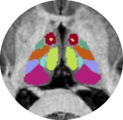
Purpose: Thalamic nuclei are largely invisible in conventional MRI due to poor contrast. Thalamus Optimized Multi-Atlas Segmentation (THOMAS) provides automatic segmentation of 12 thalamic nuclei using white-matter-nulled (WMn) MPRAGE sequence at 7T. Application of THOMAS to Magnetization Prepared 2 Rapid Gradient Echo (MP2RAGE) sequence acquired at 7T has been investigated in this study. Methods: 8 healthy volunteers and 5 pediatric-onset multiple sclerosis patients were recruited at the Children's Hospital of Philadelphia and scanned at Siemens 7T with WMn-MPRAGE and multi-echo MP2RAGE (ME-MP2RAGE) sequences. White-matter-nulled contrast was synthesized (MP2-SYN) from T1 maps from ME-MP2RAGE sequence. Thalamic nuclei were segmented using THOMAS joint label fusion algorithm from WMn-MPRAGE and MP2-SYN datasets. THOMAS pipeline was modified to use majority voting to segment the bias corrected MP2-UNI images. Thalamic nuclei from MP2-SYN and MP2-UNI images were evaluated against corresponding nuclei obtained from WMn-MPRAGE images using dice coefficients, volume similarity indices (VSI) and distance between centroids. Results: For MP2-SYN, dice > 0.85 and VSI > 0.95 was achieved for the 5 larger nuclei and dice > 0.6 and VSI > 0.7 was achieved for the 7 smaller nuclei. The dice and VSI were slightly higher whilst the distance between centroids were smaller for MP2-SYN compared to MP2-UNI, indicating improved performance using the synthesized WMn image. Discussion: THOMAS algorithm can successfully segment thalamic nuclei in routinely acquired bias-free MP2RAGE images with essentially equivalent quality when evaluated against WMn-MPRAGE, hence has wider applicability in studies focused on thalamic involvement in aging and disease.
Quantitative MRI modalities, such as diffusion tensor imaging (DTI) or magnetization transfer imaging (MTI) are sensitive to the neuronal effects of aging of the cerebral white matter (WM), but lack the specificity for myelin content. Myelin water imaging (MWI) is highly specific for myelin and may be more sensitive for the detection of changes in myelin content inside the cerebral WM microstructure. In this multiparametric imaging study, we evaluated the performance of myelin water fraction (MWF) estimates as a marker for myelin alterations during normal-aging. Multiparametric MRI data derived from DTI, MTI and a novel, recently-proposed MWF-map processing and reconstruction algorithm were acquired from 54 healthy subjects (aged 18-79 years) and region-based multivariate regression analysis was performed. MWFs significantly decreased with age in most WM regions (except corticospinal tract) and changes of MWFs were associated with changes of radial diffusivity, indicating either substantial alterations or preservation of myelin content in these regions. Decreases of fractional anisotropy and magnetization transfer ratio were associated with lower MWFs in commissural fiber tracts only. Mean diffusivity had no regional effects on MWF. We conclude that MWF estimates are sensitive for the assessment of age-related myelin alterations in the cerebral WM of normal-aging brains.
 Ravinder Reddy合作论文数Department of Radiology, Perelman School of Medicine, University of Pennsylvania31
 Wilson Neil合作论文数15
 Blake Benyard合作论文数11
 Anshuman Swain合作论文数Univ Maryland, Dept Biol, College Pk, MD 20742 USA11
 Jens Fiehler合作论文数Center for Radiology and Endoscopy, Universitatsklinikum Hamburg-Eppendorf;Department of Neuroradiological diagnostics and intervention, Universitatsklinikum Hamburg-Eppendorf8
 Ashish Raj合作论文数Brain Networks Laboratory, Department of Radiology & Biomedical Imaging, University of California, San Francisco7Jan Sedlacik合作论文数6Tobias D Faizy合作论文数5
 Susan A. Gauthier合作论文数Feil Family Brain and Mind Research Institute, Weill Cornell Medicine, Cornell University5
 Thanh Nguyen合作论文数Department of Radiology, Weill Cornell Medicine, Cornell University5
 Mohammad Haris合作论文数Perelman School of Medicine, University of Pennsylvania5Susanne Siemonsen合作论文数5Hari Hariharan合作论文数5
 Narayan Datt Soni合作论文数4
 Paul S Jacobs合作论文数Perelman School of Medicine, University of Pennsylvania4
 Gabriel Broocks合作论文数Department of Diagnostic and Interventional Neuroradiology, University Medical Center Hamburg-Eppendorf4Christian Thaler合作论文数4Janpatrick Stellmann合作论文数4
 John A. Detre合作论文数Department of Neurology, Perelman School of Medicine, University of Pennsylvania4
 Christoph Heesen合作论文数4Ryan Armbruster合作论文数3
 Hannes Leischner合作论文数3
 Abigail Cember合作论文数B1 Stellar Chance Labs, Univ Penn3
 Deepa S. Thakuri合作论文数Perelman School of Medicine, the University of Pennsylvania3
 Fabian Flottmann合作论文数3
 Shana Mccormack合作论文数3
 Timothy Vartanian合作论文数Department of Neurobiology;Medical School, Harvard;Department of Neurobiology, Harvard Medical School3
 Mark Elliott合作论文数School of Psychological Sciences and Health, University of Strathclyde3
 Andre Kemmling合作论文数2Halvor Juul合作论文数2Kevin D'Aquilla合作论文数2Yiran Li合作论文数Temple Univ, Dept Elect & Comp Engn, Philadelphia, PA 19122 USA2
 Helge Kniep合作论文数2
 Jawed Nawabi合作论文数2
 Stuart B. Prenner合作论文数Department of Medicine, Perelman School of Medicine, University of Pennsylvania2
 Payman Zamani合作论文数Department of Medicine, Perelman School of Medicine, University of Pennsylvania2
 Daniel P. Kelly合作论文数Department of Medicine, Perelman School of Medicine, University of Pennsylvania2
 Harry Ischiropoulos合作论文数Leonard and Madlyn Abramson Pediatric Research Center, Children’s Hospital of Philadelphia, Research Institute;Department of Pediatrics, Perelman School of Medicine, University of Pennsylvania2
 David Roalf合作论文数Brain Behavior Laboratory, Department of Psychiatry, Perelman School of Medicine, University of Pennsylvania2
 Jeremy J. Heit合作论文数Stanford Neuroscience Health Center, Stanford University2
 Datta Ritobrato合作论文数University of Pennsylvania2
 Kenneth B. Margulies合作论文数Margulies Laboratory, Perelman School of Medicine, University of Pennsylvania2
 Uta Hanning合作论文数Department of Clinical Radiology, University of Münster2
 Julio A. Chirinos合作论文数School of Medicine, University of Pennsylvania2
 David C. Poole合作论文数Departments of Anatomy and Physiology, College of Veterinary Medicine, Kansas State University;Department of Kinesiology, College of Health and Human Sciences, Kansas State University2
 Ze Wang合作论文数Center for Advanced Imaging Research, School of Medicine, University of Maryland;Department of Diagnostic Radiology and Nuclear Medicine, School of Medicine, University of Maryland2
 Gerhard Schön合作论文数Center for Experimental Medicine, University Medical Center Hamburg-Eppendorf2Aditya Rao合作论文数1Patrick Borchert合作论文数1




