Objective: Retrograde transfemoral artery catheterization is the most common way of implanting a percutaneous aortic valve. But in some cases, this access cannot be used and the subclavian artery access may represent an alternative to the femoral route, even offering certain advantages. This article describes prosthetic aortic valve implantation using the subclavian arterial approach and reports the findings.Methods: The valve prosthesis is a self-expandable, nitinol-based device (CoreValve; Medtronic Inc. Minneapolis, Minn). The axillary or subclavian artery was exposed with a small incision. Rapid ventricular pacing was used to reduce cardiac output while a routine aortic balloon valvuloplasty was performed. Then, an 18F sheath was inserted into the axillary artery down into the ascending aorta. By using this method, a prosthesis was implanted in 17 patients (aged 71 +/- 11 years) whose surgical risk was deemed excessive because of severe comorbidity and in whom transfemoral catheterization was considered unfeasible or at risk of severe complications.Results: Subclavian arterial injury did not occur in any patient. The postprocedural aortic valve area increased from 0.6 +/- 0.3 cm(2) to 1.44 +/- 0.35 cm(2). A transient ischemic attack occurred in 1 patient. Two patients experienced transitory brachial plexus deficit. There were no intraprocedural deaths. Two deaths occurred in the 30-day follow-up period.Conclusions: This initial experience suggests that subclavian transarterial aortic valve implantation, in selected high-risk patients, is feasible and safe with satisfactory short-term outcomes. (J Thorac Cardiovasc Surg 2011;141:487-91)
An infected aneurysm of the thoracic aorta due to mycobacterium tuberculosis is an unusual entity for which the classical treatment is antituberculosis chemotherapy and open-chest surgery. Recent improvements in endovascular treatments have led to their proposed use for infected aneurysms in patients for whom open surgery poses too high a risk. We report on a 68-year-old man with a tuberculous aortic aneurysm who had been treated with an endoprosthesis and antituberculosis chemotherapy. His clinical and radiological follow-up was uneventful and led to the discontinuation of pharmacological treatment after 16 months. However, a recurrence of the infection led to a fatal aortic rupture 4 months after discontinuation of therapy.
Mathias H. Aazami Torkel Aberg Clemens Aigner Cary W. Akins Nawwar Al-Attar Johannes M. Albes Ivan Aleksic Joseph Alex Vladimir Alexi-Meskishvili Christos Alexiou Ottavio R. Alfieri Zohair Y. Al-Halees Cem H. Alhan Imtiaz S. Ali Marco Alifano Nelson Alphonso Sharif Al-Ruzzeh Muhammad Muneer Amanullah Joseph J. Amato Vincenzo Ambrogi Marcello Carlo Ambrogi Gabriel Amir Luca Ampollini Robert H. Anderson Udo Christian Anegg Gianni D. Angelini Marco Anile Martine Antoine Carlo Antona Manuel J. Antunes Anelechi Anyanwu Jose I. Aramendi Jose Luis Aranda Olgun Kadir Aribas Kitipan V. Arom Hisao Asamura Athanasios C. Athanasiou Kalliopi Athanassiadi Tim Attmann Stephane Aubert Adel Khader Ayed Omer Aziz Jacques F. Azorin Jean Bachet Akin Eraslan Balci Xavier Baldo Yaron Bar-El Hendrick B. Barner David J. Barron Murat Basaran Cristina Basso Roberto Rafael Battellini Eugene Baudet Erwin P. Bauer Robert Bauernschmitt Urs Bauersfeld Yusuf Bayrak Matthias Bechtel David F. Beggs Ion P. Bellenis Jaroslav Benedik Federico Jose Benetti John R. Benfield Stefano Benussi Pascal A. Berdat Marius Berman Jose M. Bernal Marc-Andre Bernath Richard G. Berrisford Antoine A. Beuchat Friedhelm Beyersdorf Olivier Michel Bical Benjamin Bidstrup Paolo Biglioli Dietrich E. Birnbaum Gianluigi Bisleri Hartmuth B. Bittner Edward Black Antonio Bobbio Massimo Bonacchi Johannes Bonatti Pramod Bonde Philipp Bonhoeffer Andreas A. Boning Damien Bonnet Robert Stuart Bonser Michael A. Borger Ahmad Boseila Torsten Bossert Wolfgang Bothe Dietmar Bothig Tomaso Bottio Denis Bouchard Edward Thomas Brackenbury Michael Brandt William J. Brawn Pierre-Yves Brichon Christian Pierre Brizard Ivan Bruschweiler Alan J. Bryan Eric E. Buchser Gerald D. Buckberg Eric Gordon Butchart Jose M. Caffarena Antonio M. Calafiore Ayten K. Cangir Massimo Caputo Paolo Carbognani John Alfred Carr Thierry Carrel Angelo Carretta Raymond Cartier Aaron R. Casha Thierry Caus Alfredo Giuseppe Cerillo Stepan Cerny Alfredo Cesario Juan C. Chachques David J. Chambers Dominique Chapelle Pierre-Guy Chassot Nilanjan Chaudhuri Sylvain Chauvaud Olivier Chavanon Massimo Chello Yih-Sharng Chen Rene Chiolero Ray Chu-Jeng Chiu Cliff K. Choong Emmanuel Choukroun Thomas Decker Christensen Jan T. Christenson Paola Ciriaco Ali Civelek Massimiliano Codispoti Dennis V. Cokkinos Juan V. Comas John V. Conte Massimo Conti Graham J. Cooper Antonio F. Corno Joseph S. Coselli Maurizio Cotrufo Jacques Cotting Elvio Covino Jeremiah N. Cox Jochen T. Cremer John C. Criscione Martin Czerny Sabine H. Dabritz Willem Daenen Mark H.D. Danton Otto E. Dapunt John Dark Hiroshi Date Tirone E. David Michele De Bonis Tiziano De Giacomo Marc R. de Leval Sergio A. de Oliveira Ruggero De Paulis Marc de Perrot Raffaele De Simone Marek Andrzej Deja Alain Francois Delabays Dominique Delay Roland G. Demaria Murat M. Demirtas Thomas Deneke Joseph J. DeRose Paola D’Errigo Mohan P. Devbhandari Roberto Di Bartolomeo Duccio Di Carlo Roberto Di Donato Marco Di Eusanio Verdi J. Di Sesa Giovanni Dialetto Interactive CardioVascular and Thoracic Surgery 6 (2007) 143–146
A surgical procedure that reduces the recurrence of post-infarction posterior ventricular septal defects is described. This technique is based on a double ventriculotomy without an infarctectomy, the use of two patches, and glue, which is applied between the two patches. Excellent results have been obtained in 18 consecutives patients with this simple and reliable technique. 2006 Published by European Association for Cardio-Thoracic Surgery. All rights reserved.
A surgical procedure that reduces the recurrence of post-infarction posterior ventricular septal defects is described. This technique is based on a double ventriculotomy without an infarctectomy, the use of two patches, and glue, which is applied between the two patches. Excellent results have been obtained in 18 consecutives patients with this simple and reliable technique.
BACKGROUND AND AIM OF THE STUDY Following aortic valve replacement (AVR), a residual transprosthetic gradient can develop which has detrimental long-term effects, especially with regard to left ventricular mass regression and subsequent mortality. The Advanced Performance-ATS valve (AP-ATS) was developed to overcome this potential patient-prosthesis mismatch. In the present study, the early postoperative transprosthetic gradient was determined after AVR with ATS valves 18 to 25, to confirm the promise of superior hemodynamic performance of the AP series and to define respective indications. METHODS This prospective study included 302 consecutive patients operated on for AVR between December 1997 and December 2000. In all patients, body surface area (BSA), associated with mean transprosthetic gradient and paravalvular leaks (measured echocardiographically) was monitored, between the 5th and 8th postoperative days. RESULTS A significantly lower mean transprosthetic gradient was found for size 22 and 24 valves, compared with size 23 and 25 valves. Results for size 18 and 20 valves were in concordance with those obtained for size 22 and 24 valves. For size 23 valves the mean gradient was significantly higher when the BSA was >1.9 m2, but the use of size 22 valves in these patients overcame this potential patient-prosthesis mismatch. No patient was reoperated on for paravalvular leakage, and no greater degree of paravalvular leakage was found with AP-ATS than with Standard ATS valves. CONCLUSION These results confirm the global good hemodynamic performances of Standard ATS and especially of the AP-ATS valves. However, the significantly lower mean gradient found in the AP-ATS valves recommends their use in patients with larger BSA.
Objectives: Prosthetic heart valve obstruction (PHVO) is a potentially fatal complication of heart valve replacement with mechanical substitute mainly due to thrombosis. The purpose of this report is to present a single-center experience of 136 consecutive patients operated on between 1978 and 2001. Methods: The diagnosis of PHVO was mainly assessed by fluoroscopy and/or echocardiography. Thrombosed valves were bileaflet (82), tilting disc (47) and ball cage (7) valves; of these, 90 were in mitral, 38 in aortic, six in aortic and mitral position, and two in tricuspid position. The mean interval between the first implantation and valve thrombosis was 7.4 +/- 6.6 years (range I day to 28 years); in 37 patients preoperative medical therapy (fibrinolysis in 21, and heparin alone in 16) was unsuccessful. Results: Operative procedures included valve re-replacement in 104 cases and declotting-pannus excision in 32 cases. Early hospital mortality was 10.3% (14 patients), all in NYHA class III or IV, and one patient suffered a perioperative cerebral embolic event. Surgery was then successful in 121 of 136 patients (89%), but during a 3.15-year follow-up, prosthetic heart valve thrombosis recurred in ten out of 122 survivors (8.1%). Conclusion: From this experience, it can be concluded that for most PHVO, early operation is currently effective and safe, especially in patients in stable hemodynamic condition preoperatively. (C) 2003 Elsevier B.V. All rights reserved.
Objective: Repair of post infarction ventricular septal defect (VSD) is still a challenging procedure with a high risk of recurrence of the VSD and subsequent mortality. The aim of this retrospective study was to assess if technical change in the surgical procedure was followed by an improvement in recurrence of the VSD and operative results. Method: This retrospective study from 197 1 to 2001 included 85 patients operated on early (< 15 days) after the occurrence of a post infarction VSD. Double patch technique was introduced in 1986. A total of 44 variables were studied by a uni- and multivariate analysis. Results: Hospital death occur-red in 36 patients. Significant factors for hospital mortality included: preoperative and evolution of the clinical status, right ventricular function and type of repair (one or two patches). Moreover, no recurrence was observed in patients repaired with the double patch technique (P = 0.09). None of the studied variables were significant for long term survival. Concomitant CABG was not associated with higher hospital mortality and long-term survival rate was similar in patients with or without concomitant CABG. Conclusion: The use of the double patch technique and glue by avoiding recurrence of the VSD played a role in the reduction of the hospital mortality. This technique has to be recommended in the early repair of post infarction VSD. Concomitant CABG can be done safely to control the added risk of an associated coronary artery lesion. (C) 2002 Elsevier Science B.V. All rights reserved.
Background. Postintubation tracheobronchial rupture is usually responsible for unstable intraoperative or postoperative conditions, and its management is discussed. We insist on conservative treatment as a viable alternative after late diagnosis of postintubation tracheobronchial rupture.Methods. We conducted a retrospective study including 14 consecutive patients treated between April 1981 and July 1998.Results. Twelve tracheobronchial ruptures occurred after intubation for general surgery and two after thoracic surgery. In all cases, the tear consisted of a linear laceration of the posterior membranous wall of the tracheobronchial tree ranging from 2 to 6 cm. One death occurred in a very weak patient unfit to undergo a redo operation for surgical repair. Seven patients were treated conservatively and cured without sequelae. Six patients underwent surgical repair, of whom 2 were diagnosed and repaired intraoperatively.Conclusions. Aggressive surgical repair is not always mandatory after delayed diagnosis of iatrogenic tracheobronchial rupture. Conservative treatment must often be considered, except after lung resection. (C) 2000 by The Society of Thoracic Surgeons.
Mathias H. Aazami Torkel Aberg Clemens Aigner Cary W. Akins Nawwar Al-Attar Johannes M. Albes Ivan Aleksic Joseph Alex Vladimir Alexi-Meskishvili Christos Alexiou Ottavio R. Alfieri Zohair Y. Al-Halees Cem H. Alhan Imtiaz S. Ali Marco Alifano Nelson Alphonso Sharif Al-Ruzzeh Muhammad Muneer Amanullah Joseph J. Amato Vincenzo Ambrogi Marcello Carlo Ambrogi Gabriel Amir Luca Ampollini Robert H. Anderson Udo Christian Anegg Gianni D. Angelini Marco Anile Martine Antoine Carlo Antona Manuel J. Antunes Anelechi Anyanwu Jose I. Aramendi Jose Luis Aranda Olgun Kadir Aribas Kitipan V. Arom Hisao Asamura Athanasios C. Athanasiou Kalliopi Athanassiadi Tim Attmann Stephane Aubert Adel Khader Ayed Omer Aziz Jacques F. Azorin Jean Bachet Akin Eraslan Balci Xavier Baldo Yaron Bar-El Hendrick B. Barner David J. Barron Murat Basaran Cristina Basso Roberto Rafael Battellini Eugene Baudet Erwin P. Bauer Robert Bauernschmitt Urs Bauersfeld Yusuf Bayrak Matthias Bechtel David F. Beggs Ion P. Bellenis Jaroslav Benedik Federico Jose Benetti John R. Benfield Stefano Benussi Pascal A. Berdat Marius Berman Jose M. Bernal Marc-Andre Bernath Richard G. Berrisford Antoine A. Beuchat Friedhelm Beyersdorf Olivier Michel Bical Benjamin Bidstrup Paolo Biglioli Dietrich E. Birnbaum Gianluigi Bisleri Hartmuth B. Bittner Edward Black Antonio Bobbio Massimo Bonacchi Johannes Bonatti Pramod Bonde Philipp Bonhoeffer Andreas A. Boning Damien Bonnet Robert Stuart Bonser Michael A. Borger Ahmad Boseila Torsten Bossert Wolfgang Bothe Dietmar Bothig Tomaso Bottio Denis Bouchard Edward Thomas Brackenbury Michael Brandt William J. Brawn Pierre-Yves Brichon Christian Pierre Brizard Ivan Bruschweiler Alan J. Bryan Eric E. Buchser Gerald D. Buckberg Eric Gordon Butchart Jose M. Caffarena Antonio M. Calafiore Ayten K. Cangir Massimo Caputo Paolo Carbognani John Alfred Carr Thierry Carrel Angelo Carretta Raymond Cartier Aaron R. Casha Thierry Caus Alfredo Giuseppe Cerillo Stepan Cerny Alfredo Cesario Juan C. Chachques David J. Chambers Dominique Chapelle Pierre-Guy Chassot Nilanjan Chaudhuri Sylvain Chauvaud Olivier Chavanon Massimo Chello Yih-Sharng Chen Rene Chiolero Ray Chu-Jeng Chiu Cliff K. Choong Emmanuel Choukroun Thomas Decker Christensen Jan T. Christenson Paola Ciriaco Ali Civelek Massimiliano Codispoti Dennis V. Cokkinos Juan V. Comas John V. Conte Massimo Conti Graham J. Cooper Antonio F. Corno Joseph S. Coselli Maurizio Cotrufo Jacques Cotting Elvio Covino Jeremiah N. Cox Jochen T. Cremer John C. Criscione Martin Czerny Sabine H. Dabritz Willem Daenen Mark H.D. Danton Otto E. Dapunt John Dark Hiroshi Date Tirone E. David Michele De Bonis Tiziano De Giacomo Marc R. de Leval Sergio A. de Oliveira Ruggero De Paulis Marc de Perrot Raffaele De Simone Marek Andrzej Deja Alain Francois Delabays Dominique Delay Roland G. Demaria Murat M. Demirtas Thomas Deneke Joseph J. DeRose Paola D’Errigo Mohan P. Devbhandari Roberto Di Bartolomeo Duccio Di Carlo Roberto Di Donato Marco Di Eusanio Verdi J. Di Sesa Giovanni Dialetto Interactive CardioVascular and Thoracic Surgery 6 (2007) 143–146
Intraoperative transesophageal echocardiography (TOE) is being employed increasingly during cardiac surgery. The complication rate attributable to TOE is very low. We report the case of a 71 year old man cardiac laceration due to the TOE during cardiac surgery for by-pass graft and valve replacement. 3 months after the surgery the patient is doing well. We need to keep in mind that TOE could be a possible source of complication.
Background: Left ventricle dysfunction and co-morbidities are responsible for in increased complications after CABG. OPCAB would be an interesting alternative in very high risk patient. Methods : inclusion was decided with at least two of the following criteria : Euroscore > 9, severe LV, recent MI, terminal renal failure, lung dysfunction, Peripheral Vascular Disease, BMI > 30. Patients have been operated with the Octopus® (Medtronic) system. Results : 89 patients, mean age 69.2 ± 10.2 years, with 74 % of men, have been operated. High risk score has been determined by: Euroscore 9.8, LV function 37.7 ± 12 %, MI 57 %, renal failure 46 %, lung dysfunction 38 %, PVD 38 %, obesity 34 %. Mean graft per patient is 2.08 ± 0.84. 3 % of patients were subsequently treated with PTCA for incomplete revascularization. 14.6 % have needed a combined surgery. Early mortality was 3 %. Intensive care unit stay was 2.67 days. Early reported complications were : Low out syndrome 3 %, MI 1 %, Stroke 1 %, kidney support 6 %. Graft permeability was systematically analyzed. Conclusion : OPCAB strategy seems to be effective in very high patients reducing early complication and multi organ failure.
 Labrousse Louis合作论文数5Francesco Madonna合作论文数3Claude Deville合作论文数3C Deville合作论文数3Laurent Barandon合作论文数3Joseph J. Amato合作论文数2Eugène Baudet合作论文数2F Madonna合作论文数2Xavier Baldo合作论文数2Willem Daenen合作论文数2erwin p bauer合作论文数2
 Imtiaz Ali合作论文数Prince Mohammad bin Fahd University2Demaria Roland G合作论文数2
 Matthias Bechtel合作论文数2Munos Emmanuel合作论文数2
 Otto Dapunt合作论文数Klinikum Oldenburg2
 Thierry Carrel合作论文数Department of Cardiac Surgery, University Hospital Zürich2
 Ottavio Alfieri合作论文数Cardiochirurgia, San Raffaele Hospital2David Beggs合作论文数2
 John Dark合作论文数Translational and Clinical Research Institute, Faculty of Medical Sciences, Newcastle University2
 John V Conte合作论文数Johns Hopkins School of Medicine;Geisinger Health System;Penn State University2
 Gabriel Amir合作论文数2
 Pascal Leprince合作论文数Department of Cardiothoracic Surgery, La Pitié Salpêtrère Hospital, Pierre and Marie Curie University2John Alfred Carr合作论文数2Alan J. Bryan合作论文数2
 Marco Anile合作论文数University of Rome2
 Paola Ciriaco合作论文数2Jan T. Christenson合作论文数2Ali Civelek合作论文数Department of Cardiovascular Surgery, Marmara University School of Medicine2Pierre-Guy Chassot合作论文数2
 Massimo Bonacchi合作论文数Heart and Vessel Department, Careggi Teaching Hospital2Torsten Bossert合作论文数2Edward Black合作论文数2
 Hendrick Barner合作论文数School of Medicine, Saint Louis University2Jacques Cotting合作论文数2Tim Attmann合作论文数2
 Jose Luis Aranda合作论文数2Graham Cooper合作论文数Cardiothoracic Surgery, Northern General Hospital2
 Thierry Caus合作论文数2Kalliopi Athanassiadi合作论文数2
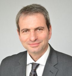 Marius Berman合作论文数Department of Cardiothoracic Surgery;Rabin Medical Center;Department of Cardiothoracic Surgery, Rabin Medical Center2Dietrich E. Birnbaum合作论文数2Antoine Beuchat合作论文数2
 Jean Bachet合作论文数2
 Ivan Aleksic合作论文数2
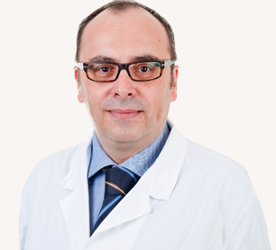 Massimo Chello合作论文数Policlinico Universitario Campus Bio-Medico2Olivier Chavanon合作论文数Institution: Grenoble University Hospital2
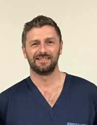 Gianluigi Bisleri合作论文数Division of Cardiac Surgery, University of Brescia Medical School2
 Luca Ampollini合作论文数Department of Surgery, University of Parma2




