Complement C1q is part of the C1 macromolecular complex that mediates the classical complement activation pathway: a major arm of innate immune defense. C1q is composed of A, B, and C chains that require post-translational prolyl 4-hydroxylation of their N-terminal collagen-like domain to enable the formation of the functional triple helical multimers. The prolyl 4-hydroxylase(s) that hydroxylate C1q have not previously been identified. Recognized prolyl 4-hydroxylases include collagen prolyl-4-hydroxylases (CP4H) and the more recently described prolyl hydroxylase domain (PHD) enzymes that act as oxygen sensors regulating hypoxia-inducible factor (HIF). We show that several small-molecule prolyl hydroxylase inhibitors that activate HIF also potently suppress C1q secretion by human macrophages. However, reducing oxygenation to a level that activates HIF does not compromise C1q hydroxylation. In vitro studies showed that a C1q A chain peptide is not a substrate for PHD2 but is a substrate for CP4H1. Circulating levels of C1q did not differ between wild-type mice or mice with genetic deficits in PHD enzymes, but were reduced by prolyl hydroxylase inhibitors. Thus, C1q is hydroxylated by CP4H, but not the structurally related PHD hydroxylases. Hence, reduction of C1q levels may be an important off-target side effect of small molecule PHD inhibitors developed as treatments for renal anemia.
that stabilize hypoxia-inducible factor Serafim Kiriakidis, Simon S. Hoer, Natalie Burrows, Gloria Biddlecome, Moddasar N. Khan, Cyrille C. Thinnes, Christopher J. Schofield, Norma Rogers, Marina Botto, Ewa Paleolog and Patrick H. Maxwell Kennedy Institute of Rheumatology, NDORMS, University of Oxford, Oxford, UK; School of Clinical Medicine, Cambridge Institute for Medical Research, Cambridge Biomedical Campus, University of Cambridge, Cambridge, UK; Oncology Research, AMGEN, Thousand Oaks, California, USA; Cobra Biologics, Keele, UK; Department of Chemistry, University of Oxford, Oxford, UK; and Centre for Complement and Inflammation Research (CCIR), Imperial College London, London, UK
Key points Combining nitric oxide (NO)‐mediated increased blood flow with angiopoietin‐1–Tie2 receptor signalling induces arteriolargenesis – the formation of arterioles from capillaries – in a model of physiological angiogenesis. This NO–Tie‐mediated arteriolargenesis requires endogenous vascular endothelial growth factor (VEGF) signalling. Inhibition of VEGF signalling increases pericyte coverage in microvessels. Together these findings indicate that generation of functional neovasculature requires close titration of NO–Tie2 signalling and localized VEGF induction, suggesting that the use of exogenous VEGF expression as a therapeutic for neovascularization may not be successful. AbstractSignalling through vascular endothelial growth factor (VEGF) receptors and the tyrosine kinase with IgG and EGF domains‐2 (Tie2) receptor by angiopoietins is required in combination with blood flow for the formation of a functional vascular network. We tested the hypothesis that VEGF and angiopoietin‐1 (Ang1) contribute differentially to neovascularization induced by nitric oxide (NO)‐mediated vasodilatation, by comparing the phenotype of new microvessels in the mesentery during induction of vascular remodelling by over‐expression of endothelial nitric oxide synthase in the fat pad of the adult rat mesentery during inhibition of angiopoietin signalling with soluble Tie2 (sTie2) and VEGF signalling with soluble Fms‐like tyrosine kinase receptor‐1 (sFlt1). We found that NO‐mediated angiogenesis was blocked by inhibition of VEGF with sFlt1 (from 881 ± 98% increase in functional vessel area to 279 ± 72%) and by inhibition of angiopoietin with sTie2 (to 337 ± 67%). Exogenous angiopoietin‐1 was required to induce arteriolargenesis (8.6 ± 1.3% of vessels with recruitment of vascular smooth muscle cells; VSMCs) in the presence of enhanced flow. sTie2 and sFlt1 both inhibited VSMC recruitment (both 0%), and VEGF inhibition increased pericyte recruitment to newly formed vessels (from 27 ± 2 to 54 ± 3% pericyte ensheathment). We demonstrate that a fine balance of VEGF and angiopoietin signalling is required for the formation of a functional vascular network. Endogenous VEGF signalling prevents excess neovessel pericyte coverage, and is required for VSMC recruitment during increased nitric oxide‐mediated vasodilatation and angiopoietin signalling (NO–Tie‐mediated arteriogenesis). Therapeutic vascular remodelling paradigms may therefore require treatments that modulate blood flow to utilize endogenous VEGF, in combination with exogenous Ang1, for effective neovascularization.
Factor-inhibiting hypoxia-inducible factor (HIF)-1 (FIH-1) is an asparaginyl beta-hydroxylase enzyme that was initially found to hydroxylate the HIF-alpha, preventing its transcriptional activity and leading to adaptive responses to hypoxia. More recently, other substrates, such as neurogenic locus notch homolog (Notch), have been found to be alternative FIH targets, but the biologic relevance of this regulation was never investigated. Given the key function of Notch in angiogenesis, we here investigate the role of FIH/Notch signaling in endothelial cells. We report that FIH-1 silencing in HUVECs results in reduced growth and increased apoptosis. The knockdown of FIH is associated with increased Notch2 activity, leading to enhanced expression of the Notch target hairy/enhancer-of-split related with YRPW motif protein 1 (Hey-1). Consistent with recent findings showing that Notch2 suppresses survivin (a key inhibitor of apoptosis), FIH targeting in HUVECs leads to selective repression of survivin in endothelial cells, thus promoting cell apoptosis and growth arrest. Our data support the concept that FIH-1 may interact with Notch2 and repress its activity, thereby playing a critical role in controlling the survival of vascular endothelial cells. These findings might pave the way toward novel, antiangiogenic strategies in disorders that are characterized by excessive vascular growth, such as cancer and rheumatoid arthritis.
Hypoxia may contribute to the pathogenesis of various diseases of the vascular wall. Hypoxia-inducible factors (HIFs) are nuclear transcriptional factors that regulate the transcription of genes that mediate cellular and tissue homeostatic responses to altered oxygenation. This article reviews the published literature on and discusses the role of the HIF pathway in diseases involving the vascular wall, including atherosclerosis, arterial aneurysms, pulmonary hypertension, vascular graft failure, chronic venous diseases, and vascular malformation.PubMed was searched with the terms "hypoxia-inducible factor" or "HIF" and "atherosclerosis," "carotid stenosis," "aneurysm," "pulmonary artery hypertension," "varicose veins," "venous thrombosis," "graft thrombosis," and "vascular malformation."In atherosclerotic plaque, HIF-1α was localized in macrophages and smooth muscle cells bordering the necrotic core. Increased HIF-1α may contribute to atherosclerosis through alteration of smooth muscle cell proliferation and migration, angiogenesis, and lipid metabolism. The expression of HIF-1α is significantly elevated in aortic aneurysms compared with nonaneurysmal arteries. In pulmonary hypertension, HIF-1α contributes to the increase of intracellular K(+) and Ca(2+) leading to vasoconstriction of pulmonary smooth muscle cells. Alteration of the HIF pathway may contribute to vascular graft failure through the formation of intimal hyperplasia. In chronic venous disease, HIF pathway dysregulation contributes to formation of varicose veins and venous thromboembolism. However, whether the activation of the HIF pathway is protective or destructive to the venous wall is unclear. Increased activation of the HIF pathway causes aberrant expression of angiogenic factors contributing to the formation and maintenance of vascular malformations.Pathologic vascular wall remodelling of many common diseases of the blood vessels has been found to be associated with altered activity of the HIF pathway. Therefore, understanding the role of the HIF pathway in diseases of the vascular wall is important to identify novel therapeutic strategies in the management of these pathologies.
Background Colorectal cancer (CRC) is characterised by hypoxia, which activates gene transcription through hypoxia-inducible factors (HIF), as well as by expression of epidermal growth factor (EGF) and EGF receptors, targeting of which has been demonstrated to provide therapeutic benefit in CRC. Although EGF has been demonstrated to induce expression of angiogenic mediators, potential interactions in CRC between EGF-mediated signalling and the hypoxia/HIF pathway remain uncharacterised. Methods PCR-based profiling was applied to identify angiogenic genes in Caco-2 CRC cells regulated by hypoxia, the hypoxia mimetic dimethyloxallylglycine (DMOG) and/or EGF. Western blotting was used to determine the role of HIF-1alpha, HIF-2alpha and MAPK cell signalling in mediating the angiogenic responses. Results We identified a total of 9 angiogenic genes, including angiopoietin-like (ANGPTL) 4, ephrin (EFNA) 3, transforming growth factor (TGF) β1 and vascular endothelial growth factor (VEGF), to be upregulated in a HIF dependent manner in Caco-2 CRC cells in response to both hypoxia and the hypoxia mimetic dimethyloxallylglycine (DMOG). Stimulation with EGF resulted in EGFR tyrosine autophosphorylation, activation of p42/p44 MAP kinases and stabilisation of HIF-1α and HIF-2α proteins. However, expression of 84 angiogenic genes remained unchanged in response to EGF alone. Crucially, addition of DMOG in combination with EGF significantly increased expression of a further 11 genes (in addition to the 9 genes upregulated in response to either DMOG alone or hypoxia alone). These additional genes included chemokines (CCL-11/eotaxin-1 and interleukin-8), collagen type IV α3 chain, integrin β3 chain, TGFα and VEGF receptor KDR. Conclusion These findings suggest that although EGFR phosphorylation activates the MAP kinase signalling and promotes HIF stabilisation in CRC, this alone is not sufficient to induce angiogenic gene expression. In contrast, HIF activation downstream of hypoxia/DMOG drives expression of genes such as ANGPTL4, EFNA3, TGFβ1 and VEGF. Finally, HIF activation synergises with EGF-mediated signalling to additionally induce a unique sub-group of candidate angiogenic genes. Our data highlight the complex interrelationship between tumour hypoxia, EGF and angiogenesis in the pathogenesis of CRC.
Hypoxia may contribute to the pathogenesis of various diseases of the vascular wall. Hypoxia-inducible factors (HIFs) are nuclear transcriptional factors that regulate the transcription of genes that mediate cellular and tissue homeostatic responses to altered oxygenation. This article reviews the published literature on and discusses the role of the HIF pathway in diseases involving the vascular wall, including atherosclerosis, arterial aneurysms, pulmonary hypertension, vascular graft failure, chronic venous diseases, and vascular malformation.PubMed was searched with the terms "hypoxia-inducible factor" or "HIF" and "atherosclerosis," "carotid stenosis," "aneurysm," "pulmonary artery hypertension," "varicose veins," "venous thrombosis," "graft thrombosis," and "vascular malformation."In atherosclerotic plaque, HIF-1α was localized in macrophages and smooth muscle cells bordering the necrotic core. Increased HIF-1α may contribute to atherosclerosis through alteration of smooth muscle cell proliferation and migration, angiogenesis, and lipid metabolism. The expression of HIF-1α is significantly elevated in aortic aneurysms compared with nonaneurysmal arteries. In pulmonary hypertension, HIF-1α contributes to the increase of intracellular K(+) and Ca(2+) leading to vasoconstriction of pulmonary smooth muscle cells. Alteration of the HIF pathway may contribute to vascular graft failure through the formation of intimal hyperplasia. In chronic venous disease, HIF pathway dysregulation contributes to formation of varicose veins and venous thromboembolism. However, whether the activation of the HIF pathway is protective or destructive to the venous wall is unclear. Increased activation of the HIF pathway causes aberrant expression of angiogenic factors contributing to the formation and maintenance of vascular malformations.Pathologic vascular wall remodelling of many common diseases of the blood vessels has been found to be associated with altered activity of the HIF pathway. Therefore, understanding the role of the HIF pathway in diseases of the vascular wall is important to identify novel therapeutic strategies in the management of these pathologies.
During tumour progression, oxygen tension in the microenvironment surrounding tumour cells is reduced, resulting in hypoxia. It is well established that cancer cells resist the negative effects of hypoxia by inducing angiogenesis predominantly via the activity of transcription factor hypoxia-inducible factor-1 (HIF-1). However, more recently HIF-1α has also been linked to increased invasive potential, although the molecular mechanisms remain to be defined. Invasive cancer cells are thought to employ membrane protrusions, termed invadopodia, to achieve matrix degradation. While many invadopodia components have been identified, signalling pathways that link extracellular stimuli to invadopodia formation remain largely unknown. Indeed, the relationship between invadopodia formation and HIF-1α has not been explored. We now report that HIF-1α is a driver of invadopodia formation. Furthermore, we have identified an important, direct and novel link between the Rho family activator β-PIX, HIF-1α and invadopodia formation. Indeed, we find that β-PIX expression is essential for invadopodia formation. In conclusion, we identify a new HIF-1α mechanistic pathway and suggest that β-PIX is a novel downstream signalling mediator during invadopodia formation.
The study aimed to investigate the viability of a varicose vein (VV) organ culture model by assessing cell death pattern. To assess pattern of cell death with time, VV organ cultures were incubated for up to 14 days with regular medium changed. To assess viability, cell death of VV organ cultures treated with sodium azide and their untreated counterparts was assayed. Increased cell death was measured in VV organ cultures from day 0 to 2. Cell death decreased gradually after day 2 and plateaued from day 8 to 14. VV organ cultures treated with sodium azide demonstrated significantly more cell death in tissue (P = 0.001). Cell death measured in cultures treated with sodium azide continued to increase until day 7. In conclusion, this study demonstrated the viability of a VV organ culture model with most cell death occurred within the first two days and then declined to a relatively low level.
The vascular endothelial lining of blood vessels plays a key 'target-effector' role in vivo, integrating the body's response to inflammatory cytokines, chemokines and growth factors (derived from both endothelial cells themselves and from other cells such as leukocytes and fibroblasts), to allow leukocyte activation, adhesion and extravasation from the flowing blood into underlying tissue. Endothelial proliferation, through the process of angiogenesis, results in an increased cell surface area for these events to occur, and further functions to deliver oxygen and nutrients, and to remove waste products. In addition to playing an important role in physiology, the endothelium is thus an active participant in inflammatory pathologies. One of the best understood diseases in which inflammation and angiogenesis play a part is rheumatoid arthritis (RA). Blockade of the inflammatory cascade in RA has significant consequences for the vasculature, highlighting the links between reducing endothelial activation and therapeutic benefit in chronic inflammatory disorders.
OBJECTIVE:Rheumatoid arthritis (RA) is characterized by hypoxia and the expression of hypoxia-inducible transcription factors (HIFs), which coordinate cellular responses to hypoxia. The objective of this study was to analyze the expression and regulation of prolyl hydroxylase domain (PHD) enzymes and factor-inhibiting HIF-1α (FIH-1), which regulate cellular HIF levels, and to study the roles of these enzymes in RA fibroblast-like synoviocytes (RA FLS).METHODS:The expression of PHD and FIH and downstream target genes was assessed by quantitative polymerase chain reaction and Western blotting. A small interfering RNA (siRNA) approach and an in vitro endothelial cell angiogenesis assay were used to analyze the roles of HIF hydroxylases.RESULTS:In human RA FLS, knockdown of PHD-2, but not knockdown of PHD-1 or FIH-1, dramatically augmented HIF-1α expression, modestly increased HIF-2α protein expression under normoxic conditions, and up-regulated HIF-dependent gene expression. In contrast, silencing of PHD-3 up-regulated HIF-2α but reduced HIF-1α, thereby decreasing the expression of HIF-regulated genes. A similar effect of PHD-2 knockdown was observed in osteoarthritis FLS (OA FLS) but not in nondiseased primary human dermal fibroblasts. These findings correlated with the induction of in vitro angiogenesis by supernatants from RA FLS and OA FLS transfected with siPHD-2 but not by supernatants from nondiseased fibroblasts or from siPHD-3-transfected cells.CONCLUSION:Our data suggest that PHD-2 is the major hydroxylase regulating HIF levels and the expression of angiogenic genes in arthritic cells. PHD-2 appears to regulate responses relevant to arthritis via HIF-α, highlighting the major importance of this enzyme in hypoxia- and angiogenesis-dependent inflammatory diseases such as RA.
Background: Doxycycline and micronized purified flavonoid fraction (MPFF) modulate vein wall remodeling that may be associated with hypoxia in varicose veins (VVs), vein graft stenosis, and deep venous thrombosis. We recently reported that in vitro exposure of non-VV (NVVs) and VVs to hypoxic conditions activates the hypoxia-inducible factor (HIF) pathway. This study investigated the in vitro effects of doxycycline and MPFF on the HIF pathway in hypoxic NVVs and VVs.Methods: Six NVVs and six VVs obtained from surgery were used to prepare vein organ cultures, which were exposed to hypoxia (1% O-2), with and without MPFF (10(-5) mol/L) or doxycycline (5 mu g/mL) for 16 hours. The veins were analyzed for HIF-1 alpha, HIF-2 alpha, and their target gene expression, with real-time polymerase chain reaction and Western blot. The differences between gene expressions were tested with one-way analysis of variance with repeated measures, followed by the Dunnett test for multiple comparisons. P < .05 was considered significant.Results: Treatment of NVV organ cultures exposed to hypoxia with doxycycline or MPFF did not significantly alter the expression of HIF-1 alpha and HIF-2 alpha messenger (m) RNA and protein compared with untreated. Doxycycline also did not significantly affect the expression of HIF-1 alpha and HIF-2 alpha mRNA and protein in VVs exposed to hypoxia compared with untreated VVs. However, MPFF significantly reduced the expression of HIF-1 alpha but not HIF-2 alpha mRNA in VVs exposed to hypoxia compared with untreated VVs. Interestingly, the reduction of the expression of HIF-1 alpha mRNA in VVs by MPFF was not reflected at the protein level. The mRNA expression of HIF target genes, namely glucose transporter-1, carbonic anhydrase-9, vascular endothelial growth factor, B-cell lymphoma 2/adenovirus E1B 19-kDa protein-interacting protein 3, prolyl hydroxylase domain-2, and prolyl hydroxylase domain-3, was not significantly altered in NVVs and VVs exposed to hypoxia and treated with doxycycline or MPFF compared with those untreated.Conclusions: Doxycycline and MPFF at a concentration corresponding to a therapeutic dose do not alter the activation of the HIF pathway in NVV and VV organ cultures exposed to hypoxia. Our findings suggest vein wall remodeling actions in NVVs and VVs are likely not HIF-dependent. (J Vasc Surg 2012;56:1069-77.)
INTRODUCTION:Dysregulated angiogenesis is implicated in the pathogenesis of rheumatoid arthritis (RA). To provide a more profound understanding of arthritis-associated angiogenesis, we evaluated the expression of angiogenesis-modulating genes at onset, peak and declining phases of collagen-induced arthritis (CIA), a well-established mouse model for RA.METHODS:CIA was induced in DBA/1 mice with type II collagen. Functional capillary density in synovial tissue of knee joints was determined by intravital fluorescence microscopy. To assess the ability of arthritic joint homogenates to induce angiogenesis, an endothelial chemotaxis assay and an in vivo matrigel plug assay were employed. The temporal expression profile of angiogenesis-related genes in arthritic paws was analysed by quantitative real-time RT-PCR using an angiogenesis focused array as well as gene specific PCR. Finally, we investigated the therapeutic effect of a monoclonal antibody specifically blocking the binding of VEGF to neuropilin (NRP)-1.RESULTS:Although arthritic paw homogenates displayed angiogenic activity in vitro and in vivo, and synovia of arthritic paws appeared highly vascularised on histological examination, the functional capillary density in arthritic knee synovia was significantly decreased, whereas capillary diameter was increased. Of the 84 genes analysed, 41 displayed a differential expression in arthritic paws as compared to control paws. Most significant alterations were seen at the peak of clinical arthritis. Increased mRNA expression could be observed for VEGF receptors (Flt-1, Flk-1, Nrp-1, Nrp-2), as well as for midkine, hepatocyte growth factor, insulin-like growth factor-1 and angiopoietin-1. Signalling through NRP-1 accounted in part for the chemotactic activity for endothelial cells observed in arthritic paw homogenates. Importantly, therapeutic administration of anti-NRP1B antibody significantly reduced disease severity and progression in CIA mice.CONCLUSIONS:Our findings confirm that the arthritic synovium in murine CIA is a site of active angiogenesis, but an altered balance in the expression of angiogenic factors seems to favour the formation of non-functional and dilated capillaries. Furthermore, our results validate NRP-1 as a key player in the pathogenesis of CIA, and support the VEGF/VEGF receptor pathway as a potential therapeutic target in RA.
The importance of inflammation in rheumatoid arthritis (RA) is well understood. This knowledge has resulted in the development of anti-inflammatory therapies--either broadly acting (such as steroids) or more specific approaches (such as antibodies against TNF)--with biologic therapies (including TNF inhibitors) revolutionizing the treatment of RA. However, what is less well appreciated in RA are the links between inflammation, blood-vessel formation (angiogenesis) and cellular responses to changes in oxygen tension. Inadequate oxygenation, termed hypoxia, is thought to drive the increase in synovial angiogenesis that occurs in RA, through expression of hypoxia-inducible molecules, including vascular endothelial growth factor (VEGF). This process promotes further infiltration of inflammatory cells and production of inflammatory mediators, perpetuating synovitis. This Review highlights the molecular pathways activated by hypoxia, and how these pathways might interact with inflammatory signaling to promote and maintain synovitis in RA, with a particular focus on the response of macrophages to hypoxia in the context of RA. Successful treatment of RA, for example with anti-TNF antibodies, reduces levels of proangiogenic factors, including VEGF, and leads to normalization of the vasculature. These processes emphasise the close links between hypoxia, angiogenesis and inflammation in this disease and supports the concept that angiogenesis blockade could be of therapeutic benefit in RA.
INTRODUCTION:Hypoxia and T-helper cell 1 (Th1) cytokine-driven inflammation are key features of rheumatoid arthritis (RA) and contribute to disease pathogenesis by promoting angiogenesis. The objective of our study was to characterise the angiogenic gene signature of RA fibroblast-like synoviocytes (FLS) in response to hypoxia, as well as Th1 and T-helper cell 2 (Th2) cytokines, and in particular to dissect out effects of combined hypoxia and cytokines on hypoxia inducible transcription factors (HIFs) and angiogenesis.METHODS:Human angiogenesis PCR arrays were used to screen cDNA from RA FLS exposed to hypoxia (1% oxygen) or dimethyloxalylglycine, which stabilises HIFs. The involvement of HIF isoforms in generating the angiogenic signature of RA FLS stimulated with hypoxia and/or cytokines was investigated using a DNA-binding assay and RNA interference. The angiogenic potential of conditioned media from hypoxia-treated and/or cytokine-treated RA FLS was measured using an in vitro endothelial-based assay.RESULTS:Expression of 12 angiogenic genes was significantly altered in RA FLS exposed to hypoxia, and seven of these were changed by dimethyloxalylglycine, including ephrin A3 (EFNA3), vascular endothelial growth factor (VEGF), adipokines angiopoietin-like (ANGPTL)-4 and leptin. These four proangiogenic genes were dependent on HIF-1 in hypoxia to various degrees: EFNA3 >ANGPTL-4 >VEGF >leptin. The Th1 cytokines TNFα and IL-1β induced HIF-1 but not HIF-2 transcription as well as activity, and this effect was additive with hypoxia. In contrast, Th2 cytokines had no effect on HIFs. IL-1β synergised with hypoxia to upregulate EFNA3 and VEGF in a HIF-1-dependent fashion but, despite strongly inducing HIF-1, TNFα suppressed adipokine expression and had minimal effect on EFNA3. Supernatants from RA FLS subjected to hypoxia and TNFα induced fewer endothelial tubules than those from FLS subjected to TNFα or hypoxia alone, despite high VEGF protein levels. The Th2 cytokine IL-4 strongly induced ANGPTL-4 and angiogenesis by normoxic FLS and synergised with hypoxia to induce further proangiogenic activity.CONCLUSION:The present work demonstrates that Th1 cytokines in combination with hypoxia are not sufficient to induce angiogenic activity by RA FLS despite HIF-1 activation and VEGF production. In contrast, Th2 cytokines induce angiogenic activity in normoxia and hypoxia, despite their inability to activate HIFs, highlighting the complex relationships between hypoxia, angiogenesis and inflammation in RA.
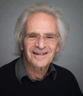 Marc Feldmann合作论文数Kennedy Institute of Rheumatology, Medical Sciences Division, University of Oxford;Somerville College, University of Oxford42
 Serafim Kiriakidis合作论文数Nuffield Department of Orthopaedics, Rheumatology and Musculoskeletal Sciences, University of Oxford38
 Claudia Monaco合作论文数The Kennedy Institute of Rheumatology, University of Oxford15
 Peter Taylor合作论文数St Peter's College, University of Oxford;Medical Sciences Division, University of Oxford10
 Alun Davies合作论文数Division of Surgery, Department of Surgery & Cancer, Faculty of Medicine, Imperial College London10
 Evangelos Andreakos合作论文数Kennedy Institute of Rheumatology10
 Leigh Madden合作论文数Department of Sport, Health and Exercise Science, University of Hull8Helene Larsen合作论文数7Luke L. Gompels合作论文数Kennedy Institute of Rheumatology, Imperial College London7R.N. Maini合作论文数7Fionula M. Brennan合作论文数6Ann Sandison合作论文数Charing Cross Hospital6Mohammed A. Akhavani合作论文数6Miotla J M合作论文数5Brian M. Foxwell合作论文数5Lorraine E. Harry合作论文数5Yvonne Raatz合作论文数4H Tchórzewski合作论文数4
 Muz Barbara合作论文数Department of Radiation Oncology, Washington University in Saint Louis School of Medicine4
 Dorian Haskard合作论文数National Heart & Lung Institute, Imperial College London;Faculty of Medicine, Imperial College London;Royal Postgraduate Medical School4
 Richard Williams合作论文数Nuffield Department of Orthopaedics, Rheumatology and Musculoskeletal Sciences, University of Oxford4
 Ngayu Thairu合作论文数Faculty of Medicine, Imperial College London4Norbert V. Kang合作论文数4
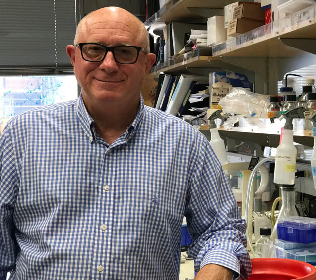 H. Michael Shepard合作论文数Biologics21.NET4Jennifer Grosjean合作论文数4Percy F. Sumariwalla合作论文数4
 Jagdeep Nanchahal合作论文数Department of Plastic and Reconstructive Surgery, Imperial College School of Medicine4Chung S. Lim合作论文数4Moddasar N. Khan合作论文数3Branavan Sivakumar合作论文数3Peter Dawson合作论文数3K Zeman合作论文数3Ballara S C合作论文数3Afuwape A O合作论文数3
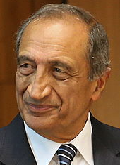 Ravinder N. Maini合作论文数Kennedy Institute of Rheumatology;Imperial College London3
 Tonia Vincent合作论文数Medical Sciences Division, University of Oxford;Centre for OA Pathogenesis, University of Oxford3Tak Loon Khong合作论文数Kennedy Institute of Rheumatology, Imperial College London3Michael Pearse合作论文数3Sylvia Young合作论文数3
 Ngee Han Lim合作论文数The Kennedy Institute of Rheumatology, Imperial College of London3James G. Carter合作论文数2John Bainbridge合作论文数2Richard McCloskey合作论文数2Simon S. Hoer合作论文数2ellen mcconnell合作论文数2Kay McNamee合作论文数2Damian Crawford合作论文数2
 Maria J C Machado合作论文数2




