CobE, a protein implicated in vitamin B12 biosynthesis, from Pseudomonas aeruginosa has been overexpressed in Escherichia coli, purified and crystallized using hanging-drop vapour diffusion. The crystals belong to the primitive orthorhombic space group P2(1)2(1)2(1), with unit-cell parameters a = 31.86, b = 41.07, c = 87.41 A. The diffraction extends to a resolution of 1.9 A. There is one molecule per asymmetric unit and the estimated solvent content is 35%. SeMet-labelled CobE has been prepared and crystallizes under the same conditions as the native protein with diffraction to 1.7 A. The anomalous measurements will be used for phasing.
One of the most intriguing steps during cobalamin (vitamin B12) biosynthesis is the ring contraction process that leads to the extrusion of one of the integral macrocyclic carbon atoms from the tetrapyrrole-derived framework. The aerobic cobalamin pathway requires the action of a monooxygenase called CobG (precorrin-3B synthase), which generates a hydroxylactone intermediate that is subsequently ring-contracted by CobJ. However, in the photosynthetic bacterium Rhodobacter capsulatus, which harbors an aerobic-like pathway, there is no cobG in the main cobalamin biosynthetic operon although it does contain an additional uncharacterized gene called orf663. To demonstrate the involvement of Orf663 in cobalamin synthesis, the first dedicated 10 genes of the B12 pathway (including orf663), encoding enzymes for the transformation of uroporphyrinogen III into hydrogenobyrinic acid (HBA), were sequentially cloned into a plasmid to generate an artificial operon, which, when transformed into Escherichia coli, endowed the host with the ability to make HBA. Deletion of orf663 from this operon prevented HBA synthesis, demonstrating that it was essential for corrin construction. HBA synthesis was restored to this recombinant strain either by returning orf663 or by substituting it with cobG. Recombinant overproduction of Orf663, now renamed CobZ, allowed the characterization of a novel cofactor-rich protein, housing two Fe-S centers, a flavin, and a heme group, which like B12 itself is a modified tetrapyrrole. A mechanism for Orf663 (CobZ) in cobalamin biosynthesis is proposed.
The cobaltochelatase required for the synthesis of vitamin B12 (cobalamin) in the archaeal kingdom has been identified as CbiX through similarity searching with the CbiX from Bacillus megaterium. However, the CbiX proteins in the archaea are much shorter than the CbiX proteins found in eubacteria, typically containing less than half the number of amino acids in their primary structure. For this reason the shorter CbiX proteins have been termed CbiXS and the longer versions CbiXL. The CbiXS proteins from Methanosarcina barkeri and Methanobacter thermoautotrophicum were overproduced in Escherichia coli as recombinant proteins and characterized. Through complementation studies of a defined chelatase-deficient strain of E. coli and by direct in vitro assays the function of CbiXS as a sirohydrochlorin cobaltochelatase has been demonstrated. On the basis of sequence alignments and conserved active site residues we suggest that CbiXS may represent a primordial chelatase, giving rise to larger chelatases such as CbiXL, SirB, CbiK, and HemH through gene duplication and subsequent variation and selection. A classification scheme for chelatases is proposed.
In Bacillus megaterium, the hemAXBCDL genes were isolated and were found to be highly similar to the genes from Bacillus subtilis that are required for the conversion of glutamyl-tRNA into uroporphyrinogen III. Overproduction and purification of HemC (porphobilinogen deaminase) and -D (uroporphyrinogen III synthase) allowed these enzymes to be used for the in vitro synthesis of uroporphyrinogen III from porphobilinogen. A second smaller cluster of three genes (termed sirABC) was also isolated and found to encode the enzymes that catalyse the transformation of uroporphyrinogen III into sirohaem on the basis of their ability to complement a defined Escherichia coli (cysG) mutant. The functions of SirC and -B were investigated by direct enzyme assay, where SirC was found to act as a precorrin-2 dehydrogenase, generating sirohydrochlorin, and SirB was found to act as a ferrochelatase responsible for the final step in sirohaem synthesis. CbiX, a protein found encoded within the main B. megaterium cobalamin biosynthetic operon, shares a high degree of similarity with SirB and acts as the cobaltochelatase associated with cobalamin biosynthesis by inserting cobalt into sirohydrochlorin. CbiX contains an unusual histidine-rich region in the C-terminal portion of the protein, which was not found to be essential in the chelation process. Sequence alignments suggest that SirB and CbiX share a similar active site to the cobaltochelatase, CbiK, from Salmonella enterica.
CbiX is a cobaltochelatase required for the biosynthesis of vitamin B-12 and is found in Archaea as a short form (CbiX(S) containing 120 - 145 amino acids) and in some bacteria as a longer version ( CbiX(L) containing 300 - 350 amino acids). Purification of either recombinant Bacillus megaterium or Synechocystis CbiXL in Escherichia coli, which is facilitated by the presence of a naturally occurring histidine-rich region of the protein, results in the isolation of a dark brown protein solution. The UV/ visible spectrum of the protein is consistent with the presence of a redox group, and the lack of definition within the spectrum is suggestive of a 4Fe-4S center. The presence of an iron-sulfur center was confirmed by EPR analysis of the proteins, which produces a pseudoaxial spectrum with g values at 2.04, 1.94, and 1.90. The EPR spectrum was absent at 70 K, an observation that is diagnostic of a 4Fe-4S center. Redox potentiometry coupled with optical spectroscopy allowed the midpoint potential of the redox center to be determined for the CbiXL from both B. megaterium and Synechocystis. Sequence analysis of CbiXL proteins reveals only two conserved cysteine residues within the CbiXL proteins, which are part of an MXCXXC motif. Mutagenesis of the two cysteines leads to loss of both the EPR spectrum and UV/ visible spectral features of the Fe-S center in the protein, clearly indicating that these residues are involved in ligating the cofactor to the apoprotein possibly in a butterfly arrangement. The potential physiological role of the iron-sulfur center is discussed.
Vitamin B12, or cobalamin, is one of the most structurally complex small molecules made in Nature. Major progress has been made over the past decade in understanding how this synthesis is accomplished. This review covers some of the most important findings that have been made and provides the reader with a complete description of the transformation of uroporphyrinogen III into adenosylcobalamin (AdoCbl). 183 references are cited.
All tetrapyrroles are synthesized through a branched pathway, and although each tetrapyrrole receives unique modifications around the ring periphery, they all share the unifying feature of a central metal ion. Each pathway maintains a unique metal ion chelatase, and several tertiary structures have been determined, including those of the protoporphyrin ferrochelatase from both human and Bacillus subtilus, and the cobalt chelatase CbiK. These enzymes exhibit strong structural similarity and appear to function by a similar mechanism. Met8p, from Saccharomyces cerevisiae, catalyses ferrochelation during the synthesis of sirohaem, and the structure reveals a novel chelatase architecture whereby both ferrochelation and NAD+-dependent dehydrogenation take place in a single bifunctional active site. Asp-141 appears to participate in both catalytic reactions. The final common biosynthetic step in tetrapyrrole biosynthesis is the generation of uroporphyrinogen by uroporphyrinogen III synthase, whereby the D ring of hydroxymethylbilane is flipped during ring closure to generate the asymmetrical structure of uroporphyrinogen III. The recently derived structure of uroporphyrinogen III synthase reveals a bi-lobed structure in which the active site lies between the domains.
A search for genes encoding enzymes involved in cobalamin (vitamin B12) production in the commercially important organism Propionibacterium freudenreichii (P. shermanii) has resulted in the isolation of an additional 14 genes encoding enzymes responsible for 17 steps of the anaerobic B12 pathway in this organism. All of the genes believed to be necessary for the biosynthesis of adenosylcobinamide from uroporphyrinogen III have now been isolated except two (cbiA and an as yet unidentified gene encoding cobalt reductase). Most of the genes are contained in two divergent operons, one of which, in turn, is closely linked to the operon encoding the B12-dependent enzyme methylmalonyl-CoA mutase. The close linkage of the three genes encoding the subunits of transcarboxylase to the hemYHBXRL gene cluster is reported. The functions of the P. freudenreichii B12 pathway genes are discussed, and a mechanism for the regulation of cobalamin and propionic acid production by oxygen in this organism is proposed.
Sirohaem is a tetrapyrrole‐derived prosthetic group that is required for the essential assimilation of sulfur and nitrogen into all living systems as part of the sulfite and nitrite reductase systems. The final two steps in the biosynthesis of sirohaem involve a β‐NAD+‐dependent dehydrogenation of precorrin‐2 to generate sirohydrochlorin followed by ferrochelation to yield sirohaem. In Saccharomyces cerevisiae, Met8p is a bifunctional enzyme that carries out both of these reactions. Here, we report the 2.2 Å resolution crystal structure of Met8p, which adopts a novel fold that bears no resemblance to the previously determined structures of cobalt‐ or ferro‐chelatases. Analysis of mutant proteins suggests that both catalytic activities share a single active site, and that Asp141 plays an essential role in both dehydrogenase and chelatase processes.
Conference Abstract| June 01 2002 Structural diversity in metal ion chelation, and the structure of uro'gen III synthase H. L. Schubert; H. L. Schubert 1Dept. of Biochemistry, University of Utah, Salt Lake City, UT, 84132-3201, USA Search for other works by this author on: This Site PubMed Google Scholar E. Raux; E. Raux 3Dept. of Biochemistry, Queen Mary College, London, E1 4NS, UK Search for other works by this author on: This Site PubMed Google Scholar M. A. A. Mathews; M. A. A. Mathews 1Dept. of Biochemistry, University of Utah, Salt Lake City, UT, 84132-3201, USA Search for other works by this author on: This Site PubMed Google Scholar J. D. Phillips; J. D. Phillips 1Dept. of Biochemistry, University of Utah, Salt Lake City, UT, 84132-3201, USA Search for other works by this author on: This Site PubMed Google Scholar K. S. Wilson; K. S. Wilson 2Structural Biology Laboratory, University of York, York, YO10 5DD, UK Search for other works by this author on: This Site PubMed Google Scholar C. P. Hill; C. P. Hill 1Dept. of Biochemistry, University of Utah, Salt Lake City, UT, 84132-3201, USA Search for other works by this author on: This Site PubMed Google Scholar M. J. Warren M. J. Warren 3Dept. of Biochemistry, Queen Mary College, London, E1 4NS, UK Search for other works by this author on: This Site PubMed Google Scholar Biochem Soc Trans (2002) 30 (3): A48. https://doi.org/10.1042/bst030a048 Views Icon Views Article contents Figures & tables Video Audio Supplementary Data Peer Review Share Icon Share Twitter LinkedIn Cite Icon Cite Get Permissions Citation H. L. Schubert, E. Raux, M. A. A. Mathews, J. D. Phillips, K. S. Wilson, C. P. Hill, M. J. Warren; Structural diversity in metal ion chelation, and the structure of uro'gen III synthase. Biochem Soc Trans 1 June 2002; 30 (3): A48. doi: https://doi.org/10.1042/bst030a048 Download citation file: Ris (Zotero) Reference Manager EasyBib Bookends Mendeley Papers EndNote RefWorks BibTex toolbar search Search Dropdown Menu toolbar search search input Search input auto suggest filter your search All ContentAll JournalsBiochemical Society Transactions Search Advanced Search This content is only available as a PDF. © 2002 Biochemical Society2002 Article PDF first page preview Close Modal You do not currently have access to this content.
Sirohaem, the prosthetic group of assimilatory sulfite and nitrite reductases, is a modified tetrapyrrole that belongs to the same fraternity of metallo-prosthetic groups as haem, chlorophyll, cobalamin and coenzyme F430 [Warren & Scott (1990), Trends Biochem Sci. 15, 486-491]. In Saccharomyces cerevisiae, the last step in the biosynthesis of sirohaem involves Met8p, a bifunctional enzyme responsible for both the NAD(+)-dependent dehydrogenation of the corrin ring and ferrochelation. Optimization of the protein storage buffer according to the results of crystallization trials resulted in a more monodisperse protein solution. Crystals were grown that diffracted to 2.1 A.
The ability of Porphyromonas gingivalis to biosynthesize tetrapyrroles de novo has been investigated. Extracts of the bacterium do not possess activity for 5-aminolevulinic-acid dehydratase or porphobilinogen deaminase, two key enzymes involved in the synthesis of uroporphyrinogen III. Similarly, it was not possible to detect any genetic evidence for these early enzymes with the use of degenerate polymerase chain reaction. However, the bacterium does appear to harbor some of the enzymes for cobalamin biosynthesis since cobyric acid, a pathway intermediate, was converted into cobinamide. Furthermore, degenerate polymerase chain reaction with primers to cbiP, which encodes cobyric-acid synthase, produced a fragment with a high degree of identity to Salmonella typhimurium cbiP. Indeed, the recently released genome sequence data confirmed the presence of cbiP together with 14 other genes of the cobalamin pathway. A number of these genes were cloned and functionally characterized. Although P. gingivalis harbors all the genes necessary to convert precorrin-2 into cobalamin, it is missing the genes for the synthesis of precorrin-2. Either the organism has a novel pathway for the synthesis of precorrin-2, or more likely, it has lost this early part of the pathway. The remainder of the pathway may be being maintained to act as a salvage route for corrin synthesis.
The biosynthesis of cobalamin (vitamin B12) is described, revealing how the concerted action of around 30 enzyme-mediated steps results in the synthesis of one of Nature's most structurally complex ‘small molecules’. The plethora of genome sequences has meant that bacteria capable of cobalamin synthesis can be easily identified and their biosynthetic genes compared. Whereas only a few years ago cobalamin synthesis was thought to occur by one of two routes, there are apparently a number of variations on these two pathways, where the major differences seem to be concerned with the process of ring contraction. A comparison of what is currently known about these pathways is presented. Finally, the process of cobalt chelation is discussed and the structure/function of the cobalt chelatase associated with the oxygen-independent pathway (CbiK) is described.
MET1 and MET8 mutants of Saccharomyces cerevisiae can be complemented by Salmonella typhimurium cysG, indicating that the genes are involved in the transformation of uroporphyrinogen III into sirohaem. In the present study, we have demonstrated complementation of defined cysG mutants of Sal. typhimurium and Escherichia coli, with either MET1 or MET8 cloned in tandem with Pseudomonas denitrificans cobA. The conclusion drawn from these experiments is that MET1 encodes the S-adenosyl-L-methionine uroporphyrinogen III transmethylase activity, and MET8 encodes the dehydrogenase and chelatase activities (all three functions are encoded by Sal. typhimurium and E. coli cysG). MET8 was further cloned into pET14b to allow expression of the protein with an N-terminal His-tag. After purification, the functions of the His-tagged Met8p were studied in vitro by assay with precorrin-2 in the presence of NAD(+) and Co(2+), The results demonstrated that Met8p acts as a dehydrogenase and chelatase in the biosynthesis of sirohaem. Moreover, despite the fact that S. cerevisiae does not make cobalamins de novo, we have shown also that MET8 is able to complement cobalamin cobaltochelatase mutants and have revealed a subtle difference In the early stages of the anaerobic cobalamin biosynthetic pathways between Sal. typhimurium and Bacillus megaterium.
Prosthetic groups such as heme, chlorophyll, and cobalamin (vitamin B(12)) are characterized by their branched biosynthetic pathway and unique metal insertion steps. The metal ion chelatases can be broadly classed either as single-subunit ATP-independent enzymes, such as the anaerobic cobalt chelatase and the protoporphyrin IX (PPIX) ferrochelatase, or as heterotrimeric, ATP-dependent enzymes, such as the Mg chelatase involved in chlorophyll biosynthesis. The X-ray structure of the anaerobic cobalt chelatase from Salmonella typhimurium, CbiK, has been solved to 2.4 A resolution. Despite a lack of significant amino acid sequence similarity, the protein structure is homologous to that of Bacillus subtilis PPIX ferrochelatase. Both enzymes contain a histidine residue previously identified as the metal ion ligand, but CbiK contains a second histidine in place of the glutamic acid residue identified as a general base in PPIX ferrochelatase. Site-directed mutagenesis has confirmed a role for this histidine and a nearby glutamic acid in cobalt binding, modulating metal ion specificity as well as catalytic efficiency. Contrary to the predicted protoporphyrin binding site in PPIX ferrochelatase, the precorrin-2 binding site in CbiK is clearly defined within a large horizontal cleft between the N- and C-terminal domains. The structural similarity has implications for the understanding of the evolution of this branched biosynthetic pathway.
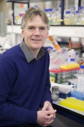 Martin Warren合作论文数Department of Biochemistry, University of Southampton31
 Heidi L. Schubert合作论文数Department of Biochemistry, University of Utah19
 Keith S Wilson合作论文数Department of Chemistry, University of York16
 A Rambach合作论文数7Claude Thermes合作论文数7A.A. Brindley合作论文数5
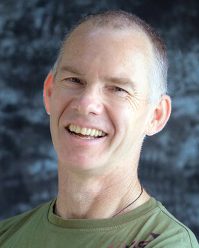 Christopher P. Hill合作论文数Department of Biochemistry, Spencer Fox Eccles School of Medicine, University of Utah4Leech H K合作论文数4A.Ian Scott合作论文数4Charles A. Roessner合作论文数4A Lanois合作论文数3S C Woodcock合作论文数3Peter Heathcote合作论文数School of Biological Sciences, Queen Mary, University of London3J. Vevodova合作论文数3Andrew Munro合作论文数Department of Chemistry, School of Natural Sciences, Faculty of Science and Engineering, The University of Manchester2McLean Kirsty J合作论文数Manchester Inst Biotechnol, Univ Manchester2Beck R合作论文数2
 John D. Phillips合作论文数Hematology Division, University of Utah School of Medicine;Hematology Division, University of Utah School of Medicine2J. M. Roper合作论文数2SEJ Rigby合作论文数2Jennifer M. Roper合作论文数2SE GHARBIA合作论文数1Ke-xue Huang合作论文数1
 Andy Lawrence合作论文数School of Chemistry, The University of Edinburgh1Sarah Peters合作论文数Division of Psychology and Mental Health, School of Health Sciences, Faculty of Biology, Medicine and Health, The University of Manchester1M Elizabeth Stroupe合作论文数1
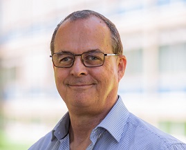 Dieter Jahn合作论文数Institut fur Mikrobiologie, Fakultat fur Lebenswissenschaften, Technische Universitat Braunschweig1
 Thomas Leustek合作论文数Department of Plant Biology, School of Environmental and Biological Sciences, Rutgers, The State University of New Jersey1McGoldrick Helen M合作论文数1Ross M. Graham合作论文数1
 Elizabeth D. Getzoff (Elizabeth Getzoff)合作论文数Department of Integrative Structural and Computational Biology, Scripps Research Institute1
 Marco Malten合作论文数1Stefano Santabarbara合作论文数Centro CNR Biologia Cellulare e Molecolare delle Piante, Università degli Studi di Milano1
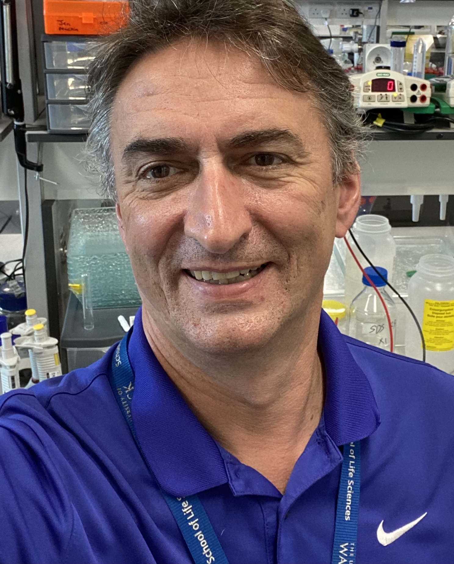 David Roper合作论文数School of Life Sciences, Faculty of Science, Engineering and Medicine, The University of Warwick1
 Nigel Robinson合作论文数Department of Chemistry, Durham University;Department of Biosciences, Durham University1N. P. J. Stamford合作论文数1Patricio J. Santander合作论文数1Haroun N. Shah合作论文数Department of Oral Microbiology;Medical College, London Hospital;Department of Oral Microbiology, London Hospital Medical College1




