Previous genetic analyses of chromosome 17 markers and NF1 (Fain et al. 1987) were extended in an attempt to order marker loci that map physically to 17cen----17q12. Three additional markers (HHH202, CRI-L581, and CRI-L946) were included in the analyses. Recombinants within the cluster of seven unordered marker loci were identified by pairwise analyses for each family and by examining the within-sibship segregation patterns for different markers. Changes in the segregation pattern for different loci define genetic breakpoints. Given that interference is complete in the region, markers with the same segregation pattern lie on one side of the breakpoint, while markers with different segregation patterns lie on opposite sides of the breakpoint. If the order of boundary markers is known, markers on each side of a breakpoint can be oriented in relation to the centromere. The order cen-(HHH202/NF1)-(EW207)-(EW203/CRI-L581)- (CRI-L946)-(HOX-2/NGFR)-qter was inferred by combining information from physical breakpoints in a panel of mouse/human hybrids and information from genetic breakpoints found in 16 NF1 families.
A total of 15 polymorphic markers were used to construct a genetic map that encompasses the NF1 locus on chromosome 17. The markers were a subset of a large collection of chromosome 17-specific probes and were selected for marker typing in NF1 families after physical localization to the pericentric region of the chromosome. Multilocus data for a total of 17 informative NF1 families and 39 other families were included in genetic analyses. No recombination was observed between NF1 and four markers, one or more of which was informative in 86% of parents. More-refined physical mapping studies demonstrated that all four of the markers are proximal to the chromosome 17 translocation breakpoints from two NF1 patients bearing balanced translocations. The region flanking the disease locus spans a distance of 1 centimorgan (cM) in males and 9 cM in females. Close flanking markers were informative in 76% of meioses. Sex differences in recombination rates in the pericentric region were highly significant statistically.
The primary aim of the insulin‐dependent diabetes mellitus (IDDM) component of Genetic Analysis Workshop 5 (GAW5) was to collect and analyze new data on DNA polymorphisms closely linked to the HLA‐D region and the insulin gene. The probes and restriction enzymes described here were used by all ten participating labs, and the data from Southern blotting were interpreted and reported according to conventions developed for the Workshop. These DNA data on members of 94 families with two or more IDDM sibs constitute the largest such sample available. The data were used in most of the analyses presented at the Workshop meeting, and are available on request.
We have developed and mapped by genetic linkage a primary set of markers for chromosome 17. The map consists of 21 loci derived from 27 probe/enzyme systems, including eight highly informative markers at loci containing a variable number of tandemly repeated DNA sequences (VNTRs). The map is continuous from the telomeric region of the short arm to the telomeric region of the long arm, covering estimated genetic distances of 218 cM in males and 279 cM in females. The average heterozygosity among all 21 loci in the population sample analyzed is 58%; 77% heterozygosity was observed among the eight VNTR markers that were highly informative. This map will make it possible to detect by linkage the location of genetic defects associated with chromosome 17 and will also provide anchor points for a high-resolution map of this chromosome. (C) 1988 Academic Press, Inc.
The gene causing von Recklinghausen neurofibromatosis, or NF1, has been more precisely localized in the pericentromeric region of chromosome 17. Narrowing of the location for the disease became possible through the identification of eight new DNA probe genetic markers in the centromeric region. Markers that closely flank the centromere also closely flank the NF1 gene. Although there was evidence against this localization in one recombinant, a review of the clinical records revealed a borderline diagnosis of NF1. Significant sex differences in recombination were observed in the pericentric region, and odds for different orders were less discriminating when sex differences were considered in multilocus analyses. The location of the NF1 gene with respect to the centromere could not be determined because recombinants between NF1 and the centromere were not detected in the set of families tested.
Linkage analysis of 15 Utah kindreds demonstrated that a gene responsible for von Recklinghausen neurofibromatosis (NF) is located near the centromere on chromosome 17. The families also gave no evidence for heterogeneity, indicating that a significant proportion of NF cases are due to mutations at a single locus. Further genetic analysis can now refine this localization and may lead to the eventual identification and cloning of the defective gene responsible for this disorder.
Our initial attempt to map NF was directed towards chromosomes 4 and 19, both of which had provided positive evidence for linkage in previous reports. This analysis showed no evidence in support of either hypothesis. Our second attempt at mapping NF was a general search of the genome, analysing a set of markers selected according to their degree of polymorphism, chromosomal location, ease of use, and availability. Data for linkage analysis were obtained from 17 multiplex families which are segregating a gene for NF. Linkage analyses were performed using PAP. Of note is the lod score of +1.17 at a recombination fraction of 0.1 between NF and the centromere of chromosome 17.
 David F. Barker合作论文数Department of Medical Informatics, University of Utah10
 David E. Goldgar合作论文数Department of Dermatology, University of Utah School of Medicine;Huntsman Cancer Institute, University of Utah7
 Huntington F. Willard合作论文数Howard Hughes Medical Institute6K Nguyen合作论文数6Mark H Skolnick合作论文数6
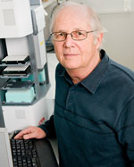 Mark F. Leppert合作论文数Department of Human Genetics, School of Medicine, University of Utah Health5Ray White合作论文数5P. Fain合作论文数4PR FAIN合作论文数4J. Kivlin合作论文数4Peter Oconnell合作论文数4J Carey合作论文数4L Cannon合作论文数3
 Tim Bishop合作论文数School of Medicine, Faculty of Medicine and Health, University of Leeds;Leeds Institute of Medical Research at St James's, University of Leeds3J.-M. Lalouel合作论文数3J S Waye合作论文数2G M Greig合作论文数2
 Leslie Leinwand合作论文数BioFrontiers Institute, University of Colorado Boulder;Department of Molecular, Cellular, and Developmental Biology, College of Arts and Sciences, University of Colorado Boulder2Margaret R. Wallace合作论文数Department of Human Genetics, University of Michigan2
 Yusuke Nakamura合作论文数Department of Medicine, University of Chicago;Department of Surgery, University of Chicago;Center for Personalized Therapeutics, The University of Chicago;Laboratory of Molecular Medicine, Institute of Medical Science, The University of Tokyo2
 Bonnie Jeanne Baty合作论文数2C Depreval合作论文数1P Avoustin合作论文数1Ma Permutt合作论文数1Sl Holbeck合作论文数1P CARTWRIGHT合作论文数1MH SKOLNICK合作论文数1Peter O'Connell合作论文数1FS COLLINS合作论文数1JM Lalouel合作论文数1K STEPHENS合作论文数1j bertrams合作论文数1K Fehsel合作论文数1M Lathrop合作论文数1
 Viktor Horvath合作论文数1
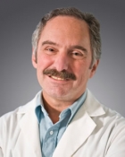 Gerald T. Nepom合作论文数Benaroya Research Institute at Virginia Mason;Immune Tolerance Network, National Institute of Allergy and Infectious Diseases1
 Christopher Mathew合作论文数King’s Global Health Institute, School of Basic & Medical Biosciences, Faculty of Life Sciences and Medicine, Kings College London;Department of Medical and Molecular Genetics, School of Basic & Medical Biosciences, Faculty of Life Sciences and Medicine, Kings College London;Sydney Brenner Institute for Molecular Bioscience, Faculty of Health Sciences, Witwatersrand University1Robinson D M合作论文数1J. Johnson合作论文数1Samuel A. Latt合作论文数1
 Francis Collins合作论文数National Human Genome Research Institute, National Institutes of Health1A. Cambon-Thomsen合作论文数1
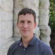 David Barker合作论文数School of Chemical Sciences, School of Biological Sciences, Faculty of Science, The University of Auckland1
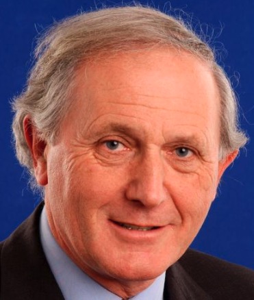 Bruce Ponder合作论文数Jesus College, University of Cambridge;Department of Oncology, University of Cambridge1Michael Litt合作论文数1S. Kondoleon合作论文数1Thomas W. Hodge合作论文数Departments of Microbiology, Medicine, Epidemiology and Surgery, University of Alabama in Birmingham University1
 Rick B. Spielman合作论文数1Mark Lathrop合作论文数1




