Uridine phosphorylase (UP) catalyzes the reversible phosphorolysis of uridine to uracil and ribose 1-phosphate and is a key enzyme in the pyrimidine-salvage pathway. Escherichia coli UP is structurally homologous to E. coli purine nucleoside phosphorylase and other members of the type I family of nucleoside phosphorylases. The structures of 5-benzylacyclouridine, 5-phenylthioacyclouridine, 5-phenylselenenylacyclouridine, 5-m-benzyloxybenzyl acyclouridine and 5-m-benzyloxybenzyl barbituric acid acyclonucleoside bound to the active site of E. coli UP have been determined, with resolutions ranging from 1.95 to 2.3 A. For all five complexes the acyclo sugar moiety binds to the active site in a conformation that mimics the ribose ring of the natural substrates. Surprisingly, the terminal hydroxyl group occupies the position of the nonessential 5'-hydroxyl substituent of the substrate rather than the 3'-hydroxyl group, which is normally required for catalytic activity. Until recently, inhibitors of UP were designed with limited structural knowledge of the active-site residues. These structures explain the basis of inhibition for this series of acyclouridine analogs and suggest possible additional avenues for future drug-design efforts. Furthermore, the studies can be extended to design inhibitors of human UP, for which no X-ray structure is available.
The development of new and effective antiprotozoal drugs has been a difficult challenge because of the close similarity of the metabolic pathways between microbial and mammalian systems. 5'-Methylthioadenosine/S-adenosylhomocysteine (MTA/AdoHcy) nucleosidase is thought to be an ideal target for therapeutic drug design as the enzyme is present in many microbes but not in mammals. MTA/AdoHcy nucleosidase (MTAN) irreversibly depurinates MTA or AdoHcy to form adenine and the corresponding thioribose. The inhibition of MTAN leads to a buildup of toxic byproducts that affect various microbial pathways such as quorum sensing, biological methylation, polyamine biosynthesis, and methionine recycling. The design of nucleosidase-specific inhibitors is complicated by its structural similarity to the human MTA phosphorylase (MTAP). The crystal structures of human MTAP complexed with formycin A and 5'-methylthiotubercidin have been solved to 2.0 and 2.1 A resolution, respectively. Comparisons of the MTAP and MTAN inhibitor complexes reveal size and electrostatic potential differences in the purine, ribose, and 5'-alkylthio binding sites, which account for the substrate specificity and reactions catalyzed. In addition, the differences between the two enzymes have allowed the identification of exploitable regions that can be targeted for the development of high-affinity nucleosidase-specific inhibitors. Sequence alignments of Escherichia coli MTAN, human MTAP, and plant MTA nucleosidases also reveal potential structural changes to the 5'-alkylthio binding site that account for the substrate preference of plant MTA nucleosidases.
The crystal structure of Acetobacter aceti PurE was determined to a resolution of 1.55 A and is compared with the known structures of the class I PurEs from a mesophile, Escherichia coli, and a thermophile, Thermotoga maritima. Analyses of the general factors that increase protein stability are examined as potential explanations for the acid stability of A. aceti PurE. Increased inter-subunit hydrogen bonding and an increased number of arginine-containing salt bridges appear to account for the bulk of the increased acid stability. A chain of histidines linking two active sites is discussed in the context of the proton transfers catalyzed by the enzyme.
Thiazole synthase is the key enzyme involved in the formation of the thiazole moiety of thiamin pyrophosphate. We have determined the structure of this enzyme in complex with ThiS, the sulfur carrier protein, at 3.15 A resolution. Thiazole synthase is a tetramer with 222 symmetry. The monomer is a (betaalpha)(8) barrel with similarities to the aldolase class 1 and flavin mononucleotide dependent oxidoreductase and phosphate binding superfamilies. The sulfur carrier protein (ThiS) is a compact protein with a fold similar to that of ubiquitin. The structure allowed us to model the substrate, deoxy-D-xylulose 5-phosphate (DXP), in the active site. This model identified Glu98 and Asp182 as new active site residues likely to be involved in the catalysis of thiazole formation. The function of these residues was probed by mutagenesis experiments, which confirmed that both residues are essential for thiazole formation and identified Asp182 as the base involved in the deprotonation at C3 of the thiazole synthase DXP imine. Comparison of the ThiS binding surface to the surface of ubiquitin identified a conserved hydrophobic patch of unknown function on ubiquitin that may be involved in complex formation between ubiquitin and one of its binding partners.
The thiO gene of Bacillus subtilis encodes an FAD-dependent glycine oxidase. This enzyme is a homotetramer with a monomer molecular mass of 42 kDa. In this paper, we demonstrate that ThiO is required for the biosynthesis of the thiazole moiety of thiamin pyrophosphate and describe the structure of the enzyme with N-acetylglycine bound at the active site. The closest structural relatives of ThiO are sarcosine oxidase and d-amino acid oxidase. The ThiO structure, as well as the observation that N-cyclopropylglycine is a good substrate, supports a hydride transfer mechanism for the enzyme. A mechanistic proposal for the role of ThiO in thiazole biosynthesis is also described.
 Steven Edward Ealick合作论文数Department of Chemistry and Chemical Biology, College of Arts & Sciences, Cornell University17
 W. Bu合作论文数7
 Tadhg Begley合作论文数Department of Chemistry, Texas A&M University5
 Pieter C Dorrestein合作论文数Departments of Pharmacology and Pediatrics, Skaggs School of Pharmacy and Pharmaceutical Sciences, University of California, San Diego;Collaborative Mass Spectrometry Innovation Center, University of California, San Diego;Institute for Metabolomics Medicine, University of California, San Diego4j e lee合作论文数3
 P. Lynne Howell合作论文数Department of Biochemistry, Temerty Faculty of Medicine, University of Toronto3
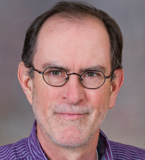 Michael K. Riscoe合作论文数Department of Molecular Microbiology and Immunology, Oregon Health & Science University3Cp Mill合作论文数2HL Zhai合作论文数2
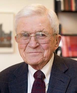 Fred McLafferty合作论文数Department of Chemistry and Chemical Biology, The College of Arts and Sciences, Cornell University2K. A. Cornell合作论文数2
 T. Joseph Kappock合作论文数2E.V. Soriano合作论文数1Tp Begley合作论文数1Kl Colabroy合作论文数1JH Park合作论文数1Johnathan R Chittuluru合作论文数1Ka Cornell合作论文数1Ke Burns合作论文数1Weiming Bu合作论文数1AM Augustine合作论文数1Ae Ransome合作论文数1
 Colabroy Keri L合作论文数Department of Chemistry, Muhlenberg College1
 Abhishek Chatterjee合作论文数Division of Surgical Oncology and Plastic Surgery, Tufts Medical Center1Janice R. Sufrin合作论文数1
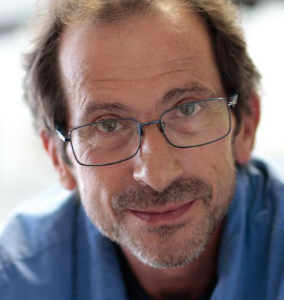 Andrei Osterman合作论文数Graduate School of Biomedical Sciences, Sanford Burnham Prebys;Infectious and Inflammatory Diseases Center, Sanford Burnham Prebys;NCI-Designated Cancer Center, Sanford Burnham Prebys1




