BACKGROUND:Voltage-gated calcium channels in small-cell lung carcinomas may initiate autoimmunity in the paraneoplastic neuromuscular disorder Lambert-Eaton syndrome. The calcium-channel subtype that is responsible is not known.METHODS:We compared the effects of antagonists of L-type, N-type, and P/Q-type neuronal calcium channels on the depolarization-dependent influx of calcium-45 in cultured carcinoma cells. Serum samples from patients with various disorders were tested for reactivity with P/Q-type channels solubilized from carcinoma and cerebellar membranes and N-type channels from cerebral cortex.RESULTS:P/Q-type calcium-channel antagonists were the most potent inhibitors of depolarization-induced 45Ca influx in cultured small-cell carcinoma cell lines. Anti-P/Q-type calcium-channel antibodies were found in serum from all 32 patients with Lambert-Eaton syndrome and a diagnosis of cancer and in 91 percent of the 33 patients with Lambert-Eaton syndrome without cancer. Anti-N-type calcium-channel antibodies were found in 49 percent of the 65 patients with the Lambert-Eaton Syndrome. Lower titers of anti-P/Q-type and anti-N-type calcium-channel antibodies were found in 54 percent of 70 patients with a paraneoplastic encephalomyeloneuropathic complication of lung, ovarian, or breast carcinoma, 24 percent of 90 patients with cancer but no evident neurologic complications, 23 percent of 78 patients with sporadic amyotrophic lateral sclerosis, and less than 3 percent of 69 patients with myasthenia gravis, epilepsy, or scleroderma.CONCLUSIONS:The high frequency of P/Q-type calcium-channel antibodies found in patients with Lambert-Eaton syndrome implies that antibodies of this specificity have a role in the presynaptic pathophysiology of this disorder.
A patient with slow muscle relaxation but without accompanying motor unit or myotonic electrical activity had a unique staircase twitch response to repeated nerve stimulation. During 1‐Hz stimulation, twitches recorded by measurement of the ankle dorsiflexor group displayed progressively increasing relaxation times with successive stimuli (37% increase) unlike the progressively decreasing relaxation time of the normal response (12–37% decrease). The response may be diagnostic of this unusual myopathy; the test methods are noninvasive and easily tolerated.
Serum autoantibodies found by radioimmunoassay in 27 of 52 patients with the Lambert-Eaton myasthenic syndrome (LES) bound specifically to a soluble omega-conotoxin binding component of a voltage-gated Ca2+ channel (VGCC) complex extracted from small cell lung carcinoma (SCC). These antibodies were not found in 43 control patients with other neurologic diseases, including myasthenia gravis, peripheral neuropathies, and amyotrophic lateral sclerosis, or in 9 patients with endocrine autoimmunity, but they were found in 2 of 21 control patients with SCC without a history of LES, 1 of whom had severe autonomic neuropathy. Seropositivity was more frequent in patients with LES who had evidence of a primary lung cancer (76%) than in those with other neoplasms or without evidence of cancer (30%). Antigens extracted from SCC tumor lines derived from patients with and without LES and from a human neuroblastoma line yielded results that were highly correlated. A control extract of colonic carcinoma (derived from a patient with LES) yielded negative results. The data implicate a tumor-associated VGCC as the autoimmunizing stimulus in a subset of patients with LES and provide the first direct evidence that the VGCC complex in SCC is a target for some LES antibodies. The serologic test described should be a useful aid in diagnosing LES.
The Lambert-Eaton myasthenic syndrome (LES) is an autoimmune presynaptic disorder of peripheral cholinergic neurotransmission in which there is often an associated small cell lung carcinoma (SCC). SCC lines established from patients with and without LES exhibit a Ca2+ influx response to depolarization by K+ that is consistent with the presence of voltage-gated Ca2+ channels. Autoantibodies antagonistic to SCC Ca2+ channel activity were found exclusively in patients with LES, independent of cancer status. Depolarization-induced uptake of 45Ca2+ by SCC lines was reduced maximally after 3-4 days of exposure to serum immunoglobulins from 14 of 19 LES patients, while 53 control immunoglobulins (including patients with SCC, other tumors, other paraneoplastic syndromes, and other neurological and autoimmune diseases) were without effect. The snail neurotoxin omega-conotoxin of subtype GVIA, which is a specific antagonist of presynaptic Ca2+ channels, inhibited K+-stimulated Ca2+ uptake in a dose-dependent manner that was essentially irreversible. Adenosine, reported to be a specific antagonist of neuronal Ca2+ channels, also impaired voltage-stimulated Ca2+ influx in SCC. Use of LES patients' IgG and omega-conotoxin in further studies of SCC may facilitate identification and purification of the LES antigen(s) and yield a quantitative serological test for diagnosing this autoimmune paraneoplastic syndrome.
Annals of the New York Academy of SciencesVolume 540, Issue 1 p. 369-371 Voltage-Dependent Ca2+ Channels in Small Cell Carcinomas Are Blocked by Autoantibodies from Patients with Lambert-Eaton Myasthenic Syndromea HENRY J. DE AIZPURUA, HENRY J. DE AIZPURUA Neuroimmunology Laboratory Mayo Clinic Rochester, Minnesota 55905Search for more papers by this authorGUY E. GRIESMANN, GUY E. GRIESMANN Neuroimmunology Laboratory Mayo Clinic Rochester, Minnesota 55905Search for more papers by this authorEDWARD H. LAMBERT, EDWARD H. LAMBERT Department of Neurology University of Minnesota Minneapolis, Minnesota 55455Search for more papers by this authorVANDA A. LENNON, VANDA A. LENNON Neuroimmunology Laboratory Mayo Clinic Rochester, Minnesota 55905Search for more papers by this author HENRY J. DE AIZPURUA, HENRY J. DE AIZPURUA Neuroimmunology Laboratory Mayo Clinic Rochester, Minnesota 55905Search for more papers by this authorGUY E. GRIESMANN, GUY E. GRIESMANN Neuroimmunology Laboratory Mayo Clinic Rochester, Minnesota 55905Search for more papers by this authorEDWARD H. LAMBERT, EDWARD H. LAMBERT Department of Neurology University of Minnesota Minneapolis, Minnesota 55455Search for more papers by this authorVANDA A. LENNON, VANDA A. LENNON Neuroimmunology Laboratory Mayo Clinic Rochester, Minnesota 55905Search for more papers by this author First published: November 1988 https://doi.org/10.1111/j.1749-6632.1988.tb27102.xCitations: 3 a This work was supported by Grants CA 37343 (VAL) and NS 23691 (EHL) from the National Institutes of Health. AboutPDF ToolsRequest permissionExport citationAdd to favoritesTrack citation ShareShare Give accessShare full text accessShare full-text accessPlease review our Terms and Conditions of Use and check box below to share full-text version of article.I have read and accept the Wiley Online Library Terms and Conditions of UseShareable LinkUse the link below to share a full-text version of this article with your friends and colleagues. Learn more.Copy URL Share a linkShare onFacebookTwitterLinkedInRedditWechat No abstract is available for this article.Citing Literature Volume540, Issue1Advances in NeuroimmunologyNovember 1988Pages 369-371 RelatedInformation
Antibodies in individual patients with the Lambert-Eaton myasthenic syndrome (LES) differ in their reactivity with mouse motor nerve terminals. Of 26 LES patients' sera injected a single time into mice, 3 caused a highly significant reduction in stimulus-dependent quantal release (m) of acetylcholine (ACh) (to 6, 33, and 42 quanta per impulse at 1 Hz, respectively; mean for 10 control sera, 100 quanta at 1 Hz). The most potent serum (LES-A) was fully effective in mice deficient in complement component C5 and in mice depleted of complement components C3----C9 by cobra venom factor. A single i.v. injection of serum reduced m in direct proportion to log dose. Responses to K+ depolarization and increasing concentrations of Ca2+ were like those observed in human LES. With LES-A serum, or its IgG, m was reduced near maximally in 1 day and plateaued in 3-4 days. Recovery began after day 8; m was in the normal range by day 20-30. Electromyographic (EMG) abnormalities were not seen until m fell below 40 quanta per impulse at 1 Hz. Below 10 quanta, clinical signs of weakness appeared, and the EMG abnormalities were those classically associated with LES: a marked reduction of compound muscle action potential to a single nerve stimulus in rested muscle, a further decrement during stimulation at slow rates, but marked facilitation during rapid repetitive stimulation.
Annals of the New York Academy of SciencesVolume 540, Issue 1 p. 516-519 Synthetic Peptide of Human Acetylcholine Receptor α-Subunit Sequence 125-147 (Methionine 144), a More Potent Autoantigen Than Its Norleucine 144 Analoga VANDA A. LENNON, VANDA A. LENNON Departments of Immunology, Neurology, Mayo Clinic Rochester, Minnesota 55905Search for more papers by this authorZHONG-XIAN HUANG, ZHONG-XIAN HUANG Department of Neurology, University of Minnesota Minneapolis, Minnesota 55455Search for more papers by this authorDANIEL J. McCORMICK, DANIEL J. McCORMICK Departments Biochemistry and Molecular Biology, Mayo Clinic Rochester, Minnesota 55905Search for more papers by this authorGUY E. GRIESMANN, GUY E. GRIESMANN Departments of Immunology, Neurology, Mayo Clinic Rochester, Minnesota 55905Search for more papers by this authorNAOKI FUJII, NAOKI FUJII Departments of Immunology, Neurology, Mayo Clinic Rochester, Minnesota 55905Search for more papers by this authorEDWARD H. LAMBERT, EDWARD H. LAMBERT Department of Neurology, University of Minnesota Minneapolis, Minnesota 55455Search for more papers by this author VANDA A. LENNON, VANDA A. LENNON Departments of Immunology, Neurology, Mayo Clinic Rochester, Minnesota 55905Search for more papers by this authorZHONG-XIAN HUANG, ZHONG-XIAN HUANG Department of Neurology, University of Minnesota Minneapolis, Minnesota 55455Search for more papers by this authorDANIEL J. McCORMICK, DANIEL J. McCORMICK Departments Biochemistry and Molecular Biology, Mayo Clinic Rochester, Minnesota 55905Search for more papers by this authorGUY E. GRIESMANN, GUY E. GRIESMANN Departments of Immunology, Neurology, Mayo Clinic Rochester, Minnesota 55905Search for more papers by this authorNAOKI FUJII, NAOKI FUJII Departments of Immunology, Neurology, Mayo Clinic Rochester, Minnesota 55905Search for more papers by this authorEDWARD H. LAMBERT, EDWARD H. LAMBERT Department of Neurology, University of Minnesota Minneapolis, Minnesota 55455Search for more papers by this author First published: November 1988 https://doi.org/10.1111/j.1749-6632.1988.tb27156.xCitations: 2 a This work was supported by grants from the National Institutes of Health (NS 15057, V.A.L.; NS 24694, D.J.M.; NS 23691, E.H.L.) and The William K. Warren Foundation. AboutPDF ToolsRequest permissionExport citationAdd to favoritesTrack citation ShareShare Give accessShare full text accessShare full-text accessPlease review our Terms and Conditions of Use and check box below to share full-text version of article.I have read and accept the Wiley Online Library Terms and Conditions of UseShareable LinkUse the link below to share a full-text version of this article with your friends and colleagues. Learn more.Copy URL Share a linkShare onFacebookTwitterLinked InRedditWechat Citing Literature Volume540, Issue1Advances in NeuroimmunologyNovember 1988Pages 516-519 RelatedInformation
Abstract: The presence of muscarinic and nicotinic acetylcholine receptors (m‐ and nAChR, respectively) in small cell carcinomas (SCC) of the lung was assessed by measurement of specific binding of (—)[3H]quinuclidinyl benzilate ([3H]QNB) and 125I‐α‐bungarotoxin, respectively. Of five SCC studied, four were originally derived from patients with the Lambert‐Eaton myasthenic syndrome, an autoimmune disease of neuromuscular transmission, and one was from a patient without evidence of neurological disease. There was no evidence of nAChR, but all tumors bound (‐)[3H]QNB in a saturable and specific manner. Dissociation constants derived from saturation isotherms ranged between 0.13 and 0.3 nM and maximum binding capacities between 35.4 and 181.7 fmol/mg protein (mean values for the five SCC). Competition studies revealed a pharmacological profile consistent with previous descriptions of mAChR. Competition by pirenzepine revealed only one class of binding sites, that with a relatively low affinity for pirenzepine. In the presence of the guanine nucleotide analogue 5′‐guanylyl imidodiphosphate, a decrease in affinity of the mAChR of SCC for oxotremorine was observed, with an increase in the pseudo‐Hill coefficient, but there was no change in the binding of atropine. The expression on SCC of mAChR, apparently of the M2 subclass, represents yet another neural differentiation marker of SCC. It is noteworthy that the expression of this marker is not restricted to patients with an autoimmune paraneoplastic syndrome involving cholinergic neurons.
Six patients in two unrelated families had a hereditary form of sustained muscle activity of peripheral nerve origin. Although varying in severity among affected family members, the disease was manifested clinically as generalized myokymia and muscle stiffness. Motor and sensory nerve conduction velocities and results of nerve biopsy were normal. Repetitive after-discharges followed each motor nerve stimulus. Needle electrode examination demonstrated spontaneous recurrent bursts of motor unit activity. The burst activity of a motor unit occurred rhythmically (0.5 to 4 bursts per second) and independently of the activity of other units. Within a burst, the firing rate was high (150 to 300 Hz). The clinical and electromyographic abnormalities improved considerably after treatment with carbamazepine or phenytoin.
Five familial cases (in two families) and one sporadic case of a new congenital myasthenic syndrome were investigated. Symptoms arise in infancy or later life. Typically, one finds selective involvement of cervical, scapular, and finger extensor muscles, ophthalmoparesis, and variable involvement of other muscles. There is a repetitive muscle action potential to single nerve stimulus in all muscles and a decremental response at 2 to 3 Hz stimulation in clinical affected muscles. Microelectrode studies reveal markedly prolonged end-plate potential (epp), miniature end-plate potential (mepp), and miniature end-plate current; normal quantum content of the epp; and a smaller than normal or low-normal mepp amplitude. Light microscopy demonstrates predominance of type I fibers, small groups of atrophic fibers, tubular aggregates and vacuoles near end-plates, abnormal end-plate configuration, and nonspecific myopathic changes. Abundant acetylcholinesterase activity is present at all end-plates, and the activity and kinetic properties of this enzyme in muscle are normal. Calcium accumulated at the end-plate in one patient. Quantitative electron microscopy shows decrease in the size of nerve terminals, increase in the density of synaptic vesicles, and reduction in the length of postsynaptic membranes. There is focal degeneration of junctional folds with corresponding loss of acetylcholine receptor, most marked in cases with the lowest mepp amplitude. There are no immune complexes at the end-plate. Fiber regions near end-plates display dilation, proliferation, and degeneration of the sarcoplasmic reticulum; nuclear, mitochondrial, and myofibrillar degeneration; and vacuoles resembling those found in periodic paralysis. A prolonged open time of the acetylcholine-induced ion channel is considered to be the basic abnormality and may account for the physiological, morphological, and clinical alterations.
 Vanda A. Lennon合作论文数Department of Immunology, Mayo Clinic;Department of Neurology, Mayo Clinic;Division of Clinical Biochemistry & Immunology, Department of Laboratory Medicine and Pathology, Mayo Clinic14
 Guy E Griesmann合作论文数5
 Andrew Engel合作论文数Mayo Clinic3
 Donald W. Mulder合作论文数University of California, Los Angeles3
 Peter J. Dyck合作论文数3D W MULDER合作论文数2Peter J Dyck合作论文数2
 Jasper R. Daube合作论文数Mayo Clinic2John N. Whitaker合作论文数2Ko Sahashi合作论文数2Manuel R. Gomez合作论文数2
 P. James B. Dyck合作论文数Department of Neurology, Mayo Clinic2G R Diessner合作论文数1J A BASTRON合作论文数1CF TORRES合作论文数1A. Bariout合作论文数1Ga Gronert合作论文数1GE Griesmann合作论文数1DA SCHOLZ合作论文数1W. Newlon Tauxe合作论文数1Wr Sherman合作论文数1RG AUGER合作论文数1a yudell合作论文数1akio ohnishi合作论文数1F.John Service合作论文数1TE BERTORINI合作论文数1Johannes Jakobsen合作论文数1Va Lennon合作论文数1Mf Sparks合作论文数1TJ Kryzer合作论文数1NAOKI FUJII合作论文数1A. Djeridane合作论文数1El Haouri M合作论文数1
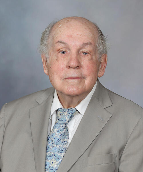 Peter J. Dyck合作论文数Department of Neurology, Mayo Clinic1Talhari Anette Chrusciak合作论文数1De Aizpurua H J合作论文数1ZHONG-XIAN HUANG合作论文数1Salhi Aicha合作论文数1Gregory Julia L合作论文数1Sayre G P合作论文数1Clapasson Andrea合作论文数1Traore Pierre合作论文数1Kryzer T J合作论文数1
 Julie Cunningham合作论文数1
 Phillip A. Low合作论文数Department of Neurology, Mayo Clinic1
 John C. Papadimitriou合作论文数Department of Pathology, University of Maryland School of Medicine1
 Earl Howard Wood合作论文数Mayo Clinic1
 Paul Iaizzo合作论文数Departments of Surgery, School of Medical, University of Minnesota1
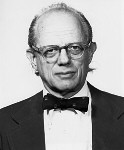 Herman Feshbach合作论文数Massachusetts Institute of Technology1




