This study investigated the impact of the Sudanese smokeless tobacco Toombak on cultivable oral bacteria, its potential antibacterial properties, and its own microbial composition. Oral swabs from 20 adult male participants, including ten long-term Toombak users and ten non-users, were analyzed through microbial inoculation and identification techniques. In addition, Toombak samples were examined for viable microorganisms, tested for antibacterial activity against selected reference strains, and the results were statistically analyzed using the Wilcoxon rank-sum test (α = 0.05). The overall bacterial counts, diversity, and species distribution showed no statistically significant differences between users and non-users, with the exception of Schaalia odontolytica (p = 0.0306) and the proportion of Gram-positive rods (p = 0.0137) expressed as percentages, which were more frequent in the control group. Toombak itself harbored mainly Bacillus spp., and no inhibitory activity against the tested bacteria was observed. While a non-significant trend toward lower bacterial counts was observed in Toombak users (1.83 × 10⁸ vs. 3.31 × 10⁸ CFU/ml), this finding should be interpreted cautiously given the exploratory design and substantial interindividual variability. Further studies using larger cohorts and culture-independent methods are warranted to better clarify its microbial impact.
The heat-not-burn tobacco product IQOS (I Quit Ordinary Smoking) has recently become widely used. However, the impact of IQOS aerosol on the oral microbiome remains unclear. The present study therefore aimed to investigate the influence of IQOS aerosol on the microbial composition of microcosm biofilms formed from human saliva using a standardized biofilm reactor. A custom-designed biofilm reactor was constructed to enable the intermittent exposure of biofilms to IQOS aerosol. Microcosm biofilms were formed on bovine enamel samples with a defined surface (19.635 mm2) with unstimulated pooled human saliva from three healthy probands being used as inoculum. Biofilm formation took place with continuous nutrient medium supply for 5 days. The biofilm in the test setup was exposed to IQOS aerosol 8 times a day for 5 min each time. A parallel test setup ensured simultaneous biofilm formation without exposure to IQOS aersol and served as a negative control. After 5 days, the microbial composition of the formed biofilms was examined by amplicon sequencing using the V1-V3 region of the 16S rRNA gene. In addition, the biofilm was visualized using scanning electron microscopy. After one week, the surfaces of the bovine enamel samples on which biofilm formation took place were similarly covered, whether under the influence of IQOS aerosol or in the negative control. The Simpson index showed significant differences (P < 0.05), while the Pielou index showed highly significant differences (0.01 < P < 0.05), as did the Shannon index (0.001 < P < 0.01) and the Richness index (P < 0.001). The β-diversity showed different clustering between the treated biofilm and the negative control, corresponding to the significantly different (p = 0.001) microbial community caused by the IQOS aerosol. The abundance of the genera Gemella, Haemophilus, Neisseria and Rothia was significantly lower in biofilms influenced by IQOS aerosol. Additionally, the abundance of different species was significantly modified by IQOS aerosol. IQOS aersol may shift oral microbial composition, even though not inhibiting biofilm growth. This highlights the need for further research into the effects on oral microbal ecology of IQOS users, as well as the development of prevention and education measures regarding the potential health risks associated with IQOS.
Caries is still one of the most common diseases in Germany and world-wide, and most adults, children and adolescents are affected during their lifetime. The primary goal of oral disease prevention is therefore maintaining healthy teeth that are either free from decay or sufficiently restored without any secondary caries. This updated S3-guideline is intended to provide scientifically sound information on basic measures and recommendations for caries prevention in the permanent dentition, based on the current state of knowledge on etiology and pathogenesis of caries. This guideline applies to all persons with permanent dentition, provides information for dentists, educators, teachers, parents and other multipliers. Its core messages can be divided into measures to be taken by the population at home, and measures recommended and carried out by staff members in dental practices. The classic caries prevention pillars (tooth brushing, fluoride use, nutritional advice and regular dental consultations) have been expanded to seven recommendations: (I) Oral hygiene, including interdental cleaning; (II) Risk-adapted recommendations for fluoride use as part of daily oral hygiene, plus use of fluoridated table salt; (III) Reducing sugar consumption. In addition, in dental practices (IV) the participation in risk-adapted and structured prevention programs should be recommended, (V) application of further highly concentrated fluoride preparations such as varnishes, (VI) application of chlorhexidine in case of orthodontic treatment or exposed root surfaces, and (VII) fissure sealing in areas at high risk of caries development should be considered. These measures can help promote lifelong dental health. The recommendations of the guideline should be implemented in individual and professional caries prevention.
OBJECTIVE:This study investigated the ecotoxic and genotoxic effects of composite components after grinding. METHODS:Samples of composite materials Ceram.x Spectra® ST (CX) and Filtek™ Supreme XTE (FS) were ground to dust in a plexiglas box with a standard diamond bur (106 µm). Aqueous (≤ 5 mg/mL) and organic (≤ 790 mg/mL) extracts were prepared to mimic wastewater samples. Ecotoxicity was assessed in luminescent bacteria (Vibrio fischeri), algae (Desmodesmus subspicatus), water flea (Daphnia magna), and fish embryo (Danio rerio). Genotoxicity was evaluated in human lung adenocarcinoma cells (A459) using the Comet assay. All statistical analyses were conducted using a significance level of α = 0.05. RESULTS:Both materials inhibited V. fischeri bioluminescence; CX aqueous eluates showed significantly stronger effects than FS at 2.5 mg/mL (p < 0.01) and 1.25 mg/mL (p < 0.05). Organic extracts were similarly potent (EC50 ≈ 0.27-0.28 mg/mL) without significant differences. In algae, CX aqueous eluates caused significantly higher growth inhibition at all concentrations than FS (p < 0.01); CX organic extracts completely suppressed growth. In Daphnia, CX aqueous eluates caused 100% immobilization, while FS showed no effect. In fish embryos, CX aqueous eluates induced up to 100% mortality (≥ 1.33 mg/mL), whereas FS caused no mortality but impaired pigmentation. Organic extracts of both materials induced embryo mortality up to 100% at 2 mg/mL. No biologically relevant genotoxicity was found. SIGNIFICANCE:Both composites exhibit concentration-dependent ecotoxic effects across aqueous organisms. Identifying responsible compounds and implementing effective wastewater treatment are crucial to reduce potential environmental risks.
Helicobacter pylori is known for colonizing the gastric mucosa and instigating severe upper gastrointestinal diseases such as gastritis, gastroduodenal ulcers, and gastric cancer. To date, there is no data available on the oral cavity as transmission site, whether H. pylori can survive in the oral cavity or in human saliva. The aim of the study was to investigate the influence of oral microorganisms and human saliva on the survival of H. pylori in human saliva. H. pylori strains KE, a motile derivate of type strain H. pylori 26695, and H. pylori SS1, a clinical isolate from a gastric biopsy, were grown in human pooled saliva (pooled from 4 healthy human donors, 0.22 μm filter-sterilized) or in BBF (Brucella browth formula; control) either as mono-cultures or in co-culture with Streptococcus mutans, Streptococcus oralis, Actinomyces naeslundii, Lacticaseibacillus casei and Candida dubliniensis. Bacterial survival of H. pylori and the oral microorganisms were investigated using colony forming units (CFU) assay and MALDI-TOF MS at baseline and after 24, 48 and 168 h. In saliva, H. pylori KE demonstrated enhanced survival in co-culture with S. mutans, A. naeslundii, and C. dubliniensis, enduring for at least 48 h. In contrast, L. casei and S. oralis inhibited H. pylori KE in saliva. H. pylori KE could not be cultured after 168 h in saliva, neither in mono- nor co-culture. In contrast, H. pylori SS1 in saliva could be cultured after 168 h in co-culture with S. mutans and C. dubliniensis, but not in mono-culture. In BBF, H. pylori KE could be cultured after 168 h with S. mutans, L. casei and C. dubliniensis, and H. pylori SS1 with L. casei and C. dubliniensis, but not with S. mutans. Notably, the co-cultured microorganisms survived at high CFU numbers similar to those of the monocultures. The study suggests that H. pylori can transiently survive in human saliva and even with presence of certain oral microorganisms. However, it may not be a permanent resident of the oral microbiota. The co-survival with oral microorganisms emphasizes the necessity for studying the role of the oral microbiota in the infectious and transmission cycle of H. pylori.
IntroductionA range of disinfectant mouthwashes are available for oral hygiene. The gold standard is Chlorhexidine digluconate (CHX), which, like other available products, cannot be used without side effects in the long term. However, in recent years, therapy with herbal products, often considered antiquated, has regained considerable interest. Therefore, the search for plant compounds as an alternative to existing oral disinfectants is meaningful.MethodsIn this study, eleven Mediterranean plant extracts were tested for their antimicrobial effect in vitro. Methanol extracts of the following plants were produced by the pharmaceutical faculty of the University of Athens: Mentha aquatica, Mentha longifolia, Sideritis euboea, Sideritis syriaca, Stachys spinosa, Satureja parnassica, Satureja thymbra, Lavandula stoechas, Achillea taygetea, Phlomis cretica, and Vaccinium myrtillus. The extracts were dissolved for microdilution experiments at concentrations ranging from 10 to 0.019 mg/ml. The oral pathogens tested were Streptococcus mutans, Streptococcus oralis, Streptococcus sobrinus, Prevotella intermedia, Fusobacterium nucleatum, Parvimonas micra, Porphyromonas gingivalis, and Candida albicans. Enterococcus faecalis, Staphylococcus aureus, and Escherichia coli were used as references.ResultsAll extracts, except the methanol extract of V. myrtillus, showed an antibacterial effect at concentrations ranging from 10 to 0.15 mg/ml. None of the extracts exhibited a significant antifungal effect. In general, the anaerobic pathogens could be inhibited and killed at lower concentrations compared to the aerobic pathogens. S. oralis also showed good susceptibility to the extracts. Additionally, the extracts' ability to inhibit biofilm formation by S. mutans was tested. L. stoechas at a concentration of 0.3 mg/ml showed a moderate inhibitory effect. The extracts of L. stoechas, S. thymbra, S. parnassica, and the methanol extract of V. myrtillus were effective at concentrations up to 1.25 mg/ml. P. cretica was able to inhibit and kill S. mutans at a concentration of 0.6 mg/ml, but its effectiveness in biofilm inhibition significantly decreased at 2.5 mg/ml.DiscussionThe study's hypothesis that all extracts would exhibit an antimicrobial effect was thus confirmed.
Oral biofilm is the main cause of pathologies affecting the hard and soft oral tissues around teeth. Its main components are the periodontal pathogens and other bacteria of the supragingival and subgingival biofilm. Different alternative strategies that could be adjuvants to the usual periodontal treatments used to eliminate biofilms are available. One of these methods is antimicrobial photodynamic therapy using VIS and water-filtered infrared-A combined with a photosensitizer. In this review, different recent studies were collected to evaluate the antimicrobial effects of antimicrobial photodynamic therapy and the effectiveness of different types of photosensitizers.
Objectives The increasing demand for alternatives to antibiotics against resistant bacteria has led to research on natural products. The aim of this study was to analyze the antimicrobial and antibiofilm activity of 16 Mediterranean herb extracts. Materials and Methods The extracts were analyzed using High Performance Thin Layer Chromatography. The minimum inhibitory concentration and minimum bactericidal concentration of the extracts from Achillea taygetea, Cistus creticus ssp. creticus, Cistus monspeliensis, Lavandula stoechas, Mentha aquatica, Mentha longifolia, Origanum vulgare, Phlomis cretica, Rosmarinus officinalis, Salvia sclarea, Satureja parnassica, Satureja thymbra, Sideritis euboea, Sideritis syriaca, Stachys spinosa, and Thymus longicaulis were determined against eight oral bacteria and fungus Candida albicans. Microtiter plate test was conducted to evaluate the antibiofilm activity against Streptococcus mutans. Results Overall, all tested extracts efficiently suppressed the growth of obligate anaerobic bacteria. When applied at concentrations ≥ 0.15 mg / ml, the extracts exhibited moderate to high antibiofilm activity comparable to that of chlorhexidine (CHX) against S. mutans. Interestingly, R. officinalis (MIC: 0.01–0.06 mg / ml) and O. vulgare (MIC: 0.04–1.25 mg / ml) demonstrated the highest antibacterial activity against oral bacteria. Additionally, R. officinalis and L. stoechas significantly inhibited S. mutans biofilm formation at 0.15 mg / ml. Conclusions The tested plant extracts can be considered as alternative natural antimicrobial and antibiofilm agents. Clinical Relevance: Mediterranean herb extracts show promise as natural alternatives to combat oral bacteria and biofilm formation, offering potential new therapies for infectious oral diseases in the context of antibiotic resistance.
Objectives The increasing demand for alternatives to antibiotics against resistant bacteria has led to research on natural products. The aim of this study was to analyze the antimicrobial and antibiofilm activity of 16 Mediterranean herb extracts.Materials and methods The extracts were analyzed using High Performance Thin Layer Chromatography. The minimum inhibitory concentration and minimum bactericidal concentration of the extracts from Achillea taygetea, Cistus creticus ssp. creticus, Cistus monspeliensis, Lavandula stoechas, Mentha aquatica, Mentha longifolia, Origanum vulgare, Phlomis cretica, Rosmarinus officinalis, Salvia sclarea, Satureja parnassica, Satureja thymbra, Sideritis euboea, Sideritis syriaca, Stachys spinosa, and Thymus longicaulis were determined against eight oral bacteria and fungus Candida albicans. Microtiter plate test was conducted to evaluate the antibiofilm activity against Streptococcus mutans.Results Overall, all tested extracts efficiently suppressed the growth of obligate anaerobic bacteria. When applied at concentrations >= 0.15 mg/ml, the extracts exhibited moderate to high antibiofilm activity comparable to that of chlorhexidine (CHX) against S. mutans. Interestingly, R. officinalis (MIC: 0.01-0.06 mg/ml) and O. vulgare (MIC: 0.04-1.25 mg/ml) demonstrated the highest antibacterial activity against oral bacteria. Additionally, R. officinalis and L. stoechas significantly inhibited S. mutans biofilm formation at 0.15 mg/ml.Conclusions The tested plant extracts can be considered as alternative natural antimicrobial and antibiofilm agents.Clinical relevance Mediterranean herb extracts show promise as natural alternatives to combat oral bacteria and biofilm formation, offering potential new therapies for infectious oral diseases in the context of antibiotic resistance.
This ex vivo study aimed to investigate surface roughness and substance loss after treatment with different professional cleaning methods and to determine whether subsequent polishing with a rubber cup and polishing paste is necessary. Samples (flat and natural surfaces) of human enamel and dentin were prepared (baseline) and treated with either a curette, air-polishing with erythritol, a rubber cup and polishing paste, or a combination thereof (treatment). Subsequently, all samples were immersed in an ultrasonic bath (ultrasonic) to remove residues from the treatment procedures. The surface roughness values sRa and sRz as well as tissue loss were measured profilometrically. Linear regression models were used to compare group differences (roughness and loss) considering the corresponding baseline value. The significance level was set at p<0.05. sRa increased significantly after treatment with curettes or air-polishing with erythritol in both enamel (p<0.001) and dentin (p<0.001) of flat samples. The same effect was observed for sRz in dentin (p<0.001) but not for enamel compared to negative control. Polishing with a rubber cup and paste alone had no significant effect on roughness values. When combined with other treatments, the effect of curette or air-polishing with erythritol dominated the effect. In enamel, none of the tested methods led to measurable tissue loss. In dentin, air-polishing with erythritol caused ≤50% tissue loss compared to the curette. Conclusively, for enamel, treatment effects on roughness were measurable but of limited clinical relevance. For dentin, air-polishing resulted in a smaller but insignificant roughness increase and less tissue loss compared to the curette. Polishing with a rubber cup and paste did not affect surface roughness. Regarding the clinical application, the use of air-polishing seems to be a less invasive procedure than using a curette; polishing with rubber cup and paste offers no advantage in terms of reducing roughness as a final procedure.
IntroductionChlorhexidine (CHX) is a commonly used antiseptic in situations of limited oral hygiene ability such as after periodontal surgery. However, CHX is also considered as a possible factor in the emergence of cross-resistance to antibiotics. The aim of this study was to analyze the changes in the oral microbiota and the prevalence of antimicrobial resistance genes (ARGs) due to CHX treatment.Materials and methodsWe analyzed the oral metagenome of 20 patients who applied a 0.2% CHX mouthwash twice daily for 4 weeks following periodontal surgical procedures. Saliva and supragingival plaque samples were examined before, directly after 4 weeks, and another 4 weeks after discontinuing the CHX treatment.ResultsAlpha-diversity decreased significantly with CHX use. The Bray–Curtis dissimilarity increased in both sample sites and mainly streptococci showed a higher relative abundance after CHX treatment. Although no significant changes of ARGs could be detected, an increase in prevalence was found for genes that encode for tetracycline efflux pumps.ConclusionCHX treatment appears to promote a caries-associated bacterial community and the emergence of tetracycline resistance genes. Future research should focus on CHX-related changes in the microbial community and whether the discovered tetracycline resistance genes promote resistance to CHX.
INTRODUCTION:The effects of air-polishing on exposed root dentin surfaces are largely unknown, as there are only few studies which show heterogeneous results. Thus, this study was to investigate roughness changes of exposed dentin surfaces after air-polishing and the influence of subsequent polishing with cup and paste. METHODS:Totally 54 teeth with exposed root dentin surfaces were treated using a split-mouth design by either air-polishing with erythritol and a rubber cup with polishing paste on the test side, or rubber cup and paste alone. Teeth were finally cleaned using a sonic tooth brush. Impressions were taken at relevant time points and replicated using epoxy resin. The resulting casts were profilometrically analysed to obtain the average surface roughness (sRa) and maximum peak-to-valley height (sRz), which are given as the mean ± standard deviation in μm. RESULTS:After air-polishing, in comparison to the baseline, there was a slight but significant increase in sRa (0.168 ± 0.143, p < 0.001), but sRz did not change (-0.471 ± 4.857, p = 0.936). Subsequent polishing with cup and paste and cleaning with a sonic toothbrush did not reduce the surface roughness (sonic toothbrush-air-polishing, sRa -0.044 ± 0.081, p = 0.218; sRz -0.551 ± 3.563, p = 0.903). CONCLUSION:The use of erythritol led to a slight increase in the roughness of the dentin surface, which was not reduced by polishing with a cup and paste. Polishing paste did not seem to conceal surface irregularities.
We would like to update the readership about the procedure that led to the correction of [...]
A range of disinfectant mouthwashes are available for oral hygiene. The gold standard is Chlorhexidine digluconate (CHX), which, like other available products, cannot be used without side effects in the long term. However, in recent years, therapy with herbal products, often considered antiquated, has regained considerable interest. Therefore, the search for plant compounds as an alternative to existing oral disinfectants is meaningful. In this study, eleven Mediterranean plant extracts were tested for their antimicrobial effect in vitro. Methanol extracts of the following plants were produced by the pharmaceutical faculty of the University of Athens: Mentha aquatica, Mentha longifolia, Sideritis euboea, Sideritis syriaca, Stachys spinosa, Satureja parnassica, Satureja thymbra, Lavandula stoechas, Achillea taygetea, Phlomis cretica, and Vaccinium myrtillus. The extracts were dissolved for microdilution experiments at concentrations ranging from 10 mg/ml to 0.019 mg/ml. The oral pathogens tested were Streptococcus mutans, Streptococcus oralis, Streptococcus sobrinus, Prevotella intermedia, Fusobacterium nucleatum, Parvimonas micra, Porphyromonas gingivalis, and Candida albicans. Enterococcus faecalis, Staphylococcus aureus, and Escherichia coli were used as references. All extracts, except the methanol extract of V. myrtillus, showed an antibacterial effect at concentrations ranging from 10 mg/ml to 0.15 mg/ml. None of the extracts exhibited a significant antifungal effect. In general, the anaerobic pathogens could be inhibited and killed at lower concentrations compared to the aerobic pathogens. S. oralis also showed good susceptibility to the extracts. Additionally, the extracts' ability to inhibit biofilm formation by S. mutans was tested. L. stoechas at a concentration of 0.3 mg/ml showed a moderate inhibitory effect. The extracts of L. stoechas, S. thymbra, S. parnassica, and the methanol extract of V. myrtillus were effective at concentrations up to 1.25 mg/ml. P. cretica was able to inhibit and kill S. mutans at a concentration of 0.6 mg/ml, but its effectiveness in biofilm inhibition significantly decreased at 2.5 mg/ml. The study's hypothesis that all extracts would exhibit an antimicrobial effect was thus confirmed.
Antimicrobial photodynamic treatment (aPDT) with visible light plus water-filtered infrared-A irradiation (VIS-wIRA) and natural single- or multi-component photosensitizers (PSs) was shown to have potent antimicrobial activity. The aim of this study was to obtain information on the antimicrobial effects of aPDT-VIS-wIRA with lingonberry extract (LE) against bacteria that play a role in oral health. Planktonic bacterial cultures of the Gram-positive E. faecalis T9, S. mutans DSM20523, S. oralis ATCC 35037 and S. sobrinus PSM 203513, the Gram-negative N. oralis 14F2 FG-15-7B, F. nucleatum ATCC 25586, and V. parvula DSM, the anaerobic F. nucleatum ATCC 25586 and V. parvula DSM 2008, and the total mixed bacteria from pooled saliva and supra- and subgingival plaques of volunteers were all treated and compared. aPDT-VIS-wIRA with LE as PS significantly (p < 0.008) reduced the growth of all tested Gram-positive, Gram-negative, as well as aerobic and anaerobic bacterial strains, whereas without irradiation no reductions were seen (p < 0.0001). NaCl, with or without irradiation, was ineffective. After treatment with CHX 0.2%, the highest killing rate (100%) was observed, and no bacteria (0 log10 CFU) were cultivable. The method also significantly reduced all of the bacteria present in saliva and in the gingival biofilms. Three-dimensional visualization of viable and non-viable microorganisms revealed that LE penetrated deeper into the cell wall layers than CHX 0.2%. LE was an appropriate PS for eradicating microorganisms with VIS-wIRA, either in their planktonic form or in saliva and gingival plaque biofilms. These results encourage further investigation in order to determine which LE compounds contribute to the photosensitizing effect and to evaluate the size of the effect on maintaining oral health.
OBJECTIVES:Low-frequency, low-intensity ultrasound is commonly utilized in various dental research fields to remove biofilms from surfaces, but no clear recommendation exists in dental studies so far. Therefore, this study aims to optimize the sonication procedure for the dental field to efficiently detach bacteria while preserving viability. MATERIALS AND METHODS:Initial biofilm was formed in vivo on bovine enamel slabs (n = 6) which were worn by four healthy participants for 4 h and 24 h. The enamel slabs covered with biofilm were then ultrasonicated ex vivo for various time periods (0, 1, 2, 4, 6 min). Colony-forming units were determined for quantification, and bacteria were identified using MALDI-TOF. Scanning electron microscopic images were taken to also examine the efficiency of ultrasonications for different time periods. RESULTS:Ultrasonication for 1 min resulted in the highest bacterial counts, with at least 4.5-fold number compared to the non-sonicated control (p < 0.05). Most bacteria were detached within the first 2 min of sonication, but there were still bacteria detached afterwards, although significantly fewer (p < 0.0001). The highest bacterial diversity was observed after 1 and 2 min of sonication (p < 0.03). Longer sonication periods negatively affected bacterial counts of anaerobes, Gram-negative bacteria, and bacilli. Scanning electron microscopic images demonstrated the ability of ultrasound to desorb microorganisms, as well as revealing cell damage and remaining bacteria. CONCLUSIONS:With the use of low-frequency, low-intensity ultrasound, significantly higher bacterial counts and diversity can be reached. A shorter sonication time of 1 min shows the best results overall. CLINICAL RELEVANCE:This standardization is recommended to study initial oral biofilms aged up to 24 h to maximize the outcome of experiments and lead to better comparability of studies.
OBJECTIVE:The aim was to investigate the effect of particles released during grinding of dental composites on human gingival keratinocytes (HGK). METHODS:Specimens from Filtek™ Supreme XTE and ceram.x® universal were prepared and ground to dust. The dust was filtered (≤ 5 µm) and the particle size distribution was examined using NANO-flex®-180° dynamic light scattering (DLS). Suspensions at five concentrations (3, 10, 30, 100 and 300 µg/mL) were prepared using keratinocyte growth medium (KGM). These suspensions, as well as a positive (CuO) and a negative control (KGM) were added to HGK. The cells treated with Filtek™ Supreme XTE suspensions were analyzed by real-time monitoring using RTCA iCELLigence™. In addition, light and scanning electron microscopic images of the exposed cells were taken. Indirect immunofluorescence staining was performed to detect the extracellular matrix protein fibronectin. RESULTS:In distilled water, DLS showed similar particles' range (171.9 nm- 2.7 µm) for both composites. In saliva, larger particles were detected (Filtek™ Supreme XTE: 243 nm-6,5 µm; ceram.x® universal: 204 nm- 4,6 µm). iCELLigence™ revealed similar results of cell growth parameters for HGK incubated with composite dust (≤ 5 µm) at different concentrations. The microscopic images indicated unaltered cell structures and formation of large agglomerates with high particle concentration (> 100 µg/mL). Exposure to composite dust resulted in upregulation of fibronectin expression. SIGNIFICANCE:Grinding of dental composite materials generates dust particles of different sizes. The particle size distribution seems to be more influenced by the suspending medium than the material itself. While cell growth of HGK seem not to be affected by the particles, an upregulation of fibronectin in the intercellular space concomitant by increasing particle concentration may indicate an increase of cell migration/mobility.
The aim of the study was to evaluate the effects of erosion and abrasion on resin-matrix ceramic CAD/CAM materials [CERASMART (GC); VITA ENAMIC (VITA Zahnfabrik); Lava Ultimate (3 M)] in comparison to feldspar ceramic (VITABLOCS Mark II, VITA Zahnfabrik) and resin composite materials (ceram.x universal, Dentsply Sirona). Daily brushing and acid exposure were simulated using a brushing apparatus and a solution of 0.5 vol% citric acid. Microhardness, surface roughness, and substance loss were measured at baseline and after simulation of 1 and 3 years of function. All materials showed a decrease in microhardness after 3 years and an increase in surface roughness (Ra) after 1 and 3 years. The Ra increase was statistically significantly lower for the resin-matrix ceramics than for feldspar ceramic and similar to composite material. After 3 years, only feldspar ceramic showed no significant substance loss. In conclusion, resin-matrix ceramics demonstrate reduced roughening compared to feldspar ceramics, potentially improving restoration longevity by preventing plaque buildup, but differences in abrasion resistance suggest the need for further material-specific research. Future research should aim to replicate clinical conditions closely and to transition to in vivo trials.
Background Antimicrobial resistance (AMR) is an ever-growing threat to modern medicine and, according to the latest reports, it causes nearly twice as many deaths globally as AIDS or malaria. Elucidating reservoirs and dissemination routes of antimicrobial resistance genes (ARGs) are essential in fighting AMR. Human commensals represent an important reservoir, which is underexplored for the oral microbiota. Here, we set out to investigate the resistome and phenotypic resistance of oral biofilm microbiota from 179 orally healthy (H), caries active (C), and periodontally diseased (P) individuals (TRN: DRKS00013119, Registration date: 22.10.2022). The samples were analysed using shotgun metagenomic sequencing combined, for the first time, with culture technique. A selection of 997 isolates was tested for resistance to relevant antibiotics. Results The shotgun metagenomics sequencing resulted in 2,069,295,923 reads classified into 4856 species-level OTUs. PERMANOVA analysis of beta-diversity revealed significant differences between the groups regarding their microbiota composition and their ARG profile. The samples were clustered into three ecotypes based on their microbial composition. The bacterial composition of H and C samples greatly overlapped and was based on ecotypes 1 and 2 whereas ecotype 3 was only detected in periodontitis. We found 64 ARGs conveying resistance to 36 antibiotics, particularly to tetracycline, macrolide-lincosamide-streptogramin, and beta-lactam antibiotics, and a correspondingly high prevalence of phenotypic resistance. Based on the microbiota composition, these ARGs cluster in different resistotypes, and a higher prevalence is found in healthy and caries active than in periodontally diseased individuals. There was a significant association between the resistotypes and the ecotypes. Although numerous associations were found between specific antibiotic resistance and bacterial taxa, only a few taxa showed matching associations with both genotypic and phenotypic analyses. Conclusions Our findings show the importance of the oral microbiota from different niches within the oral cavity as a reservoir for antibiotic resistance. Additionally, the present study showed the need for using more than one method to reveal antibiotic resistance within the total oral biofilm, as a clear mismatch between the shotgun metagenomics method and the phenotypic resistance characterization was shown.
Objectives: The purpose of this study was to examine and evaluate a Serious Game developed for oral prophylaxis of children concerning its impact on the user’s state of knowledge, individual responsibility, attitude towards oral hygiene, and player experience in a randomized controlled study through qualitative analysis. Material and Methods: Thirty school children were randomly assigned to play either a dental Serious Game or a control game. Before and after playing, all children were interviewed with a predefined questionnaire. After the transcription of the interviews, qualitative content analysis was performed using a category system. Results: Both games were evaluated very positively, while only the experimental group showed a noticeable increase in knowledge regarding caries and the use of fluorides. The individual level of game-difficulty had major influence on the evaluation and winning had positive effects on the self-efficacy of the children. Conclusions: The investigated dental Serious Game was able to improve oral health- and oral hygiene related self-efficacy while being highly attractive to children. Serious Games may be cost-effective tools for oral prophylaxis in children. Further studies with quantitative designs examining clinical effects are needed.
 Ali Al-Ahmad合作论文数110
 Thomas Attin合作论文数Clinic of Conservative and Preventive Dentistry, Center of Dental Medicine, University of Zurich76
 Kirstin Vach合作论文数52
 Lamprini Karygianni合作论文数Department of Operative Dentistry and Periodontology, Albert-Ludwigs-University50
 Kielbassa Andrej M合作论文数38Altenburger Markus Jörg合作论文数University Medical Hospital and Dental School, Albert-Ludwigs-University Freiburg36Annette Wittmer合作论文数36Wrbas Karl-Thomas合作论文数35Olga Polydorou合作论文数Dental School and Hospital, University Medical Center Freiburg32Wolfgang Buchalla合作论文数31
 Annette Anderson合作论文数30Joachim Klimek合作论文数23
 Jorg F Schirrmeister合作论文数22Edgar Schäfer合作论文数21
 Petra Hahn合作论文数20Marie Follo合作论文数Department of Medicine I, Medical Center – University of Freiburg and Faculty of Medicine, University Hospital Freiburg, Freiburg, Germany20
 Lussi Adrian合作论文数University of Bern;University of Innsbruck18HG Schaller合作论文数17
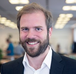 Woelber Johan P合作论文数Department of Operative Dentistry and Periodontology, University Freiburg Medical Center17Auschill Thorsten M合作论文数16
 Pascal Tomakidi合作论文数16
 Fabian Cieplik合作论文数Department of Operative Dentistry and Periodontology, University Medical Center Regensburg Regensburg15Arweiler Nicole B合作论文数15
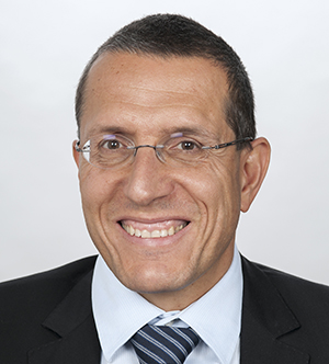 Anton Sculean合作论文数Department of Periodontology, School of Dental Medicine, Faculty of Medicine, University of Bern12
 Martin Wolkewitz合作论文数Institute of Medical Biometry and Statistics, Universitätsklinikum Freiburg;Institute of Medical Biometry and Medical Informatics, University of Freiburg12
 Christian Hannig合作论文数Poliklinik fur Zahnerhaltung, Universitatsklinikum Carl Gustav Carus Dresden12Argyropoulou Aikaterini合作论文数11
 Jorg Philipp Tchorz合作论文数11Alexios-Leandros Skaltsounis合作论文数11
 Susanne Proksch合作论文数11
 Thorsten Steinberg合作论文数10
 Nicole B. Arweiler合作论文数Department of Periodontology, Philipps University of Marburg10Petra Ratka-Kruger合作论文数10Christian Tennert合作论文数10A. Wittmer合作论文数9
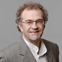 Klaus Kümmerer合作论文数Institute of Sustainable Chemistry, Leuphana University of Lüneburg;International Research and Education Sustainable Collaborative Centre, Leuphana University of Lüneburg8M. Wiedmann-Al-Ahmad合作论文数8
 Simon Schulz合作论文数8
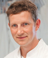 Tim Maisch合作论文数Zentrum für Seltene Erkrankungen, Universitätsklinikum Würzburg8Auschill Thorsten Mathias合作论文数7
 Karl-Anton Hiller合作论文数Department of Conservative Dentistry and Periodontology, University of Regensburg Medical Center;Universität Regensburg;University Hospital Regensburg7Altenburger Markus J合作论文数6
 Nadine Schlueter合作论文数Department of Conservative and Preventive Dentistry;Dental Clinic;Justus Liebig University;Department of Conservative and Preventive Dentistry, Justus Liebig University6Armin König合作论文数6J Schulte-Mönting合作论文数5
 David Auer合作论文数5Xiaojun Mao合作论文数5Elamin Fadil合作论文数Bart's and The London School of Medicine and Dentistry, Queen Mary University of London;Khartoum Centre for Research and Medical Training;El Razi Dental School;El Razi Dental School, Queen Mary University of London5Daniel Jonas合作论文数Department of Environmental Health Sciences, University Hospital Freiburg5




