The thyroid-stimulating hormone receptor (TSHR) is a promising molecular target for thyroid cancer imaging and therapy. Our previous work has demonstrated that PET imaging with the radiolabeled anti-TSHR human monoclonal antibody K1-70 enables assessment of TSHR expression in thyroid cancer. However, full-length antibody-based radiopharmaceuticals exhibit delayed systemic clearance and increased off-target radiation burden, resulting in suboptimal pharmacokinetics for immuno-PET imaging. Herein, we report the synthesis and evaluation of 64Cu-labeled anti-TSHR K1-70 antibody fragment antigen-binding (Fab) and single-chain variable fragment (scFv), for immuno-PET imaging of TSHR in thyroid cancer mouse models. These smaller formats enabled rapid tumor targeting and favorable pharmacokinetics with high tumor-to-background contrast. Two radiotracers, named 64Cu-NOTA-TSHR-Fab and 64Cu-NOTA-TSHR-scFv, were prepared by conjugating TSHR-Fab or TSHR-scFv to p-SCN-Bn-NOTA, followed by radiolabeling with 64Cu, achieving high radiochemical purity (>99%). The specificity and binding affinity of each radiotracer were determined by cellular uptake and binding assays using TSHR-positive THJ529T cells and corresponding wild-type controls. Both radiotracers exhibited specific, nanomolar binding affinity to TSHR-positive cells. Immuno-PET imaging, ex vivo biodistribution, and blocking studies of each radiotracer were performed in NSG mice bearing subcutaneous TSHR-positive THJ529T tumor xenografts at various time points (1, 4, 18, and 24 h postinjection). In comparative in vivo evaluations, 64Cu-NOTA-TSHR-Fab showed rapid and superior TSHR-specific tumor accumulation compared with 64Cu-NOTA-TSHR-scFv, evident as early as 1 h postinjection. Both radiotracers demonstrated rapid pharmacokinetics and low background signal, but with high renal uptake. This head-to-head comparison of small-size antibody fragment-based radiotracers for TSHR-targeted immuno-PET imaging identifies 64Cu-NOTA-TSHR-Fab as a promising immuno-PET radiotracer for in vivo detection of TSHR expression in thyroid cancer and for guiding TSHR-targeted therapy.
Background. Cardiovascular disease is a leading cause of death among women with breast cancer, and the 2026 ACC/AHA dyslipidemia guideline endorses coronary artery calcium (CAC) scoring to guide statin therapy before cardiotoxic treatment. Breast cancer patients routinely undergo staging 18F-fluorodeoxyglucose PET/CT, whose low-dose CT visualizes the coronary arteries, thus enabling CAC quantification at no additional cost or radiation. Methods. In this single-center retrospective study, consecutive women with newly diagnosed breast cancer undergoing staging 18F-FDG PET/CT (2009?2021) had semi-automated Agatston CAC scoring performed on the low-dose CT and were stratified by CAC presence (CAC-P) versus absence (CAC-A). We assessed a composite of cardiac diagnostic testing (stress testing, coronary CT angiography, invasive angiography), clinical events, and reclassification of statin eligibility per ACC/AHA guideline thresholds in a prevention-eligible subgroup. Results. Among 276 women (mean age 55.5 years; median follow-up 7.1 years), CAC was present in 68 (25%) but was clinically reported in only 5.4%. CAC-P was associated with more cardiac testing (34% vs 12%; age-adjusted hazard ratio 2.75, 95% CI 1.43?5.28) and, though underpowered, with more atherosclerotic events (7.4% vs 1.4%), but not with the all-cause composite. In the prevention-eligible subgroup (n=39), CAC scoring would have changed statin eligibility in 64%, initiating therapy in 62% of CAC-P women and supporting de-prescribing in 67% of CAC-A women. Conclusions. CAC can be feasibly quantified from staging PET/CT in women with breast cancer and would frequently reclassify statin eligibility at no additional cost or radiation, yet is rarely reported.
Cerebral glucose hypometabolism (CGHM) involves reduced brain glucose uptake on FDG-PET imaging and occurs in neurological and systemic diseases. Its reversibility remains uncertain. We describe a 40-year-old man with reversible global CGHM associated with cryptogenic organizing pneumonia (COP) and COVID-19. His neurocognitive symptoms improved rapidly following steroid-induced hyperglycemia, with FDG-PET demonstrating complete CGHM resolution after two months. His medical background included primary refractory transformed follicular lymphoma diagnosed 30 months earlier, managed with multiple treatment regimens including CAR-T therapy. After post-CAR-T relapse, combination therapy with polatuzumab vendotin, bendamustine, and obinutuzumab achieved complete remission, which was maintained when CGHM developed. This case establishes that CGHM can be temporary and reversible, highlighting a previously unrecognized pathway through which systemic illness may produce neurocognitive impairment. The rapid cognitive recovery coinciding with steroid-induced hyperglycemia and COP treatment is significant. Additional investigation is warranted to determine CGHM's significance in diverse pathological conditions and determine therapeutic interventions.
BACKGROUND:Functional pituitary adenomas (FPAs), especially those "MRI-occult" on 1.5T/3T and smaller than 10 mm, present diagnostic and therapeutic challenges, leading to repeated investigations, invasive procedures, and suboptimal outcomes. This review examines the potential clinical use and limitations of 7T MRI and amino-acid (AA)-PET with tracers like L-[methyl-11C]methionine (MET) or O-(2-[18F]fluoroethyl)-L-tyrosine (FET) for detecting MRI-occult FPAs. MATERIALS AND METHODS:A systematic search of Medline, Embase, and Web of Science was performed up to August 2025, following the Preferred Reporting Items for Systematic Reviews and Meta-Analyses guidelines. Studies evaluating 7T MRI and AA-PET for MRI-occult PAs were included and independently reviewed by three authors. Eighteen studies met inclusion criteria: 4 evaluated the 7T MRI, 8 MET-PET, three FET-PET, and three hybrid/multi-tracer approaches. RESULTS:7T MRI offers superior spatial resolution, increased signal-to-noise ratio, and improved tissue contrast. AA-PET provides high tumor-to-background contrast, facilitating MRI-occult PAs localization. CONCLUSIONS:Despite these advances, small cohort sizes, heterogeneous protocols, high costs, and limited accessibility limit the current evidence base. Ultra-high-field 7T MRI and AA-PET represent emerging complementary imaging techniques for MRI-occult FPAs. Current evidence is preliminary, underscoring the need to standardize protocols, conduct prospective multicenter studies, and assess cost-effectiveness before widespread clinical adoption.
Background and ObjectivesCognitive and psychiatric syndromes after coronavirus disease 2019 (COVID-19) are common and disabling. Different studies have aimed to link objective cognitive test performance to brain structure after SARS-CoV-2 infection, but a literature synthesis showing where and how the structure relates to specific cognitive domains has not yet been produced. Therefore, we aimed to perform a systematic literature review on morphologic and cognitive changes after COVID-19 infection.MethodsThis systematic review was reported in accordance with preferred reporting items for systematic reviews and meta-analyses (PRISMA). We included all published studies involving adult patients with confirmed SARS-CoV-2 infection, with brain imaging analysis (either magnetic resonance imaging [MRI] and 18F-fludeoxyglucose positron emitting tomography [18F-FDG-PET]) and objective assessment of cognitive changes up to July 29, 2024, focusing on studies reporting correlations between both features. The Newcastle-Ottawa Scale was used to assess the risk of bias.ResultsTwenty-one peer-reviewed studies published with 1,483 patients fulfilled the inclusion criteria. Of those, 9 were cross-sectional studies and 12 were cohorts. Age and sex ratios varied considerably across studies. MRI and 18F-FDG-PET revealed varying results regarding the neuroanatomical and metabolic changes following COVID-19 infection. MRI findings demonstrated significant associations between neuroanatomical alterations and cognitive function, particularly in attention, memory, and executive domains. White fiber density correlated positively with cognitive testing scores, while reductions in fractional anisotropy in the inferior longitudinal fasciculus were associated with executive dysfunction. Resting-state functional connectivity changes were associated with cognitive performance. 18F-FDG-PET studies revealed metabolic alterations in patients with post-COVID-19 infection, finding hypometabolism in frontal, temporal, and limbic regions correlating with cognitive dysfunction. On the other hand, several studies reported no significant associations between brain morphology or cerebral blood flow and cognitive outcomes.DiscussionAcross the included studies, results indicate that patients after COVID-19 infection exhibit structural, functional, and metabolic brain alterations that correlate with cognitive deficits, particularly in attention, memory, and executive function. Longitudinal findings suggest that these changes may persist, with disease severity perhaps influencing the extent of impairment. However, inconsistencies across studies highlight the need for further research to clarify the underlying mechanisms, permanence of changes, and long-term cognitive implications.
Oncocytic thyroid carcinoma (OTC) is often aggressive and refractory to radioiodine therapy, which is the current standard of care for metastatic thyroid cancer. The thyroid-stimulating hormone receptor (TSHR) regulates thyroid function and metabolism and is highly expressed in the thyroid gland and most thyroid tumors including OTC. Here, we report positron emission tomography (PET) imaging of radiolabeled TSHR antibody for detecting tumoral TSHR expression and monitoring TSHR chimeric antigen receptor (CAR) T-cell therapy response in an OTC mouse model. Radiotracer 89Zr-DFO-TSHR-Ab was prepared as previously reported for PET imaging of TSHR expression. Tissue microarray analysis confirmed TSHR expression in both normal thyroid and OTC tumors. A human OTC xenograft model was established by subcutaneous injection of XTC.UC1 cells into NSG mice. PET imaging and biodistribution studies of TSHR expression were subsequently conducted in this model to assess TSHR-expression change before and after TSHR CAR T-cell therapy. TSHR-targeted CAR T-cells were administered intravenously, and longitudinal PET/CT imaging with 89Zr-DFO-TSHR-Ab was performed at 24, 72, and 120 h postinjection to monitor tumor response. Eight weeks later, the same mice were rechallenged with XTC.UC1 cells in the contralateral flank to assess long-term therapeutic efficacy and immune memory through serial PET/CT imaging. PET imaging and biodistribution studies demonstrated that this radiotracer effectively detected TSHR-expressing OTC, with reasonable tumor uptake and imaging contrast. Following CAR T-cell therapy, TSHR PET showed significantly decreased tumor uptake, consistent with TSHR-targeted cell immunotherapy response with attenuated TSHR expression. These findings suggest 89Zr-DFO-TSHR-Ab enables noninvasive identification of OTC tumors and real-time monitoring of response to TSHR-targeted CAR T-cell therapy.
OBJECTIVES:The practice parameter was revised collaboratively by the American College of Radiology (ACR), the American Brachytherapy Society (ABS), the American College of Nuclear Medicine (ACNM), the American Radium Society (ARS), the Society of Interventional Radiology (SIR), and the Society of Nuclear Medicine and Molecular Imaging (SNMMI). This document summarizes current evidence-based guidelines for the administration of Yttrium radioembolic therapy to the liver, including training requirements, evidence-based guidelines for administration, and safe practice for administration. METHODS:This practice parameter was revised according to the process described under the heading The Process for Developing ACR Practice Parameters and Technical Standards on the ACR website ( https://www.acr.org/ClinicalResources/Practice-Parameters-and-Technical-Standards ) by the Committee on Practice Parameters-Interventional and Cardiovascular Radiology of the ACR Commission on Interventional and Cardiovascular, Committee on Practice Parameters and Technical Standards-Nuclear Medicine and Molecular Imaging of the ACR Commission on Nuclear Medicine and Molecular Imaging and the Committee on Practice Parameters-Radiation Oncology of the ACR Commission on Radiation Oncology in collaboration with the ABS, the ACNM, the ARS, the SIR, and the SNMMI. RESULTS:This review seeks not to be a comprehensive discussion of radiotherapy to the liver, but rather, seeks to provide a parameter for safe and effective therapy. We discuss the qualifications of physicians involved in this therapy, basic indications, contraindications, procedural work-up, safe-handling, and regulatory requirement for the administration of selective internal radiation therapy to patients that are likely to benefit. The goal of this document is not to define which patients are best treated by these therapies, as this is best determined for individual patients after multidisciplinary review. A consistent and evidence-based approach to therapy, however, would benefit all patients who are offered this therapy. This document seeks to provide a framework for current best practices for the administration of the 2 currently available radioembolization devices. CONCLUSIONS:As Yttrium-90 radiotherapy to the liver occupies a growing role in the treatment of primary and metastatic liver cancer, this review seeks to assist clinicians of all involved specialties to optimize the efficacy and safety of these procedures.
Advanced thyroid cancers are aggressive and often refractory to the current standard of care. The thyroid-stimulating hormone receptor (TSHR) is highly expressed in thyroid cancers and rarely expressed outside the thyroid, making it a viable target for developing radiotheranostics for imaging and therapy of advanced thyroid cancer. This study reports the radiosynthesis and preclinical evaluation of a 64Cu-labeled human antibody for positron emission tomography (PET) imaging of TSHR expression in advanced thyroid cancer mouse models. Human anti-TSHR recombinant antibody K1-70 (TSHR-Ab) was labeled with copper-64, yielding [64Cu]Cu-NOTA-TSHR-Ab with a radiochemical yield of 46.89 ± 3.74%, radiochemical purity of 98.77 ± 0.89%, and specific activity >212 GBq/μmol (n = 5). In vitro studies on TSHR-positive (THJ529TTSHR+) and wild-type (THJ529TWT) cells demonstrated the radiotracer's high specificity and nanomolar binding affinity for THJ529TTSHR+ cells, with a dissociation constant (Kd) of 4.74 nM and an inhibition constant (Ki) of 0.92 nM. ImmunoPET imaging in mice bearing dual-flank tumors (THJ529TWT and THJ529TTSHR+) at multiple time points (1, 2, 4, 18, 24, and 48 h) postinjection (p.i.) revealed rapid tumor targeting and high uptake in TSHR-positive thyroid tumors (SUVmax: 3.63 ± 0.42, 3.82 ± 0.44, and 4.09 ± 0.56 at 18, 24, and 48 h p.i., respectively). Co-injection studies with varying doses of unlabeled TSHR-Ab (0, 25, 50, 100 μg) demonstrated that the coinjection significantly reduced background signals, especially in the spleen, liver, and bone, with a dose of 25 μg effectively reducing off-target signals without affecting tumor uptake. Biodistribution and immunohistochemistry analyses supported these immunoPET imaging results. Furthermore, a comparison study with traditional [18F]FDG PET imaging showed that [64Cu]Cu-NOTA-TSHR outperformed [18F]FDG in tumor detection. In conclusion, [64Cu]Cu-NOTA-TSHR-Ab is a promising radiotracer for PET imaging of TSHR-positive advanced thyroid cancers, with the potential to guide and monitor TSHR-targeted therapies. Further clinical evaluation of [64Cu]Cu-NOTA-TSHR-Ab could provide valuable insights for patient stratification and optimization of anti-TSHR treatments.
Mesenchymal stromal cell (MSC) therapy has regenerative potentials to treat various pathological conditions including neurological diseases. MSCs isolated from various organs can differentiate into specific cell types to repair organ damages. However, their paracrine mechanisms are predicted to predominantly mediate their immunomodulatory, proangiogenic, and regenerative properties. While preclinical studies highlight the significant potential of MSC therapy in mitigating neurological damage from stroke and traumatic brain injury, the variability in clinical trial outcomes may stem from the inherent heterogeneity of somatic MSCs. Accumulating evidence has demonstrated that induced pluripotent stem cells (iPSCs) are an ideal alternative resource for the unlimited expansion and biomanufacturing of MSCs. Thus, we investigated how iPSC-derived MSCs (iMSCs) influence properties of iPSC-derived neurons. Our findings demonstrate that the secretome from iMSCs possesses neurotrophic effects, improving neuronal survival and promoting neuronal outgrowth and synaptic activity in vitro. Additionally, the iMSCs enhance metabolic activity via mitochondrial respiration in neurons, both in vitro and in mouse models. Glycolytic pathways also increased following the administration of iMSC secretome to iPSC-derived neurons. Consistently, in vivo experiments showed that intravenous administration of iMSCs compensated for the elevated energetic demand in male mice with irradiation-induced brain injury by restoring synaptic metabolic activity during acute brain damage. 18 F-FDG PET imaging also detected an increase in brain glucose uptake following iMSC administration. Together, our results highlight the potential of iMSC-based therapy in treating neuronal damage in various neurological disorders, while paving the way for future research and potential clinical applications of iMSCs in regenerative medicine.
Following lung cancer, prostate cancer is the leading cause of cancer death in men. High-risk localized tumor burden or metastatic disease often progresses, refractory to initial treatment regimens. There is ongoing development of technology to appropriately identify high-risk patients, stage them correctly, and offer appropriate treatments to obtain the best clinical outcomes. Prostate cancer-specific membrane antigen (PSMA) is a transmembrane glutamate carboxypeptidase, which helps regulate folate absorption, and its overexpression is pathologically directly proportional and associated with prostate cancer. Increased PSMA expression is a known independent risk factor for poorer survival, and most metastatic lesions in CRPC are PSMA positive. Over the last decade, several PSMA-based PET radiopharmaceuticals have demonstrated superior sensitivities and specificities compared to traditional imaging methods. These outcomes have been demonstrated by several large clinical trials. As the data emerges, these diagnostics are being integrated into standard of care protocol to facilitate nuanced identification of malignant lesions. PSMA is also being targeted through several therapeutics, including radioligands and immunotherapies such as CAR-T, BiTEs, and ADCs. This review will discuss the landscape of PSMA-based theranostics in the context of prostate cancer.
Abstract Metastatic castration-resistant prostate cancer (mCRPC) is an aggressive malignancy with poor outcomes. To investigate novel therapeutic strategies, we characterized three new metastatic prostate cancer patient derived-tumor xenograft (PDTX) models and developed 3D spheroids from each to investigate molecular targeted therapy combinations including CDK4/6 inhibitors (CDK4/6i) with AKT inhibitors (ATKi). Metastatic prostate cancer tissue was collected and three PDTX models were established and characterized using whole-exome sequencing. PDTX 3D spheroids were developed from these three PDTXs to show resistance patterns and test novel molecular-targeted therapies. CDK4/6i's were combined with AKTi's to assess synergistic antitumor response to prove our hypothesis that blockade of AKT overcomes drug resistance to CDK4/6i. This combination was evaluated in PDTX three-dimensional (3D) spheroids and in vivo experiments with responses measured by tumor volumes, PSA, and Ga-68 PSMA-11 PET-CT imaging. We demonstrated CDK4/6i's with AKTi's possess synergistic antitumor activity in three mCRPC PDTX models. These models have multiple unique pathogenic and deleterious genomic alterations with resistance to single-agent CDK4/6i's. Despite this, combination therapy with AKTi's was able to overcome resistance mechanisms. The IHC and Western blot analysis confirmed on target effects, whereas tumor volume, serum PSA ELISA, and radionuclide imaging demonstrated response to therapy with statistically significant SUV differences seen with Ga-68 PSMA-11 PET-CT. These preclinical data demonstrating antitumor synergy by overcoming single-agent CDK 4/6i as well as AKTi drug resistance provide the rational for a clinical trial combining a CDK4/6i with an AKTi in patients with mCRPC whose tumor expresses wild-type retinoblastoma 1.
592 Background: The introduction of peptide receptor radionuclide therapy (PRRT) using Lutetium-177 (177Lu) DOTATATE has led to a paradigm shift in the treatment of neuroendocrine tumors (NETs). Hematological toxicity is a well-recognized adverse effect (AE) of 177Lu DOTATATE. Most cytopenias are transient, with an estimated incidence of 10-25%, typically mild to moderate in severity (grade G1-2). The most dreaded AE of PRRT is therapy-related myeloid neoplasm (t-MN), with an estimated 2-8% incidence and a higher incidence reported in patients who also received prior chemotherapy. We sought to evaluate the hematological safety of 177Lu PRRT in the setting of NETs with extensive (i.e., innumerable) bone metastases. Methods: We retrospectively reviewed the medical records of all Mayo Clinic patients (pts) with extensive/innumerable osseous metastases, defined as >50% skeletal involvement by positron emission tomography (PET) DOTATATE, who were treated with 177Lu DOTATATE. Pts characteristics and laboratory results were collected before, during, and after 177Lu DOTATATE treatment. Hematotoxicity was graded according to the NCI-CTCAE v5. Results: Out of 27 pts, 13(48%) developed cytopenia(s) of any grade after treatment with one or more cycles of 177Lu DOTATATE. In total, there were 13(48%) pts with anemia, 13(48%) with thrombocytopenia, and 6(22%) with neutropenia. Six (22%) pts had severe hematological toxicity (G3-4). Twelve months post-PRRT, 7(26%) pts continued to experience cytopenia(s) of any grade, of whom 4(15%) had G3-4 hematotoxicity. One pt developed t-MN, and two pts had myelophthisis on bone marrow biopsy from infiltrating NET. 16(59%) pts received cytotoxic chemotherapy before PRRT, and 7(25.9%) developed subsequent cytopenia(s). Overall, 15(55%) pts completed four cycles, while 3 pts didn’t complete four cycles of 177Lu DOTATATE due to hematotoxicity. Conclusions: Pts with advanced NETs and extensive/innumerable bone metastases treated with PRRT could be at higher risk for myelosuppression than historical controls. Our study highlights the importance of carefully monitoring and assessing hematological parameters in pts being considered for 177Lu DOTATATE with extensive/innumerable bone metastasis. [Table: see text]
Breast carcinomas are well known for expression of estrogen receptor (ER) however there are other malignancies that are also express ER, possibly confounding the diagnostic interpretation of 16α-[18F]fluoro-17β-estradiol (FES; Cerianna GE HealthCare) in patients with both ER+ breast carcinomas and other malignancies. We present a case of a woman with prior history of both ER+ breast carcinoma and pulmonary adenocarcinoma with subsequent identification of an FES pulmonary nodule that was proven on histopathology to be consistent with an ER expressing pulmonary adenocarcinoma metastasis
Thyroid-stimulating hormone receptor (TSHR) is a G-protein coupled receptor that is highly expressed on benign and malignant thyroid tissues. TSHR binding and activation has long been a component of thyroid cancer molecular imaging and radiotherapy, by promoting expression of the sodium-iodide symporter (NIS) and incorporation of I-131 into thyroid hormones. Here, we report the radiosynthesis and preclinical evaluation of a Zirconium-89 (89Zr) labeled TSHR antibody to serve as a positron emission tomography (PET) diagnostic correlate for therapeutic agents targeting TSHR without reliance on NIS. TSHR human monoclonal antibody K1-70 was conjugated to chelator desferrioxamine-p-benzyl-isothiocyanate, followed by labeling with Zr-89, yielding the radiotracer 89Zr-DFO-TSHR-Ab. The in vitro cellar uptake and binding affinity of 89Zr-DFO-TSHR-Ab were analyzed in three new TSHR stable overexpressing tumor cell lines and their corresponding wild types (WT) with low or no TSHR expression. 89Zr-DFO-TSHR-Ab PET/CT imaging of TSHR expression was evaluated in tumor mouse models bearing one TSHR-positive tumor and other negative control with or without the coinjection of antibody K1-70, and then verified by radiotracer biodistribution study and tumor immunohistochemistry (IHC). The conjugate DFO-TSHR-Ab was labeled with Zr-89 at 37 °C for 60 min and purified by PD-10 column in radiochemical yields of 68.8 ± 9.9
 Hancheng Cai合作论文数Keck School of Medicine, University of Southern California15
 Sharma Akash合作论文数Mallinckrodt Institute of Radiology14
 John A. Katzenellenbogen合作论文数Department of Chemistry, College of Liberal Arts & Sciences, University of Illinois Urbana-Champaign11
 David Schuster合作论文数Department of Radiology and Imaging Sciences, School of Medicine, Emory University;Winship Cancer Institute, Emory University11
 John A. Copland III合作论文数Comprehensive Cancer Center, Mayo Clinic11
 Michael J. Welch合作论文数Department of Radiology, Washington University School of Medicine in St. Louis;Department of Biomedical Engineering, James McKelvey School of Engineering, Washington University School of Medicine in St. Louis8
 Terry L. Sharp合作论文数7Adam M Kase合作论文数Div Hematol Oncol, Mayo Clin6Winston W Tan合作论文数6Han W. Tun合作论文数Hematology, Mayo Clinic6Laura Marlow合作论文数5Daniel P. Wickland合作论文数5Brandy H Edenfield合作论文数5Erin E. Miller合作论文数5Joachim L. Petit合作论文数5Nathalie Meurice合作论文数5Yumei Zhou合作论文数5Savir-Baruch Bital合作论文数Department of Radiology and Imaging Sciences, Emory University5
 Akin Akintayo合作论文数5
 Mark Goodman合作论文数Department of Radiology and Imaging Sciences, School of Medicine, Emory University5Joshua Knight合作论文数5
 Jonathan Mcconathy合作论文数5Michael T. Barrett合作论文数5
 Carmen Dence合作论文数5Justyna J. Gleba合作论文数4Olayinka A. Abiodun-Ojo合作论文数Department of Internal Medicine, Piedmont Athens Regional Medical Center4
 Madiha Iqbal合作论文数4
 Kathryn Fowler合作论文数Department of Radiology, University of California San Diego;Jacobs Medical Center, University of California San Diego;Hillcrest Medical Center, University of California San Diego4
 Jeffrey Olson合作论文数Winship Cancer Institute, Emory University;Department of Neurosurgery, Emory University School of Medicine4
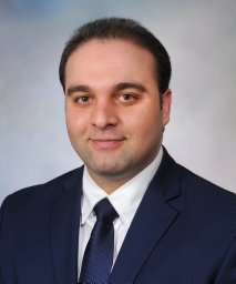 Moustafa Muhamad Alhaj合作论文数Department of Internal Medicine, Mayo Clinic4Farrokh Dehdashti合作论文数4Jonathon A. Nye合作论文数Department of Radiology, Emory University School of Medicine4
 Geoffrey S Johnson合作论文数3Amy Barone合作论文数3Hayden Leeds合作论文数3Michael Grimaldi合作论文数3Albert Chau合作论文数3
 Mariam Aboian合作论文数Neuroradiology, Radiology & Biomedical Imaging, Yale School of Medicine, Yale University3
 Karen Gauvain合作论文数3
 Thor R. Halfdanarson合作论文数School of Medicine, Mayo Clinic;Comprehensive Cancer Center, Mayo Clinic3
 Bradley C. Carthon合作论文数Winship Cancer Institute, Emory University;Department of Hematology and Medical Oncology, School of Medicine, Emory University3Starr Jason合作论文数3
 Mehmet Asim Bilen合作论文数Winship Cancer Institute, Emory University;Department of Hematology and Medical Oncology, School of Medicine, Emory University3Teoh Eugene J合作论文数3
 Ataei Fatemeh合作论文数3
 Maria Rosana Ponisio合作论文数Mallinckrodt Institute of Radiology, Washington University School of Medicine3
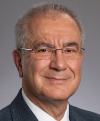 Omer Kucuk合作论文数Department of Hematology and Medical Oncology, School of Medicine, Emory University3
 Manoj Kumar Jain合作论文数Dept. of Computer Science & Engg
Indian Institute of Technology3
Manoj Kumar Jain合作论文数Dept. of Computer Science & Engg
Indian Institute of Technology3 Joshua B. Rubin合作论文数School of Medicine, Washington University in St. Louis;Division of Hematology & Oncology, Washington University in St. Louis3




