In two previous studies, based on human olfactory ecto-mesenchymal stem cells (OE-MSC) of 11 patients with autism spectrum disorders (ASD) and 11 healthy individuals, we demonstrated that the lower abundance of the enzyme MOCOS (MOlybdenum COfactor Sulfurase) and its associated lower expression of the long non-coding RNA, COSMOC, induces neurotransmission and synaptic defects as well as an exacerbated oxidative stress sensitivity. To move a step further, we assessed whether these defects were associated to a disturbed mitochondrial homeostasis. For that purpose, we used cellular and molecular techniques to quantitfy mitochondrial metabolism and biogenesis, ATP production and cell respiration in OE-MSCs from the 8 ASD patients of the cohort that display the most severe symptoms. We show here that OE-MSCs from ASD patients, when compared to control individuals, display (i) a reduced expression/abundance of glycolysis-associated transcripts and metabolites, (ii) an overall reduced ATP, mainly due to the impaired glycolysis, (iii) a reduced basal cell respiration and (iv) a modified mitochondrial network. These results are in accordance with some of our previously published data and may explain some of the symptoms - stress, overarousal, seizures, increased or decreased muscle tone, fatigue-observed in autism spectrum disorders.
Damage to the peripheral nerve impairs quality of life. Despite the advances in surgery to facilitate nerve regeneration, complete recovery remains elusive. Human olfactory ecto-mesenchymal stem cells (OEMSC) have potential for the treatment of peripheral nerve injury through the release of extracellular vesicles (EV). The current research investigates the therapeutic effects of a venous bridge, filled with freshly purified or cryoconserved OEMSC-derived EVs after a peroneal nerve loss of substance. Injured peroneal nerve was bridged with a vein into which freshly purified or cryoconserved EVs were injected or not. Nerve repair was analyzed by measuring locomotor function, muscle mechanical properties, muscle mass, axon number, and myelination. The EVs significantly increased locomotor recovery, maintained the contractile phenotype of the target muscle, and augmented the number of growing axons. These results demonstrate that EVs display a positive effect on peripheral nerve regeneration, representing an alternative to cellular therapies for peripheral nerve repair. ### Competing Interest Statement The authors have declared no competing interest.
Damage to the peripheral nerve significantly impairs quality and way of life. Despite the slow self-regeneration of the peripheral nervous system and advances in surgery, complete recovery after nerve injury remains elusive especially when there is loss of substance. Human olfactory ecto-mesenchymal stem cells (OEMSC) have potential for the treatment of peripheral nerve injury through the secretion of bioactive factors, such as proteins, cytokines, chemokines as well as the release of extracellular vesicles (EV). The current research investigates the therapeutic effects of a venous bridge, filled with freshly purified or cryoconserved OEMSC-derived EVs after a peroneal nerve loss of substance. A 7 mm defect of peroneal nerve was bridged with a vein into which freshly purified or cryoconserved OEMSC-derived EVs were injected or not. These groups were compared with unoperated rats (Control) and autografted rats with the nerve sutured in inverted position (Gold Standard). Three months after surgery, nerve repair was analyzed by measuring locomotor function, muscle mechanical properties, muscle mass, axon number, and myelination. We observed that OEMSC-derived EVs significantly (i) increased locomotor recovery, (ii) partially maintained the contractile phenotype of the target muscle, and (iii) augmented the number of growing axons. Freshly purified EVs exerted a better recovery than the Gold Standard. These results demonstrate that EVs display a positive effect on peripheral nerve regeneration, similarly to what has been observed with OEMSCs, the cells from which they originate. They represent an alternative to cellular therapies for peripheral nerve repair.
In two previous studies, based on human olfactory ecto-mesenchymal stem cells (OE-MSC) of 11 patients with autism spectrum disorders (ASD) and 11 healthy individuals, we demonstrated that the lower abundance of the enzyme MOCOS (MOlybdenum COfactor Sulfurase) and its associated lower expression of the long non-coding RNA, COSMOC, induces neurotransmission and synaptic defects as well as an exacerbated oxidative stress sensitivity. To move a step further, we assessed whether these defects were associated to a disturbed mitochondrial homeostasis. For that purpose, we used cellular and molecular techniques to quantitfy mitochondrial metabolism and biogenesis, ATP production and cell respiration in OE-MSCs from the 8 ASD patients of the cohort that display the most severe symptoms. We show here that OE-MSCs from ASD patients, when compared to control individuals, display i) a reduced expression/abundance of glycolysis-associated transcripts and metabolites, ii) an overall reduced ATP, mainly due to the impaired glycolysis, iii) a reduced basal cell respiration and iv) a modified mitochondrial network. These results are in accordance with some of our previously published data and may explain some of the symptoms – stress, overarousal, seizures, increased or decreased muscle tone, fatigue - observed in autism spectrum disorders.
Sulfur is an important element that is incorporated into many biomolecules in humans. The incorporation and transfer of sulfur into biomolecules is, however, facilitated by a series of different sulfurtransferases. Among these sulfurtransferases is the human mercaptopyruvate sulfurtransferase (MPST) also designated as tRNA thiouridine modification protein (TUM1). The role of the human TUM1 protein has been suggested in a wide range of physiological processes in the cell among which are but not limited to involvement in Molybdenum cofactor (Moco) biosynthesis, cytosolic tRNA thiolation and generation of H2S as signaling molecule both in mitochondria and the cytosol. Previous interaction studies showed that TUM1 interacts with the L-cysteine desulfurase NFS1 and the Molybdenum cofactor biosynthesis protein 3 (MOCS3). Here, we show the roles of TUM1 in human cells using CRISPR/Cas9 genetically modified Human Embryonic Kidney cells. Here, we show that TUM1 is involved in the sulfur transfer for Molybdenum cofactor synthesis and tRNA thiomodification by spectrophotometric measurement of the activity of sulfite oxidase and liquid chromatography quantification of the level of sulfur-modified tRNA. Further, we show that TUM1 has a role in hydrogen sulfide production and cellular bioenergetics.
BackgroundOlfactory ecto-mesenchymal stem cells (OE-MSC) are mesenchymal stem cells derived from the lamina propria of the nasal mucosa. They display neurogenic and immunomodulatory properties and were shown to induce recovery in animal models of spinal cord trauma, hearing loss, Parkinsons’s disease, amnesia, and peripheral nerve injury. As a step toward clinical practice, we sought to (i) devise a culture protocol that meets the requirements set by human health agencies and (ii) assess the efficacy of stem cells on neuron differentiation.MethodsNasal olfactory mucosa biopsies from three donors were used to design and validate the good manufacturing process for purifying stem cells. All processes and procedures were performed by expert staff from the cell therapy laboratory of the public hospital of Marseille (AP-HM), according to aseptic handling manipulations. Premises, materials and air were kept clean at all times to avoid cross-contamination, accidents, or even fatalities. Purified stem cells were cultivated for 24 or 48 h and conditioned media were collected before being added to the culture medium of the neuroblastoma cell line Neuro2a.ResultsCompared to the explant culture-based protocol, enzymatic digestion provides higher cell numbers more rapidly and is less prone to contamination. The use of platelet lysate in place of fetal calf serum is effective in promoting higher cell proliferation (the percentage of CFU-F progenitors is 15.5%), with the optimal percentage of platelet lysate being 10%. Cultured OE-MSCs do not show chromosomal rearrangement and, as expected, express the usual phenotypic markers of mesenchymal stem cells. When incorporated in standard culture medium, the conditioned medium of purified OE-MSCs promotes cell differentiation of Neuro2a neuroblastoma cells.ConclusionWe developed a safer and more efficient manufacturing process for clinical grade olfactory stem cells. With this protocol, human OE-MSCs will soon be used in a Phase I clinical based on their autologous transplantation in digital nerves with a neglected injury. However, further studies are required to unveil the underlying mechanisms of action.
The olfactory ecto-mesenchymal stem cell (OE-MSC) are mesenchymal stem cells originating from the lamina propria of the nasal mucosa. They have neurogenic and immune-modulatory properties and showed therapeutic potential in animal models of spinal cord trauma, hearing loss, Parkinsons’s disease, amnesia, and peripheral nerve injury. In this paper we designed a protocol that meet the requirements set by human health agencies to manufacture these stem cells for clinical applications. Once purified, OE-MSCs can be used per se or expanded in order to get the extracellular vesicles (EV) they secrete. A protocol for the extraction of these vesicles was validated and the EV from the OE-MSC were functionally tested on an in vitro model. Nasal mucosa biopsies from three donors were used to validate the manufacturing process of clinical grade OE-MSC. All stages were performed by expert staff of the cell therapy laboratory according to aseptic handling manipulations, requiring grade A laminar airflow. Enzymatic digestion provides more rapidly a high number of cells and is less likely to be contaminated. Foetal calf serum was replaced with human platelet lysate and allowed stronger cell proliferation, with the optimal percentage of platelet lysate being 10%. Cultivated OE-MSCs are sterile, highly proliferative (percentage of CFU-F progenitors was 15,5%) and their maintenance does not induce chromosomal rearrangement (karyotyping and chromosomal microarray analysis were normal). These cells express the usual phenotypic markers of OE-MSC. Purification of the EVs was performed with ultracentrifugation and size exclusion chromatography. Purified vesicles expressed the recognized markers of EVs (Minimal Information for Studies of Extracellular Vesicles (“MISEV”) guidelines) and promoted cell differentiation and neurite elongation in a model of neuroblastoma Neuro2a cell line. We developed a safer and more efficient manufacturing process for clinical-grade olfactory stem cells, these cells can now be used in humans. A phase I clinical trial will begin soon.An efficient protocol for the purification of the OE-MSC EVs have been validated. These EVs exert neurogenic properties in vitro . More studies are needed to understand the exact mechanisms of action of these EVs and prove their efficacy and safety in animal models.
Background: Bone repair induced by stem cells and biomaterials may represent an alternative to autologous bone grafting. Mesenchymal stromal/stem cells (MSCs), easily accessible in every human, are prototypical cells that can be tested, alone or with a biomaterial, for creating new osteoblasts. The aim of this study was to compare the efficiency of two biomaterials-biphasic calcium phosphate (BCP) and bioactive glass (BG)-when loaded with either adult bone marrow mesenchymal stem cells (BMMSCs) or newborn nasal ecto-mesenchymal stem cells (NE-MSCs), the latter being collected for further repair of lip cleft-associated bone loss. Materials and Methods: BMMSCs were collected from two adults and NE-MSCs from two newborn infants. An in vitro study was performed in order to determine the best experimental conditions for adhesion, viability, proliferation and osteoblastic differentiation on BCP or BG granules. Bone-associated morphological changes and gene expression modifications were quantified using histological and molecular techniques. The in vivo study was based on the subcutaneous implantation in nude mice of the biomaterials, loaded or not with one of the two cell types. Eight weeks after, bone formation was assessed using histological and electron microscopy techniques. Results: Both cell types-BMMSC and NE-MSC-display the typical stem cell surface markers-CD73+, CD90+, CD105+, nestin - and exhibit the MSC-associated osteogenic, chondrogenic and adipogenic multipotency. NE-MSCs produce less collagen and alkaline phosphatase than BMMSCs. At the transcript level, NE-MSCs express more abundantly three genes coding for bone sialoprotein, osteocalcin and osteopontin while BMMSCs produce extra copies of RunX2. BMMSCs and NE-MSCs adhere and survive on BCP and BG. In vivo experiments reveal that bone formation is only observed with BMMSCs transplanted on BCP biomaterial. Conclusion: Although belonging to the same superfamily of mesenchymal stem cells, BMMSCs and NE-MSCs exhibit striking differences, in vitro and in vivo. For future clinical applications, the association of BMMSCs with BCP biomaterial seems to be the most promising.
Animals strongly rely on chemical senses to uncover the outside world and adjust their behaviour. Chemical signals are perceived by facial sensitive chemosensors that can be clustered into three families, namely the gustatory (TASR), olfactory (OR, TAAR) and pheromonal (VNR, FPR) receptors. Over recent decades, chemoreceptors were identified in non-facial parts of the body, including the brain. In order to map chemoreceptors within the encephalon, we performed a study based on four brain atlases. The transcript expression of selected members of the three chemoreceptor families and their canonical partners was analysed in major areas of healthy and demented human brains. Genes encoding all studied chemoreceptors are transcribed in the central nervous system, particularly in the limbic system. RNA of their canonical transduction partners (G proteins, ion channels) are also observed in all studied brain areas, reinforcing the suggestion that cerebral chemoreceptors are functional. In addition, we noticed that: (i) bitterness-associated receptors display an enriched expression, (ii) the brain is equipped to sense trace amines and pheromonal cues and (iii) chemoreceptor RNA expression varies with age, but not dementia or brain trauma. Extensive studies are now required to further understand how the brain makes sense of endogenous chemicals.
Imprinting is an early sensory life experience that induces adult behaviours, such as mother recognition or homing. In a previous study, we demonstrated a striking olfactory imprinting in C. elegans that can be inherited over generations. When exposed to specific odorants during a timely controlled post-hatch period, C. elegans worms display during adulthood an enhanced migration towards these molecules. In order to unveil some of the genetic and epigenetic factors that are responsible for such a behavioural plasticity, we assessed the role of heterochronic genes using a candidate gene approach. We report here that translation of the Hunchback-Like 1 (HBL1) transcription factor in the sensory processing interneuron AIY, is a determining factor for olfactory plasticity timing in C . elegans . HBL1 may associate to the SPR1/CoREST co-repressor, the lysine demethylase SPR5/LSD1 and the histone deacetylase HDA3 to lengthen the plasticity period, whereas the translation initiation factor IFE-4 and the histone deacetylase HDA2 abridge it. We also observed that lengthened plasticity periods allow proportionally faster stable behavioral adaptation of C. elegans populations. We conclude that plasticity timing is a key factor, not only to transiently adapt individuals but also to stably adapt animal populations via multigenerational accumulation of experience.
Peripheral nerve injuries can lead to severe functional deficits. Current treatments unfortunately remain insufficient. Stem cell grafting is nevertheless a promising therapy. Our team works on olfactory stem cells, located in the nasal cavity. The latter belong to the peripheral nervous system and are easily accessible in any awake patients, (without affecting the sense of smell). Having a high mitogenic activity and differentiating potential toward neural cells, they are related to bone marrow stem cells. We validated a new production process of human olfactory stem cells from a 2 mm(2) specimen for their clinical use in human peripheral nervere generation enhancement. We recently demonstrated, using a rat model, that olfactory stem cells grafting as an adjuvant treatment to surgical repair of facial or peroneal nerves improves functional recovery due to the immunomodulatory and trophic properties of these cells. Once grafted, the olfactory cells stop proliferating and we found no migration to other peripheral organs in our animal model. These characteristics indicate a low associated carcinogenic risk. Following these positive outcomes, we will soon begin a phase I/IIa clinical trial using olfactory stem cells for sensitive peripheral injuries or neuromas. Extracellular vesicles are nanoparticles produced by cells. Their use would enable to have the beneficial paracrine effects of stemcells without their drawbacks (immunocompatibility issues and long culture time). We validated their extraction for olfactory stem cells and are currently testing their efficiency for peripheral nerve regeneration on in vitro and in vivo models. (C) 2021 l'Academie nationale de medecine. Published by Elsevier Masson SAS. All rights reserved.
Over the recent years, several methods have been experienced to repair injured peripheral nerves. Among investigated strategies, the use of natural or synthetic conduits was validated for clinical application. In this study, we assessed the therapeutic potential of vein guides, transplanted immediately or two weeks after a peroneal nerve injury and filled with olfactory ecto-mesenchymal stem cells (OEMSC). Rats were randomly allocated to five groups. A3 mm peroneal nerve loss was bridged, acutely or chronically, with a 1 cm long femoral vein and with/without OEMSCs. These four groups were compared to unoperated rats (Control group). OEMSCs were purified from male olfactory mucosae and grafted into female hosts. Three months after surgery, nerve repair was analyzed by measuring locomotor function, mechanical muscle properties, muscle mass, axon number, and myelination. We observed that stem cells significantly (i) increased locomotor recovery, (ii) partially maintained the contractile phenotype of the target muscle, and (iii) augmented the number of growing axons. OEMSCs remained in the nerve and did not migrate in other organs. These results open the way for a phase I/IIa clinical trial based on the autologous engraftment of OEMSCs in patients with a nerve injury, especially those with neglected wounds.
Background: Posttraumatic facial paralysis is a disabling condition. Current surgical management by faciofacial nerve suture provides limited recovery. To improve the outcome, the authors evaluated an add-on strategy based on a syngeneic transplantation of nasal olfactory stem cells in a rat model of facial nerve injury. The main readouts of the study were the recording of whisking function and buccal synkinesis. Methods: Sixty rats were allocated to three groups. Animals with a 2-mm facial nerve loss were repaired with a femoral vein, filled or not with olfactory stem cells. These two groups were compared to similarly injured rats but with a faciofacial nerve suture. Olfactory stem cells were purified from rat olfactory mucosa. Three months after surgery, facial motor performance was evaluated using video-based motion analysis and electromyography. Synkinesis was assessed by electromyography, using measure of buccal involuntary movements during blink reflex, and double retrograde labeling of regenerating motoneurons. Results: The authors’ study reveals that olfactory stem cell transplantation induces functional recovery in comparison to nontransplanted and faciofacial nerve suture groups. They significantly increase (1) maximal amplitude of vibrissae protraction and retraction cycles and (2) angular velocity during protraction of vibrissae. They also reduce buccal synkinesis, according to the two techniques used. However, olfactory stem cell transplantation did not improve axonal regrowth of the facial nerve, 3 months after surgery. Conclusions: The authors show here that the adjuvant strategy of syngeneic transplantation of olfactory stem cells improves functional recovery. These promising results open the way for a phase I clinical trial based on the autologous engraftment of olfactory stem cells in patients with a facial nerve paralysis.
Vitamin D is a steroid hormone implicated in the regulation of neuronal integrity and many brain functions. Its influence, as a nutrient and a hormone, on the physiopathology of the most common neurodegenerative diseases is continuously emphasized by new studies. This review addresses what is currently known about the action of vitamin D on the nervous system and neurodegenerative diseases such as Multiple Sclerosis, Alzheimer’s disease, Parkinson’s disease and Amyotrophic Lateral Sclerosis. Further vitamin D research is necessary to understand how the action of this “neuroactive” steroid can help to optimize the prevention and treatment of several neurological diseases.
Autism spectrum disorders (ASD) are complex neurodevelopmental disorders with a very large number of risk loci detected in the genome. However, at best, each of them explains rare cases, the majority being idiopathic. Genomic data on ASD derive mostly from post-mortem brain analyses or cell lines derived from blood or patient-specific induced pluripotent stem cells (iPSCS). Therefore, the transcriptional and regulatory architecture of the nervous system, particularly during early developmental periods, remains highly incomplete. To access the critical disturbances that may have occurred during pregnancy or early childhood, we recently isolated stem cells from the nasal cavity of anesthetized patients diagnosed for ASD and compared them to stem cells from gender-matched control individuals without neuropsychiatric disorders. This allowed us to discover MOCOS, a non-mutated molybdenum cofactor sulfurase-coding gene that was under-expressed in the stem cells of most ASD patients of our cohort, disturbing redox homeostasis and synaptogenesis. We now report that a divergent transcription upstream of MOCOS generates an antisense long noncoding RNA, to which we coined the name COSMOC. Surprisingly, COSMOC is strongly under-expressed in all ASD patients of our cohort with the exception of a patient affected by Asperger syndrome. Knockdown studies indicate that loss of COSMOC reduces MOCOS expression, destabilizes lipid and energy metabolisms of stem cells, but also affects neuronal maturation and splicing of synaptic genes. Impaired expression of the COSMOC/MOCOS bidirectional unit might shed new lights on the origins of ASD that could be of importance for future translational studies.
In line with experiments showing that implanted hydrogels are promising tools, we designed and injected, after a C2 spinal cord hemisection, a thermoresponsive and thermoreversible physically cross-linked poly(N-isopropylacrylamide)-poly(ethylene glycol) copolymer in order to reduce functional deficits and provide a favorable environment to axotomized axons. Nasal olfactory ecto-mesenchymal stem cells were cultured on the hydrogel in order to verify its biocompatibility. Then, inflammatory reaction (Interleukin-1β and 6, Tumor Necrosis Factor-α) was examined 15 days post-hydrogel injection. Functional recovery (postural and locomotor activities, muscle strength and tactile sensitivity) was assessed once a week, during 12 weeks. Finally, at 12 weeks post-injection, spinal reflexivity and ventilatory adjustments were measured, and the presence of glial cells and regenerated axons were determined in the injured area. Our results indicate that cells survived and proliferated on the hydrogel which, itself, did not induce an enhanced inflammation. Furthermore, we observed significant motor and sensitive improvements in hydrogel-injected animals. Hydrogel also induced H-reflex recovery close to control animals but no improved ventilatory adjustment to electrically-evoked isometric contractions. Finally, regrowing axons were visualized within the hydrogel with no glial cells colonization. Our results emphasize the effectiveness of our copolymer and its high therapeutic potential to repair the spinal cord after injury.
The discovery of novel drugs for neurodegenerative diseases has been a real challenge over the last decades. The development of patient- and/or disease-specific in vitro models represents a powerful strategy for the development and validation of lead candidates in preclinical settings. The implementation of a reliable platform modeling dopaminergic neurons will be an asset in the study of dopamine-associated pathologies such as Parkinson's disease. Disease models based on cell reprogramming strategies, using either human-induced pluripotent stem cells or transcription factor-mediated transdifferentiation, are among the most investigated strategies. However, multipotent adult stem cells remain of high interest to devise direct conversion protocols and establish in vitro models that could bypass certain limitations associated with reprogramming strategies. Here, we report the development of a six-step chemically defined protocol that drives the transdifferentiation of human nasal olfactory stem cells into dopaminergic neurons. Morphological changes were progressively accompanied by modifications matching transcript and protein dopaminergic signatures such as LIM homeobox transcription factor 1 alpha (LMX1A), LMX1B, and tyrosine hydroxylase (TH) expression, within 42 days of differentiation. Phenotypic changes were confirmed by the production of dopamine from differentiated neurons. This new strategy paves the way to develop more disease-relevant models by establishing reprogramming-free patient-specific dopaminergic cell models for drug screening and/or target validation for neurodegenerative diseases.
Olfactory receptors (ORs) are members of G protein-coupled receptor (GPCR) family and form the largest known gene superfamily in the human genome. These receptors are well known for interacting with volatile odorant molecules to lead to the initial perception of smell. Even though a large number of ORs were first described to be expressed in the nasal epithelium, ectopic expression has also been detected in non-olfactory organs, including skin. Indeed, since OR2AT4 was showed to be expressed in the human epidermis [Busse et al., 2014], these receptors have gained a growing interest for their role on skin cells. This receptor activation with Sandalore was showed to be involved in human keratinocyte re-epithelialization during wound-healing processes. However, the impact of odorant molecules on skin as well as the biological role and the involvement of ORs remained unclear. Herein, we looked at whether an olfactory receptor could play a role. Thus, the level of expression of OR2AT4 was studied at the mRNA and protein level on primary human skin cells. The effect of some volatile compounds, described to bind this receptor, was also explored. Genomic studies, cell proliferation, anti-inflammatory effect as well as an in vitro scratch assay were performed. Our results clearly showed a skin effect of volatiles compounds in addition to prove the presence of the receptor. Therefore, our works altogether supports the potential role of odorants molecules on skin through the olfactory receptor activation.
The impact of vitamin D on sensory function, including pain processing, has been receiving increasing attention. Indeed, vitamin D deficiency is associated with various chronic pain conditions, and several lines of evidence indicate that vitamin D supplementation may trigger pain relief. However, the underlying mechanisms of action remain poorly understood. We used inflammatory and non-inflammatory rat models of chronic pain to evaluate the benefits of vitamin D3 (cholecalciferol) on pain symptoms. We found that cholecalciferol supplementation improved mechanical nociceptive thresholds in monoarthritic animals and reduced mechanical hyperalgesia and cold allodynia in a model of mononeuropathy. Transcriptomic analysis of cerebrum, dorsal root ganglia, and spinal cord tissues indicate that cholecalciferol supplementation induces a massive gene dysregulation which, in the cerebrum, is associated with opioid signaling (23 genes), nociception (14), and allodynia (8), and, in the dorsal root ganglia, with axonal guidance (37 genes) and nociception (17). Among the identified cerebral dysregulated nociception-, allodynia-, and opioid-associated genes, 21 can be associated with vitamin D metabolism. However, it appears that their expression is modulated by intermediate regulators such as diverse protein kinases and not, as expected, by the vitamin D receptor. Overall, several genes-Oxt, Pdyn, Penk, Pomc, Pth, Tac1, and Tgfb1-encoding for peptides/hormones stand out as top candidates to explain the therapeutic benefit of vitamin D3 supplementation. Further studies are now warranted to detail the precise mechanisms of action but also the most favorable doses and time windows for pain relief.
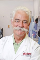 Alan Mackay-Sim合作论文数Griffith University65
 Darryl Eyles合作论文数Queensland Brain Institute, Faculty of Health, Medicine and Behavioural Sciences, The University of Queensland;Centre for RNA in Neuroscience, Faculty of Health, Medicine and Behavioural Sciences, The University of Queensland22
 Patrick Decherchi合作论文数20Michel Khrestchatisky合作论文数18Landel Véréna合作论文数CNRS, Aix Marseille Université15
 John McGrath合作论文数Greater Brisbane Clinical School, Faculty of Health, Medicine and Behavioural Sciences, University of Queensland;Queensland Brain Institute, Faculty of Health, Medicine and Behavioural Sciences, University of Queensland15
 Tanguy Marqueste合作论文数Department of Physiology, University of Manitoba Spinal Cord Research Center15
 Pascal Millet合作论文数14
 John Bianco合作论文数Princess Maxima Ctr Pediat Oncol13
 Francois S. Roman合作论文数12
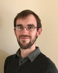 Christopher C. Perry合作论文数Department of Chemistry;Johns Hopkins Uni Versity12
 Girard Stéphane D合作论文数Aix Marseille University11Emmanuel Nivet合作论文数11
 Thomas Burne合作论文数Queensland Brain Institute, The University of Queensland10Wayne Murrell合作论文数10
 Richard D McCurdy合作论文数Ontario Veterinary College, University of Guelph10Baranger Kevin合作论文数9Jillanne Brown合作论文数8Andrew Wetzig合作论文数8
 Maria Morello合作论文数IRCCS S. Lucia, University of Rome Tor Vergata8
 Arnaud Deveze合作论文数CHU Nord Department of Otorhinolaryngology8Chris Perry合作论文数7
 Gaelle Guiraudie-Capraz合作论文数UMR 7051, Aix Marseille Univ7Duncan Mclean合作论文数7Stephane Garcia合作论文数Hôpital Nord7Bruno Gepner合作论文数7
 Charlotte Jaloux合作论文数Timone Adultes Hosp, Aix Marseille Univ6Annweiler Cedric合作论文数6Stephan Delphine合作论文数6Isabelle Virard合作论文数6Jean-Claude Stamegna合作论文数6
 Santiago Rivera合作论文数Universidad de Los Andes6Olivier Alluin合作论文数6Jean-François Chabas合作论文数5
 Maxime Bonnet合作论文数5
 Ibrahim El Chérif合作论文数CNRS, Univ Aix Marseille 25Veran Julie合作论文数5Régis Legre合作论文数5
 Yatma Gueye合作论文数5Diana Andrea Fernandes De Abreu合作论文数4
 Valery Matarazzo合作论文数4Marie-Solenne Felix合作论文数4Marie Witters合作论文数4P Gauthier合作论文数4Lavaut Marie-Noëlle合作论文数4Sbai Oualid合作论文数4
 Nicholas Hayward合作论文数Oncogenomics Laboratory, QIMR Berghofer Medical Research Institute4
 Rogerio V. Rossi合作论文数4




