Deoxygenation is driving ecological shifts across vast oceanic volumes. To understand and predict future ocean ecosystem functioning and stability, it is critical to identify the eukaryotic metabolic repertoire that enables survival under anoxia. Foraminiferan protists are one of few eukaryotic lineages that can inhabit anoxic marine sediments, environments relevant today and in Earth's past. Here, we investigate the metabolic strategies employed by a representative of an early-evolving foraminiferan group to persist anoxia. A saccamminid foraminifer inhabiting an anoxic bathyal seafloor in the Santa Barbara Basin (CA, USA) was preserved in situ. (Meta)transcriptomic analyses revealed an aerotolerant mitochondrial metabolism lacking Cytochrome c Oxidase, but expressing alternative oxidase, potentially fueled by internally released oxygen during ROS detoxification, and utilizing a TCA reductive Complex II. This foraminifer uses glutamate oxidation and aspartate-malate shuttle to generate reducing equivalents, driving ATP production via an atypical anaerobic electron-transport chain. Energy metabolism is also tightly linked to phosphate availability, facilitating substrate-level phosphorylation of high-energy intermediates. This foraminifer's competitive advantage is its anaerobic energy metabolism combined with ability to detoxify and reduce oxygen, if present. These unusual capabilities confer a blueprint for understanding how early eukaryotes may have evolved during anoxia, remained resilient during the Neoproterozoic Oxygenation Event, and likely will be ecological winners amid ongoing ocean deoxygenation.
Investigations of the metabolic capabilities of anaerobic protists advances our understanding of the evolution of eukaryotic life on Earth and for uncovering analogous extraterrestrial complex microbial life. Certain species of foraminiferan protists live in environments analogous to early Earth conditions when eukaryotes evolved, including sulfidic, anoxic and hypoxic sediment porewaters. Foraminifera are known to form symbioses as well as to harbor organelles from other eukaryotes (chloroplasts), possibly bolstering the host's independence from oxygen. The full extent of foraminiferal physiological capabilities is not fully understood. To date, evidence for foraminiferal anaerobiosis was gleaned from specimens first subjected to stresses associated with removal from in situ conditions. Here, we report comprehensive gene expression analysis of benthic foraminiferal populations preserved in situ on the euxinic (anoxic and sulfidic) bathyal seafloor, thus avoiding environmental alterations associated with sample recovery, including pressure reduction, sunlight exposure, warming, and oxygenation. Metatranscriptomics, metagenome-assembled genomes, and measurements of substrate uptake were used to study the kleptoplastidic foraminifer Nonionella stella inhabiting sulfur-oxidizing bacterial mats of the Santa Barbara Basin, off California. We show N. stella energy generation under dark euxinia is unusual because it orchestrates complex metabolic pathways for ATP production and carbon fixation through the Calvin cycle. These pathways include extended glycolysis, anaerobic fermentation, sulfide oxidation, and the presence of a membrane-bound inorganic pyrophosphatase, an enzyme that hydrolyzes inorganic pyrophosphate to actively pump protons across the mitochondrial membrane.
Prior observations suggest that foraminiferan protists use their reticulopodia (anastomosing pseudopodia) to alter sediment fabric by disrupting laminations of subtidal marine stromatolites, erasing the layered structures in an experimental setting. Because microbialites and foraminifera are found in non-marine settings, we hypothesized that foraminifera living in lakes could also disrupt layered microbialite fabric. With this aim and using a variety of multidisciplinary approaches, we conducted field surveys and an experiment on microbialites from Green Lake (GL; Fayetteville, New York State, United States), which has been studied as a Proterozoic ecosystem analog. The lake is meromictic and alkaline, receiving calcium sulfate-rich water in the monimolimnion; it supports a well-developed carbonate platform that provides access to living and relict microbialites. The living microbialites grow from early spring to autumn, forming a laminated mat at their surface (top ~5 mm), but a clotted or massive structure exists at depth (> ~ 1 cm). We observed a morphotype of “naked” foraminiferan-like protist in samples from GL microbialites and sediments; thus, considered the possibility of freshwater foraminiferan impact on microbialite fabric. Results of an experiment that seeded the cultured freshwater foraminifer Haplomyxa saranae onto the GL microbialite surface indicates via micro-CT scanning and anisotropy analysis that the introduced foraminifer impacted uppermost microbialite layering (n = 3 cores); those cores with an added inhibitor lacked changes in anisotropy for two of those three cores. Thus, it remains plausible that the much smaller, relatively common, native free-form reticulate protist, which we identified as Chlamydomyxa labyrinthuloides, can disrupt microbialite fabrics on sub-millimeter scales. Our observations do not exclude contributions of other possible causal factors.
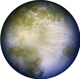
Cell viability after incubation with different electroporation buffers, Phosphate buffered saline (PBS) is used as an electroporation buffer for many eukaryotic cells. However, we observed that the PBS has a detrimental effect on the cell, causing disruption in H. palaeformis cell membrane. We then tested the tolerance of H. palaeformis to three different electroporation buffers: A-Gene Pulser Electroporation Buffer(BIO-RAD, USA) B-Iso-osmolar Buffer (Eppendorf, USA) C-Hypo-osmolar Buffer (Eppendorf, USA) Cells were incubated in each buffer independently for 10 minutes and observed for their viability under inverted microscopy. For all the three tested buffers, we noticed that once buffer was added, the cells showed instant osmic shock, include loss of motility and relative shrinking in the cell membrane (Fig. 1). After 10 minutes incubation period in each buffer, cells were transferred to their Cerophyl culture medium to recover. We observed progressive recovery of the cells. Table 1 shows the time that took the cells to completely recovered and the percentages of the recovered cells after incubation at the three different electroporation buffers. Table 1 Electroporation buffer Duration of recovery Percentage of the recovered cells Gene Pulser 10 minutes >90% Iso-osmolar 10 minutes 70% Hypo-osmolar 2-5 minutes 70% Fig.1. Light microscopy images showing the effect of the different electroporation buffer on the H. palaeformis cell membrane; PBS has detrimental effect on the cell causing disruption of cell membrane.
Certain benthic foraminifera thrive in marine sediments with low or undetectable oxygen. Potential survival avenues used by these supposedly aerobic protists include fermentation and anaerobic respiration, although details on their adaptive mechanisms remain elusive. To better understand the metabolic versatility of foraminifera, we studied two benthic species that thrive in oxygen-depleted marine sediments. Here we detail, via transcriptomics and metatranscriptomics, differential gene expression of Nonionella stella and Bolivina argentea , collected from Santa Barbara Basin, California, USA, in response to varied oxygenation and chemical amendments. Organelle-specific metabolic reconstructions revealed these two species utilize adaptable mitochondrial and peroxisomal metabolism. N. stella , most abundant in anoxia and characterized by lack of food vacuoles and abundance of intracellular lipid droplets, was predicted to couple the putative peroxisomal beta-oxidation and glyoxylate cycle with a versatile electron transport system and a partial TCA cycle. In contrast, B. argentea , most abundant in hypoxia and contains food vacuoles, was predicted to utilize the putative peroxisomal gluconeogenesis and a full TCA cycle but lacks the expression of key beta-oxidation and glyoxylate cycle genes. These metabolic adaptations likely confer ecological success while encountering deoxygenation and expand our understanding of metabolic modifications and interactions between mitochondria and peroxisomes in protists.
Goal: To determine the optimal concentration of three antibiotics, Puromycin, Geneticin (G418) and Blasticidin, that will effectively inhibit the cells growth and division of Heterometopus palaeformis. We examined the cells viability and growth rate in the presence of different concentrations of these antibiotics (Table 1, 2 &3). These antibiotics are routinely used for selecting the transformed eukaryotic cells. The choice of puromycin was based on previous transfection protocol on ciliate, T. thermophila (Iwamoto et al., 2014; Gene). The Geneticin (G418) was used by our group for selecting transformed Bodo saltans with plasmids carrying neomycin resistance gene (Neo). Procedure: -The experiments were carried in 5-ml Eppendorf tubes to cut down on the volume of the antibiotic that will be used. -The tubes were filled with 4 ml cerophyl culture medium. -Add the antibiotics at the final concentrations shown in the tables below. -In total there were 6-7 tubes per assay, including the control with no antibiotic treatment. -Transfer 1ml of cells from RAJCA culture at log phase to each of the tubes. -Inspect the cells every couple of days under inverted microscope to check for any evidence of cell toxicity and count the number of live cells. -The optimal dose of antibiotic for selection is the lowest one for which all cells have died after 7 to 12 days. A low dose is the concentration which has minimal effects on cells after 2 weeks of antibiotic treatment. Conversely, a high dose is a concentration which is highly toxic to cells within 1-3 days of starting antibiotic treatment. Results: A-Puromycin Results indicate that puromycin at concentration of 300 μg/ml kills the entire population in 10 to 12 days. Using lower puromycin concentration ( i. e., 200 μg/mL) was inefficient as the cells maintained to grow for longer that two weeks. Table 1 Puromycin (μg/ml) Cells survival (days) Exp.1 – Exp.2 300 10-12 200 16-12 100 >16->16 50 >16->16 20 >16->16 5 >16->16 0 >16->16 B-Geneticin (G418) G418 is an effective antibiotic to kill the H. palaeformis wild type cells at different concentrations. Our results show that G418 of concentration 20 μg/mL kills the entire population of cells in approximately 10 days, which is considered enough time for the selection of resistant cells after transfection. We will use this antibiotic as a second choice if needed after the puromycin. Table 2 Geneticin/ G418 (μg/ml) Cells survival (days) 200 <4 100 <4 50 6 20 10 10 14 0 >16 C-Blasticidin Blasticidin was supplemented at different concentrations to H. palaeformis cultures. However, our results show that blasticidin didn’t have any lethal effect on the cells. Although the growth rate for the cells in presence of the blasticidin were lower than the control cultures (based on cell count ). But cells remained alive and active for more than 2 weeks period at the highest concentration that we used (300 μg/ml). Therefore, we excluded Blasticidin from our list. Table 3 Blasticidin (μg/ml) Cells survival (days) 300 >16 200 >16 100 >16 50 >16 10 >16 0 >16
Developing transfection protocols for marine protists is an emerging field that will allow the functional characterization of protist genes and their roles in organism responses to the environment. We developed a CRISPR/Cas9 editing protocol for Bodo saltans, a free-living kinetoplastid with tolerance to both marine and freshwater conditions and a close non-parasitic relative of trypanosomatids. Our results show that SaCas9/single-guide RNA (sgRNA) ribonucleoprotein (RNP) complex-mediated disruption of the paraflagellar rod 2 gene (BsPFR2) was achieved using electroporation-mediated transfection. The use of CRISPR/Cas9 genome editing can increase the efficiency of targeted homologous recombination when a repair DNA template is provided. Our sequence analysis suggests two mechanisms for repairing double-strand breaks in B. saltans are active; homologous-directed repair (HDR) utilizing an exogenous DNA template that carries an antibiotic resistance gene and likley non-homologous end joining (NHEJ). However, HDR was only achieved when a single (vs. multiple) SaCas9 RNP complex was provided. Furthermore, the biallelic knockout of BsPFR2 was detrimental for the cell, highlighting its essential role for cell survival because it facilitates the movement of food particles into the cytostome. Our Cas9/sgRNA RNP complex protocol provides a new tool for assessing gene functions in B. saltans and perhaps similar protists with polycistronic transcription.
Developing transfection protocol for Bodo saltans, using SaCas9/sgRNA ribonucleoprotein (RNP) complex in conjunction with DNA repair template to disrupt the Paraflagellar rod 2 gene (BsPFR2) and increase the efficiency of targeted homologous recombination when a repair template DNA is provided. The exogenous repair template is double stranded DNA and it consists of eGFP fused with the drug selection gene nptII/neo and flanked by 500 bp of the untranslated regions (UTRs) upstream and downstream of the targeted BsPFR2 as homologous repair arms.
Developing transfection protocol for Bodo saltans, using SaCas9/sgRNA ribonucleoprotein (RNP) complex in conjunction with DNA repair template to disrupt the Paraflagellar rod 2 gene (BsPFR2) and increase the efficiency of targeted homologous recombination when a repair template DNA is provided. The exogenous repair template is double stranded DNA and it consists of eGFP fused with the drug selection gene nptII/neo and flanked by 500 bp of the untranslated regions (UTRs) upstream and downstream of the targeted BsPFR2 as homologous repair arms.
Developing transfection protocol for Bodo saltans, using SaCas9/sgRNA ribonucleoprotein (RNP) complex in conjunction with DNA repair template to disrupt the Paraflagellar rod 2 gene (BsPFR2) and increase the efficiency of targeted homologous recombination when a repair template DNA is provided. The exogenous repair template is double stranded DNA and it consists of eGFP fused with the drug selection gene nptII/neo and flanked by 500 bp of the untranslated regions (UTRs) upstream and downstream of the targeted BsPFR2 as homologous repair arms.
Developing transfection protocol for Bodo saltans, using SaCas9/sgRNA ribonucleoprotein (RNP) complex in conjunction with DNA repair template to disrupt the Paraflagellar rod 2 gene (BsPFR2) and increase the efficiency of targeted homologous recombination when a repair template DNA is provided. The exogenous repair template is double stranded DNA and it consists of eGFP fused with the drug selection gene nptII/neo and flanked by 500 bp of the untranslated regions (UTRs) upstream and downstream of the targeted BsPFR2 as homologous repair arms.
Oceanic deoxygenation is increasingly affecting marine ecosystems; many taxa will be severely challenged, yet certain nominally aerobic foraminifera (rhizarian protists) thrive in oxygen-depleted to anoxic, sometimes sulfidic, sediments uninhabitable to most eukaryotes. Gene expression analyses of foraminifera common to severely hypoxic or anoxic sediments identified metabolic strategies used by this abundant taxon. In field-collected and laboratory-incubated samples, foraminifera expressed denitrification genes regardless of oxygen regime with a putative nitric oxide dismutase, a characteristic enzyme of oxygenic denitrification. A pyruvate:ferredoxin oxidoreductase was highly expressed, indicating the capability for anaerobic energy generation during exposure to hypoxia and anoxia. Near-complete expression of a diatom's plastid genome in one foraminiferal species suggests kleptoplasty or sequestration of functional plastids, conferring a metabolic advantage despite the host living far below the euphotic zone. Through a unique integration of functions largely unrecognized among "typical" eukaryotes, benthic foraminifera represent winning microeukaryotes in the face of ongoing oceanic deoxygenation.
Diverse microbial ecosystems underpin life in the sea. Among these microbes are many unicellular eukaryotes that span the diversity of the eukaryotic tree of life. However, genetic tractability has been limited to a few species, which do not represent eukaryotic diversity or environmentally relevant taxa. Here, we report on the development of genetic tools in a range of protists primarily from marine environments. We present evidence for foreign DNA delivery and expression in 13 species never before transformed and for advancement of tools for eight other species, as well as potential reasons for why transformation of yet another 17 species tested was not achieved. Our resource in genetic manipulation will provide insights into the ancestral eukaryotic lifeforms, general eukaryote cell biology, protein diversification and the evolution of cellular pathways.
An amendment to this paper has been published and can be accessed via a link at the top of the paper.
Stable transfection of B. saltans has been achieved using plasmid for EF 1 alpha C terminal tagging.
Bodo saltans kill curve protocol using G418 (Gentamicin) Gentamycin exhibits toxicity toward both eukaryotic and prokaryotic cells by disrupting ribosome function, thereby blocking the elongation step in protein synthesis. G418 is most commonly used as a selection agent for eukaryotic cells genetically engineered to express a neomycin resistance gene (NeoR), which is encoded by either transposon Tn601 (903) or Tn5. The resistant cells grow in medium containing G418, and may be used to establish stably transfected cell lines as all the non-resistant cells die due to G418 toxicity, typically within 6 – 14 days. To establish kill curve assay for saltans various concentrations of G418 were tested. The concentration range from 1 to 50 μg/mL for selection of eukaryotic, and then higher concentrations can be used for maintaining stable transfected cell line. Kill Curve Assay Harvestthe B. saltans cellsfromaculture that is atpeakdensity (1 – 3.0 x 10⁵) by centrifugation at 800xgfor5min., discard the supernatant (medium). Replace the growth medium with fresh medium containing 0 – 50 μg/mL. For each concentration, test in triplicate. Using 6 wells plates, replace the medium in the wells every 3 – 4 days using fresh medium with the appropriate G418 concentration. Perform a daily visual inspection for evidence of toxicity, also cell count using hemocytometer. Note that the optimal dose of G418 for selection is the lowest one for which all cells have died after one week. A low dose is the concentration which has minimal effects on cells after 2 weeks of antibiotic selection. Conversely, a high dose is a concentration which is highly toxic to cells within 2-3 days of starting antibiotic selection. 5- The results indicate that a G418 concentration of 2 μg/mL kill the entire B. saltans population in 12 days which is considered enough time for selecting the resistant cells after transfection. G418 Sensitivity for B. saltans Cell Selection In all of our plasmids constructs we included the NeoR gene (Neomycine resistant gene) to select our transfected cells. Twenty-four hours after electroporation, the growth medium of transfected cells were supplemented with G418 antibiotic Solution (2 μg/mL) based on the kill curve results (above). 3. Replace the G418-containing medium every 3 – 5 days and examine cells for visual toxicity. Most non-transfected (non-resistant) cells will die within 10- 12 days, leaving the transfected cells to expand. Once cells grow to high confluence, they may be maintained, or frozen as a polyclonal line or plated by limited dilution to select for single clones.
Medium recipe: ATCC medium: 802 Sonneborn's Paramecium medium Solution 1 Rye grass Cerophyll: Cerophyll*...................2.5 g Distilled water..............1.0 L Add cerophyll to distilled water and boil for 5 minutes. Add 100 ml distilled water to compensate for evaporation. Filter through Whatman #1 filter paper and add 0.5 g Na2HPO4. Autoclave for 15 minutes at 121C. saltans food (K. pneumoniae, or E. coli). I used only the K. pneumoniae so far Agar Medium for Klebsiella pneumoniae ATCC-BAA-1705: Agar........................20.0 g Yeast extract................4.0 g Glucose......................0.16 g Distilled water............800.0 ml Dispense in 5 ml amounts. Autoclave for 25 minutes at 121C. Slant. Bacterium, grown on solution 2, is added to solution 1 (Just add very little, few colonies) and incubated at 30C for 24 hours prior to inoculation with Bodo saltans. Cerophyl powder that works best for the saltans is the powder from Pines. CultureMaintenance: Preparethe bacterizedBodo saltans medium as described above. InoculateaT25tissuecultureflask(50ml) containing20 to 25 ml of fresh medium with 1 to 2 ml from Bodo culture thatisatornearpeakdensity Incubatehorizontally at18 to 22°C(room temperature can work fine) withcapscrewedonnot very tightly. 4. Subcultureevery7 to 10days. I usually subculture 3 to 4 flasks every week Cryopreservation: HarvestandPreservation: Harvestcellsfromaculturethatisatpeakdensitybycentrifugationat800xgfor5min. 2. Adjusttheconcentrationofcellsto2x106to107 /mLinfreshmedium (Important step, even for transfection). Preparea20%(v/v)solutionofsterileDMSOinfresh Bodomedium. Add2.0mLofDMSOtoanicecoldtube PlacethetubeoniceandallowtheDMSOtosolidify(~5min)andthenadd8.0mLoficecold medium. InvertseveraltimestodissolvetheDMSO. Allowtowarmtoroomtemperature MixthecellpreparationandtheDMSOinequalportions.Thus,thefinalconcentrationwillbe106 to107and10%(v/v)DMSO.ThetimefromthemixingofthecellpreparationandDMSOstocksolution before thefreezingprocessisbegunshouldbenolessthan15minandnolongerthan30min. Dispensein0.5mLaliquotsinto1.0mLto2.0mLsterileplasticscrewcappedcryules(specialplastic vialsforcryopreservation). Placethevialsinacontrolledratefreezingunit.Fromroomtemperaturecoolat1°C/minto -40°C.If thefreezingunitcancompensatefortheheatoffusion,maintainrateat 1°C/minthroughtheheatoffusion.At- 40°Cplungeintoliquidnitrogen.Alternatively,placethevialsinNalgene1°Cfreezing apparatus.Placetheapparatusat- 80°Cfor1.5to2hoursandthenplungeampulesintoliquid nitrogen.(Thecoolingrateinthisapparatusisapproximately1°C/min.) Toestablishaculturefromthefrozenstateplaceanampuleinawaterbathsetat+35°C.Immersethe ampuletoaleveljustabovethesurfaceofthefrozenmaterial.Donotagitatetheampule. Immediatelyafterthawing,donotleaveinthewaterbath,asepticallyremovethecontentsofthe ampuleandinoculateaT25tissuecultureflaskcontaining10mLofBodomediumbacterized withKlebsiellapneumoniaesubsp.pneumoniae(ATCC®700831). Incubatehorizontallywiththecapscrewedontightlyat22°C
 Virginia Edgcomb合作论文数Woods Hole Oceanographic Institution20
 Roberto Docampo合作论文数Department of Cellular Biology, Franklin College of Arts and Sciences, University of Georgia13
 Peter R. Girguis合作论文数Department of Organismic & Evolutionary Biology, Harvard University13
 Enrique Lara合作论文数WSL Wetlands Research Group, Swiss Federal Research Institute12
 Zhu-Hong Li合作论文数Univ Georgia, Ctr Trop & Emerging Global Dis, Athens, GA 30602 USA10
 Edward Mitchell合作论文数Laboratory of Soil Biology, Institute of Biology, Faculty of Science, University of Neuchatel9
 Joan Bernhard合作论文数Department of Geology and Geophysics, Woods Hole Oceanographic Institution7David J Beaudoin合作论文数5not provided Jennifer合作论文数4not provided Cullen合作论文数4not provided Peter合作论文数4not provided Paulo合作论文数4Christopher Powers合作论文数4Utter Daniel R合作论文数Josephine Bay Paul Center for Comparative Molecular Biology and Evolution, Marine Biological Laboratory Woods Hole, MA, USA;Department of Microbiology, The Forsyth Institute Cambridge;Josephine Bay Paul Center for Comparative Molecular Biology and Evolution, The Forsyth Institute Cambridge4Anush Kosakyan合作论文数4
 Milcho Todorov合作论文数Institute of Zoology;Bulgarian Academy of Sciences;Institute of Zoology, Bulgarian Academy of Sciences4
 Thierry J Heger合作论文数Laboratory of Ecological Systems (ECOS),, École Polytechnique Fédérale de Lausanne (EPFL),4
 Ying Zhang合作论文数3
 Colleen M Hansel合作论文数Woods Hole Oceanographic Institution;School of Sustainability, Stanford University3
 Paulo A. Garcia合作论文数School of Biomedical Engineering and Sciences, Wake Forest University3
 Helena L. Filipsson合作论文数Earth Sciences Centre, Göteborg University3
 Scott D. Wankel合作论文数Stable Isotope Laboratory;US Geological Survey;Stable Isotope Laboratory, US Geological Survey3Pia A. Elustondo合作论文数2José A. Fernández Robledo合作论文数2CLEMENTE THOMAS E合作论文数2
 Rachele Cesaroni合作论文数2Natalia Janowicz合作论文数2Cristina Aresté合作论文数2Lisa Sudek合作论文数2Susana A Breglia合作论文数2Ambar Kachale合作论文数2Eleanna Kazana合作论文数2Mariana Rius合作论文数2Kodai Fukuba合作论文数2not provided Virginia合作论文数2Yu Zheng合作论文数2
 Jun Yang (杨军)合作论文数Aquatic EcoHealth Group, Key Laboratory of Urban Environment and Health, Institute of Urban Environment, Chinese Academy of Sciences;University of Chinese Academy of Sciences2Isabel Nimmo合作论文数2Yuu Ishii合作论文数2
 Brittany N Sprecher合作论文数2Ellen Nisbet合作论文数2
 Imen Lassadi合作论文数2
 Binnypreet Kaur合作论文数Inst Parasitol, Czech Acad Sci2
 Anna M. G. Novak Vanclova合作论文数2WANG Lu (王路)合作论文数2Valérie Vergé合作论文数Univ Pierre & Marie Curie Paris 06, Sorbonne Univ2
 Lev Tsypin合作论文数Department of Molecular Genetics and Cell Biology, University of Chicago2April Woods合作论文数2
 Amanda Hopes合作论文数2




