Ca2+ signaling in cells begins with the opening of Ca2+ channels in either the plasma membrane (PM) or endoplasmic reticulum (ER), leading to a sharp increase in the physiologically low (<100 nM) cytosolic Ca2+ level. The temporal and spatial regulation of Ca²+ is crucial for the precise activation of key biological processes. In the apicomplexan parasite Toxoplasma gondii, which infects approximately one-third of the global population, Ca²+ signaling governs essential aspects of the parasite’s infection cycle. T. gondii relies on Ca²+ signals to regulate pathogenic traits, with several Ca²+-signaling components playing critical roles. Ca2+ entry from the extracellular environment has been demonstrated in T. gondii for both, extracellular parasites, exposed to high Ca2+, and intracellular parasites, which acquire Ca²+ from host cells during host Ca²+ signaling events. Active egress, an essential step of the parasite’s infection cycle, is preceded by a large increase in cytosolic Ca2+, most likely initiated by release from intracellular stores. However, extracellular Ca2+ is also necessary to reach a cytosolic Ca2+ threshold required for timely egress. In this study, we investigated the mechanism of intracellular Ca²+ store replenishment and identified a central role for the SERCA-Ca2+-ATPase in maintaining Ca²+ homeostasis within the ER and in other organelles. We demonstrate mitochondrial Ca2+ uptake, which occurs by transfer of Ca2+ from the ER, likely through membrane contact sites. Our findings suggest that the T. gondii ER plays a key role in sequestering and redistributing Ca²+ to intracellular organelles following Ca²+ influx at the PM.
Millions of people are infected with Toxoplasma gondii , and the available treatment for toxoplasmosis is not ideal. Most of the drugs currently used are only effective for the acute infection, and treatment can trigger serious side effects requiring changes in the therapeutic approach.
ABSTRACTApicomplexan parasites cause persistent mortality and morbidity worldwide through diseases including malaria, toxoplasmosis, and cryptosporidiosis. Ca2+ signaling pathways have been repurposed in these eukaryotic pathogens to regulate parasite-specific cellular processes governing the transition between the replicative and lytic phases of the infectious cycle. Despite the presence of conserved Ca2+-responsive proteins, little is known about how specific signaling elements interact to impact pathogenesis. We mapped the Ca2+-responsive proteome of the model apicomplexan T. gondii via time-resolved phosphoproteomics and thermal proteome profiling. The waves of phosphoregulation following PKG activation and stimulated Ca2+ release corroborate known physiological changes but identify specific proteins operating in these pathways. Thermal profiling of parasite extracts identified many expected Ca2+-responsive proteins, such as parasite Ca2+-dependent protein kinases. Our approach also identified numerous Ca2+-responsive proteins that are not predicted to bind Ca2+, yet are critical components of the parasite signaling network. We characterized protein phosphatase 1 (PP1) as a Ca2+-responsive enzyme that relocalized to the parasite apex upon Ca2+ store release. Conditional depletion of PP1 revealed that the phosphatase regulates Ca2+ uptake to promote parasite motility. PP1 may thus be partly responsible for Ca2+-regulated serine/threonine phosphatase activity in apicomplexan parasites.
SQ109 is an anti-tubercular drug candidate that has completed Phase IIb/III clinical trials for tuberculosis and has also been shown to exhibit potent in vitro efficacy against protozoan parasites including Leishmania and Trypanosoma cruzi spp. However, its in vivo efficacy against protozoa has not been reported. Here, we evaluated the activity of SQ109 in mouse models of Leishmania, Trypanosoma spp. as well as Toxoplasma infection. In the T. cruzi mouse model, 80% of SQ109-treated mice survived at 40 days post-infection. Even though SQ109 did not cure all mice, these results are of interest since they provide a basis for future testing of combination therapies with the azole posaconazole, which acts synergistically with SQ109 in vitro. We also found that SQ109 inhibited the growth of Toxoplasma gondii in vitro with an IC50 of 1.82 µM and there was an 80% survival in mice treated with SQ109, whereas all untreated animals died 10 days post-infection. Results with Trypanosoma brucei and Leishmania donovani infected mice were not promising with only moderate efficacy. Since SQ109 is known to be extensively metabolized in animals, we investigated the activity in vitro of SQ109 metabolites. Among 16 metabolites, six mono-oxygenated forms were found active across the tested protozoan parasites, and there was a ~6× average decrease in activity of the metabolites as compared to SQ109 which is smaller than the ~25× found with mycobacteria.
Apicomplexan parasites cause persistent mortality and morbidity worldwide through diseases including malaria, toxoplasmosis, and cryptosporidiosis. Ca2+ signaling pathways have been repurposed in these eukaryotic pathogens to regulate parasite-specific cellular processes governing the replicative and lytic phases of the infectious cycle, as well as the transition between them. Despite the presence of conserved Ca2+-responsive proteins, little is known about how specific signaling elements interact to impact pathogenesis. We mapped the Ca2+-responsive proteome of the model apicomplexan Taxoplasma gondii via time-resolved phosphoproteomics and thermal proteome profiling. The waves of phosphoregulation following PKG activation and stimulated Ca2+ release corroborate known physiological changes but identify specific proteins operating in these pathways. Thermal profiling of parasite extracts identified many expected Ca2+-responsive proteins, such as parasite Ca2+-dependent protein kinases. Our approach also identified numerous Ca2+-responsive proteins that are not predicted to bind Ca2+, yet are critical components of the parasite signaling network. We characterized protein phosphatase 1 (PP1) as a Ca2+-responsive enzyme that relocalized to the parasite apex upon Ca2+ store release. Conditional depletion of PP1 revealed that the phosphatase regulates Ca2+ uptake to promote parasite motility. PP1 may thus be partly responsible for Ca2+-regulated serine/threonine phosphatase activity in apicomplexan parasites.
Developing transfection protocols for marine protists is an emerging field that will allow the functional characterization of protist genes and their roles in organism responses to the environment. We developed a CRISPR/Cas9 editing protocol for Bodo saltans, a free-living kinetoplastid with tolerance to both marine and freshwater conditions and a close non-parasitic relative of trypanosomatids. Our results show that SaCas9/single-guide RNA (sgRNA) ribonucleoprotein (RNP) complex-mediated disruption of the paraflagellar rod 2 gene (BsPFR2) was achieved using electroporation-mediated transfection. The use of CRISPR/Cas9 genome editing can increase the efficiency of targeted homologous recombination when a repair DNA template is provided. Our sequence analysis suggests two mechanisms for repairing double-strand breaks in B. saltans are active; homologous-directed repair (HDR) utilizing an exogenous DNA template that carries an antibiotic resistance gene and likley non-homologous end joining (NHEJ). However, HDR was only achieved when a single (vs. multiple) SaCas9 RNP complex was provided. Furthermore, the biallelic knockout of BsPFR2 was detrimental for the cell, highlighting its essential role for cell survival because it facilitates the movement of food particles into the cytostome. Our Cas9/sgRNA RNP complex protocol provides a new tool for assessing gene functions in B. saltans and perhaps similar protists with polycistronic transcription.
Two-pore channels (TPCs) are a ubiquitous family of cation channels that localize to acidic organelles in animals and plants to regulate numerous Ca 2+ -dependent events. Little is known about TPCs in unicellular organisms despite their ancient origins. Here, we characterize a TPC from Toxoplasma gondii , the causative agent of toxoplasmosis. TgTPC is a member of a novel clad of TPCs in Apicomplexa, distinct from previously identified TPCs and only present in coccidians. We show that TgTPC localizes not to acidic organelles but to the apicoplast, a non-photosynthetic plastid found in most apicomplexan parasites. Conditional silencing of TgTPC resulted in progressive loss of apicoplast integrity, severely affecting growth and the lytic cycle. Isolation of TPC null mutants revealed a selective role for TPCs in replication independent of apicoplast loss that required conserved residues within the pore-lining region. Using a genetically-encoded Ca 2+ indicator targeted to the apicoplast, we show that Ca 2+ signals deriving from the ER but not from the extracellular space are selectively transmitted to the lumen. Deletion of the TgTPC gene caused reduced apicoplast Ca 2+ uptake and membrane contact site formation between the apicoplast and the ER. Fundamental roles for TPCs in maintaining organelle integrity, inter-organelle communication and growth emerge.
The tuberculosis drug candidate SQ109 targets the trehalose monomycolate transporter MmpL3 in Mycobacterium tuberculosis and also has activity against other pathogens. We found related proteins in 22 protozoa, including Trypanosoma cruzi and Entamoeba histolytica, as well as in archaea and other bacteria, including the fatty acid transporter, FarE. We show these proteins, alpha-MMPL proteins, adopt similar structures to that of MsMmpL3 having two sets (P1 and P2) of conserved active site/H+-transporter Asp, Tyr and Phe residues in “pentad” motifs (DYxxF) that can bind to the SQ109 ethylenediamine and adamantyl moieties. Based on structural comparisons with MsMmpL3, we find that there are superimposable transmembrane and H+-transporter structures in much larger proteins, beta-MMPLs, found in apicomplexan parasites, fungi, plants and animals. They also contain double “pentad” motifs in which the P1 Asp is totally conserved, but the P2 Asp may also be a Glu, and the P2 Phe seen in the alpha-MMPLs is a His that H-bonds to the P1 and P2 Asp/Glu residues. There are also 5 conserved Ser/Thr residues that extend the H-bond/H+-transporter network with 2 interacting directly with the P1 Asp, and the His. We propose that all MMPL proteins are involved in proton motive force-mediated lipid (phospholipid, glycolipid, sterol, fatty acid) transport, and that SQ109 may target some pathogens directly, by binding to the P1/P2 motifs. Overall, the results are of general interest since they indicate that there are two major classes of lipid transporters: alpha-MMPL proteins found in many bacteria and protozoa, and much larger, beta-MMPL proteins, found in fungi, apicomplexa, plants and animals and, in some cases, they are potential drug targets.
Abstract Toxoplasma gondii , an obligate intracellular parasite, is capable of invading virtually any nucleated cell. Ca 2+ signaling is universal and both T. gondii and its mammalian host cell will utilize Ca 2+ signaling to stimulate diverse cellular functions. Egress of T. gondii from the host cell is an essential step for the infection cycle of T. gondii and a cytosolic Ca 2+ increase initiates the Ca 2+ signaling cascade that culminates in stimulation of motility and egress. In this work we demonstrate that intracellular T. gondii is capable of taking up Ca 2+ from the host cytoplasm when this concentration is increased during host signaling events. Both intracellular and extracellular Ca 2+ sources are important to reach a threshold of cytosolic Ca 2+ needed for a successful egress. Two peaks of Ca 2+ were observed in single parasites that egressed with the second peak resulting from Ca 2+ influx. We patched infected host cells to allow a precise delivery of exact concentrations of Ca 2+ for stimulating motility and egress. Using this approach, we found that low potassium concentration modulates but do not trigger host cell egress. This is the first study using whole-cell patches to study the role of ions such as K + and Ca 2+ in T. gondii egress.
Diverse microbial ecosystems underpin life in the sea. Among these microbes are many unicellular eukaryotes that span the diversity of the eukaryotic tree of life. However, genetic tractability has been limited to a few species, which do not represent eukaryotic diversity or environmentally relevant taxa. Here, we report on the development of genetic tools in a range of protists primarily from marine environments. We present evidence for foreign DNA delivery and expression in 13 species never before transformed and for advancement of tools for eight other species, as well as potential reasons for why transformation of yet another 17 species tested was not achieved. Our resource in genetic manipulation will provide insights into the ancestral eukaryotic lifeforms, general eukaryote cell biology, protein diversification and the evolution of cellular pathways.
Apicomplexan infections cause substantial morbidity and mortality, worldwide. New, improved therapies are needed. Herein, we create a next generation anti-apicomplexan lead compound, JAG21, a tetrahydroquinolone, with increased sp3-character to improve parasite selectivity. Relative to other cytochrome b inhibitors, JAG21 has improved solubility and ADMET properties, without need for pro-drug. JAG21 significantly reduces Toxoplasma gondii tachyzoites and encysted bradyzoites in vitro, and in primary and established chronic murine infections. Moreover, JAG21 treatment leads to 100% survival. Further, JAG21 is efficacious against drug-resistant Plasmodium falciparum in vitro. Causal prophylaxis and radical cure are achieved after P. berghei sporozoite infection with oral administration of a single dose (2.5 mg/kg) or 3 days treatment at reduced dose (0.625 mg/kg/day), eliminating parasitemia, and leading to 100% survival. Enzymatic, binding, and co-crystallography/pharmacophore studies demonstrate selectivity for apicomplexan relative to mammalian enzymes. JAG21 has significant promise as a pre-clinical candidate for prevention, treatment, and cure of toxoplasmosis and malaria.
Fluctuations of the cytosolic calcium ion (Ca2+) concentration regulate a variety of cellular functions in all eukaryotes. Cells express a sophisticated set of mechanisms to balance the cytosolic Ca2+ levels and the signals that elevate Ca2+ in the cytosol are compensated by mechanisms that reduce it. Alterations in Ca2+-dependent homeostatic mechanisms are the cause of many prominent diseases in humans, such as heart failure or neuronal death. The genetic tractability of Toxoplasma gondii and the availability of genetic tools enabled the use of Genetically Encoded Calcium Indicators (GECIs) expressed in the cytoplasm, which started a new era in the studies of Toxoplasma calcium signaling. It was finally possible to see Ca2+ oscillations prior to exit of the parasite from host cells. Years after Endo et al showed that ionophores triggered egress, the assumption that oscillations occur prior to egress from host cells has been validated by experiments using GECIs. GECIs allowed the visualization of specific Ca2+ signals in live intracellular parasites and to distinguish these signals from host cell calcium fluctuations. In this chapter we present an overview describing "tried and true" methods of our lab who pioneered the first use of GECI's in Toxoplasma, including GECI choice, methodology for transfection and selection of ideal clones, their characterization, and the use of GECI-expressing parasites for fluorometric and microscopic analysis.
Toxoplasma gondii , an obligate intracellular parasite, is capable of invading virtually any nucleated cell. Ca2+ signaling is universal and both T. gondii and its mammalian host cell will utilize Ca2+ signaling to stimulate diverse cellular functions. Egress of T. gondii from the host cell is an essential step for the infection cycle of T. gondii and a cytosolic Ca2+ increase initiates the Ca2+ signaling cascade that culminates in stimulation of motility and egress. In this work we demonstrate that intracellular T. gondii is capable of taking up Ca2+ from the host cytoplasm when this concentration is increased during host signaling events. Both intracellular and extracellular Ca2+ sources are important to reach a threshold of cytosolic Ca2+ needed for a successful egress. Two peaks of Ca2+ were observed in single parasites that egressed with the second peak resulting from Ca2+ influx. We patched infected host cells to allow a precise delivery of exact concentrations of Ca2+ for stimulating motility and egress. Using this approach, we found that low potassium concentration modulates but do not trigger host cell egress. This is the first study using whole-cell patches to study the role of ions such as K+ and Ca2+ in T. gondii egress.
An amendment to this paper has been published and can be accessed via a link at the top of the paper.
Bisphosphonates are the most commonly prescribed drugs for the treatment of osteoporosis and other bone illnesses. Some of them have also shown antiparasitic activity. In search of improving the pharmacological profile of commercial bisphosphonates, our group had previously developed first row transition metal complexes with N-containing bisphosphonates (NBPs). In this work, we extended our studies to heteroleptic palladium–NBP complexes including DNA intercalating polypyridyl co-ligands (NN) with the aim of obtaining potential multi-target species. Complexes of the formula [Pd(NBP)2(NN)]·2NaCl·xH2O with NBP = alendronate (ale) or pamidronate (pam) and NN = 1,10 phenanthroline (phen) or 2,2′-bipyridine (bpy) were synthesized and fully characterized. All the obtained compounds were much more active in vitro against T. cruzi (amastigote form) than the corresponding NBP ligands. In addition, complexes were nontoxic to mammalian cells up to 50–100 µM. Compounds with phen as ligand were 15 times more active than their bpy analogous. Related to the potential mechanism of action, all complexes were potent inhibitors of two parasitic enzymes of the isoprenoid biosynthetic pathway. No correlation between the anti-T. cruzi activity and the enzymatic inhibition results was observed. On the contrary, the high antiparasitic activity of phen-containing complexes could be related to their ability to interact with DNA in an intercalative-like mode. These rationally designed compounds are good candidates for further studies and good leaders for future drug developments. Four new palladium heteroleptic complexes with N-containing commercial bisphosphonates and DNA intercalating polypyridyl co-ligands were synthesized and fully characterized. All complexes displayed high anti-T. cruzi activity which could be related to the inhibition of the parasitic farnesyl diphosphate synthase enzyme but mainly to their ability to interact DNA.
Stable transfection of B. saltans has been achieved using plasmid for EF 1 alpha C terminal tagging.
1, Preparation of Terrific Broth 800 mL distilled H2O 12g Tryptone 24g Yeast extract 4 mL Glycerol Bring volume up to 900 mL with distilled H2O Autoclave and cool to room temperature Bring volume to 1000 mL with 100 mL filter sterilized solution containing 0.17M KH2PO4(2.314g – Potassium phosphate monobasic) 0.72M K2HPO4(16.43g – Potassium phosphate dibasic trihydrate) 2, Cells Rosetta-gami 2 cells expressing SaCas9 on pET32/Lic vector. Protein Expression Grow 10 mL starter culture overnight at 37°C (LB is fine) Inoculate 1 mL in 100 mL of Terrific Broth the next morning O.D. should be between 0.2 and 0.5 after 3 hours – if higher discard culture When O.D. is between 0.5 and 0.7 induce with IPTG and move o 18°C – grow overnight The next morning spin down and purify 3, Protein Purification Binding Buffer - 1 L - pH 8.0 Elution Buffer - 200 mL - pH 8.0 Lysis buffer 40 mL biding buffer 1 EDTA-free Protease Inhibitor pellet 300 mL P8849 500 mL lysozyme 10 mL DNase 10 mL RNase - Resuspend the pellet in 12 ml Lysis buffer - 1 hour on ice at 4°C - 80 seconds 30% sonication in 10 mL aliquotes (10 seconds on, 10 seconds off) - 12,000 rpm 20 minutes 4°C - Filter the supernatant with 0.45 uM filter to remove all bacterial - Purify the SaCas9 from the filtered supernatant by using a commercial His column (by following the manufacturer’s manual)
This protocol is a modified version to improve the yield of Bodo saltans cell density in culture. The original protocol is: https://www.protocols.io/view/bodo-saltans-culture-protocol-sh6eb9e
Medium recipe: ATCC medium: 802 Sonneborn's Paramecium medium Solution 1 Rye grass Cerophyll: Cerophyll*...................2.5 g Distilled water..............1.0 L Add cerophyll to distilled water and boil for 5 minutes. Add 100 ml distilled water to compensate for evaporation. Filter through Whatman #1 filter paper and add 0.5 g Na2HPO4. Autoclave for 15 minutes at 121C. saltans food (K. pneumoniae, or E. coli). I used only the K. pneumoniae so far Agar Medium for Klebsiella pneumoniae ATCC-BAA-1705: Agar........................20.0 g Yeast extract................4.0 g Glucose......................0.16 g Distilled water............800.0 ml Dispense in 5 ml amounts. Autoclave for 25 minutes at 121C. Slant. Bacterium, grown on solution 2, is added to solution 1 (Just add very little, few colonies) and incubated at 30C for 24 hours prior to inoculation with Bodo saltans. Cerophyl powder that works best for the saltans is the powder from Pines. CultureMaintenance: Preparethe bacterizedBodo saltans medium as described above. InoculateaT25tissuecultureflask(50ml) containing20 to 25 ml of fresh medium with 1 to 2 ml from Bodo culture thatisatornearpeakdensity Incubatehorizontally at18 to 22°C(room temperature can work fine) withcapscrewedonnot very tightly. 4. Subcultureevery7 to 10days. I usually subculture 3 to 4 flasks every week Cryopreservation: HarvestandPreservation: Harvestcellsfromaculturethatisatpeakdensitybycentrifugationat800xgfor5min. 2. Adjusttheconcentrationofcellsto2x106to107 /mLinfreshmedium (Important step, even for transfection). Preparea20%(v/v)solutionofsterileDMSOinfresh Bodomedium. Add2.0mLofDMSOtoanicecoldtube PlacethetubeoniceandallowtheDMSOtosolidify(~5min)andthenadd8.0mLoficecold medium. InvertseveraltimestodissolvetheDMSO. Allowtowarmtoroomtemperature MixthecellpreparationandtheDMSOinequalportions.Thus,thefinalconcentrationwillbe106 to107and10%(v/v)DMSO.ThetimefromthemixingofthecellpreparationandDMSOstocksolution before thefreezingprocessisbegunshouldbenolessthan15minandnolongerthan30min. Dispensein0.5mLaliquotsinto1.0mLto2.0mLsterileplasticscrewcappedcryules(specialplastic vialsforcryopreservation). Placethevialsinacontrolledratefreezingunit.Fromroomtemperaturecoolat1°C/minto -40°C.If thefreezingunitcancompensatefortheheatoffusion,maintainrateat 1°C/minthroughtheheatoffusion.At- 40°Cplungeintoliquidnitrogen.Alternatively,placethevialsinNalgene1°Cfreezing apparatus.Placetheapparatusat- 80°Cfor1.5to2hoursandthenplungeampulesintoliquid nitrogen.(Thecoolingrateinthisapparatusisapproximately1°C/min.) Toestablishaculturefromthefrozenstateplaceanampuleinawaterbathsetat+35°C.Immersethe ampuletoaleveljustabovethesurfaceofthefrozenmaterial.Donotagitatetheampule. Immediatelyafterthawing,donotleaveinthewaterbath,asepticallyremovethecontentsofthe ampuleandinoculateaT25tissuecultureflaskcontaining10mLofBodomediumbacterized withKlebsiellapneumoniaesubsp.pneumoniae(ATCC®700831). Incubatehorizontallywiththecapscrewedontightlyat22°C
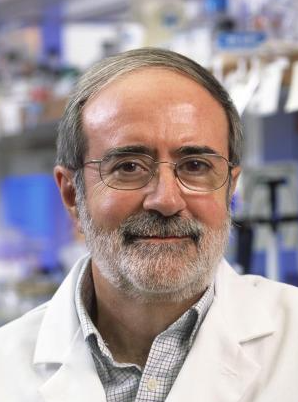 Roberto Docampo合作论文数Department of Cellular Biology, Franklin College of Arts and Sciences, University of Georgia24
 Silvia N.J. Moreno合作论文数Center for Tropical and Emerging Global Diseases, Department of Cellular Biology, Franklin College of Arts and Sciences, University of Georgia22
 Virginia Edgcomb合作论文数Woods Hole Oceanographic Institution11
 Fatma Gomaa合作论文数Department of Organismic & Evolutionary Biology, Harvard University10
 Peter R. Girguis合作论文数Department of Organismic & Evolutionary Biology, Harvard University10
 De-Pei Liu (刘德培)合作论文数Peking Union Medical College7
 Eric Oldfield合作论文数Department of Chemistry, College of Liberal Arts & Sciences, University of Illinois at Urbana-Champaign5
 Noelia Lander合作论文数Centro de Biotecnología, Instituto de Estudios Avanzados-MCT5
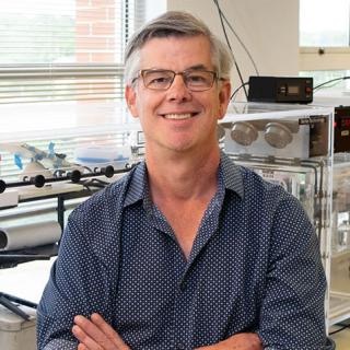 Michael Terns合作论文数Department of Biochemistry and Molecular Biology, Franklin College of Arts and Sciences, University of Georgia5
 Catherine Li合作论文数5RM Terns合作论文数4CC Liang合作论文数4
 Stephen A Vella合作论文数Univ Georgia, Ctr Trop & Emerging Global Dis, Athens, GA 30602 USA4WX Yin合作论文数3Beejan Asady合作论文数3
 Szajnman Sergio H合作论文数Facultad de Ciencias Exactas y Naturales, Universidad de Buenos Aires3Rodriguez J B合作论文数3
 Malwal Satish R合作论文数Department of Chemistry, Indian Institute of Science Education and Research3
 Niyogi Sayantanee合作论文数Department of Cellular Biology and Center for Tropical and Emerging Global Diseases, University of Georgia3Chih-Chuan Liang合作论文数3
 Veronica A. Jimenez合作论文数Department of Chemistry, Andrés Bello University;Centro de Química Teórica y Computacional, Exact Sciences School, Andrés Bello University3Vanina E. Alvarez合作论文数3
 Javier G De Gaudenzi合作论文数Instituto de Investigaciones Biotecnológicas-Instituto Tecnológico de Chascomús, CONICET-UNSAM3Miryam Andrea Hortua Triana合作论文数3Pia A. Elustondo合作论文数2José A. Fernández Robledo合作论文数2CLEMENTE THOMAS E合作论文数2
 Rachele Cesaroni合作论文数2Natalia Janowicz合作论文数2Cristina Aresté合作论文数2Lisa Sudek合作论文数2Susana A Breglia合作论文数2Ambar Kachale合作论文数2Eleanna Kazana合作论文数2Mariana Rius合作论文数2Kodai Fukuba合作论文数2
 Le Chang合作论文数2Isabel Nimmo合作论文数2Yuu Ishii合作论文数2
 Brittany N Sprecher合作论文数2Ellen Nisbet合作论文数2
 Imen Lassadi合作论文数2
 Alex W Chan合作论文数Whitehead Institute for Biomedical Research2Christina A. Moore合作论文数2
 Binnypreet Kaur合作论文数Inst Parasitol, Czech Acad Sci2
 Anna M. G. Novak Vanclova合作论文数2WANG Lu (王路)合作论文数2Valérie Vergé合作论文数Univ Pierre & Marie Curie Paris 06, Sorbonne Univ2
 Trong-Nhat Phan合作论文数Host Parasite Res Lab, Inst Pasteur Korea2




