High-density areas (HDAs) are frequently observed in follow-up CT of large-vessel occlusions after endovascular therapy. Utilizing the established ASPECTS regions, incorporating the subarachnoid space and ventricles, we developed a novel HDA score to evaluate its correlation and predictive value for hemorrhagic transformations and clinical outcomes. This retrospective, multicenter study included consecutive patients who had HDA on follow-up CT after endovascular therapy. Multivariable logistic regression and area under the receiver operating characteristic curve (AUC) analyses assessed the associations and predictive value of HDA location and score with hemorrhagic transformations and unfavorable clinical outcomes. Among the 1130 consecutive patients treated with endovascular therapy, 542 patients (326 males; median age 70 years) had HDA were finally included. Multivariable logistic regression showed that HDA location in the lentiform nucleus (OR, 1.6; 95
To evaluate the diagnostic utility of MRI-based radiomics in stratifying the risk of tumor deposits (TD) in patients with rectal cancer (RC). This study retrospectively analyzed 729 patients with RC from two institutions (January 2018–August 2024). Patients were classified into three groups according to the number of TD: no TD (TD0), 1–2 TD (TD1-2), and ≥ 3 TD (TD3+). Radiomics features were extracted from the tumor and the largest nodule within the rectal mesentery on MRI images. Predictive models were developed with the XGBoost algorithm. Model performance was evaluated using the receiver operating characteristic curve, area under the curve, confusion matrix, precision, accuracy, recall, and F1 score. Three hundred seventy-six patients were ultimately included and allocated into training, test, and validation sets. The tumor model (developed using tumor features) achieved AUCs of 0.871 (test set) and 0.848 (validation set), with corresponding accuracy, precision, recall, and F1 of 0.745/0.716, 0.764/0.688, 0.764/0.734, and 0.764/0.710, respectively. The nodule model (developed using the largest nodule) yielded AUCs of 0.839/0.804, accuracy of 0.673/0.637, precision of 0.571/0.614, recall of 0.800/0.686, and F1 of 0.667/0.648 in the test and validation sets, respectively. The fusion model, which combined tumor and nodule features, achieved enhanced performance with AUCs of 0.873/0.858, accuracy of 0.800/0.784, precision of 0.804/0.712, recall of 0.745/0.775, and F1 of 0.774/0.742, outperformed both individual models and two radiologists (accuracy 0.676/0.589). MRI-derived radiomics demonstrates significant potential for risk stratification of TD in RC. The radiomics model integrating tumor features and maximal short-axis diameter of mesorectal nodules effectively predicts three distinct quantity-based categories of TD in RC, enabling preoperative risk stratification and assisting personalized treatment planning.
Tumors display genomic and phenotypic heterogeneity, which holds prognostic significance and may influence therapy response. Radiographic imaging modalities, such as computed tomography, magnetic resonance imaging, nuclear medicine techniques, and ultrasonography, are routinely used to generate parametric maps to identify, measure, and map tumor heterogeneity from different perspectives encompassing anatomy, physiology, and metabolism. This review underscores the potential of artificial intelligence (AI)-based habitat imaging analysis, referred to as Radiomics++, in decoding intratumor heterogeneity compared to conventional radiomics. We highlight the general workflow, underlying principles, detailed methodology, and clinical applications of habitat imaging analysis to guide researchers. Validation advancements are then reviewed to verify the reliability of generated habitats by correlating radiologic phenotypes with biologic underpinnings. Furthermore, we address key challenges and opportunities in clinical translation, including data heterogeneity, model performance, and interpretability. Finally, integrating AI-defined habitats with multi-omics is anticipated to deepen our understanding of tumor evolution and advance precision medicine.
Objective:The aim of this study was to develop and validate a cascaded artificial intelligence (AI) framework using a segmentation-discrimination strategy for automated detection of ossicular chain malformations (OCM) on computed tomography scans. Methods:Patients diagnosed with OCM between January 2009 and January 2023, along with healthy controls, were retrospectively enrolled. A coarse-to-fine nnU-Net framework was applied for automated segmentation of the auditory ossicles. Separate discrimination models were developed for the malleus, incus, and stapes, and subsequently integrated to generate a patient-level model. The performance of the proposed framework was compared with deep learning networks, a volume-threshold algorithm, and by radiologists. The temporal external validation was conducted on data from multiple sites within Shaanxi Province, using the same CT vendors. The discriminatory capacity of each ossicular-level model was assessed using area under the curve (AUC), sensitivity, specificity, and accuracy. Results:A total of 2,462 temporal bone computed tomography scans (training set, n = 1,302; test set, n = 556; validation set, n = 604) were analyzed. The cascaded network demonstrated AUC values of 0.962, 0.930, and 0.931 for the malleus, incus, and stapes, respectively. At the patient level, accuracy, sensitivity, and specificity were 0.874, 0.949, and 0.800, respectively, surpassing the performance of ResNet, DenseNet, the volume-threshold algorithm, and junior radiologists, while equaling that of senior radiologists. In temporal validation, the network maintained robust performance, with AUC values of 0.972, 0.973, and 0.963, and patient-level metrics of 0.960 (accuracy), 0.974 (sensitivity), and 0.947 (specificity). Conclusion:The cascaded AI framework using a segmentation-discrimination strategy demonstrated high performance in identifying patients with diverse types of OCMs on computed tomography scans. This approach has the potential to support radiologists in the diagnostic evaluation of OCMs.
Precise brain segmentation is fundamental for quantitative neuroimaging analysis. However, most existing methods lack generalization across the human lifespan and diverse imaging modalities, limiting their utility for Comprehensive Brain Segmentation (CBS) (i.e., tissue segmentation, parcellation, and lesion labeling). To address this, we propose BrainSeg, a novel unified framework, for CBS by using large-scale datasets spanning the entire lifespan, with adaptability to diverse uni- and multimodal input scenarios without the need for retraining or finetuning. Comprehensive experiments are conducted on lifespan data ranging from 14 gestational weeks to 100 years of age, consisting of 45,998 multimodal scans from 26 datasets, which are further augmented by our proposed synthesis strategy. Systematic validation and in-depth analysis demonstrate that our BrainSeg can achieve state-of-the-art performance across all three core CBS tasks, with the averaged Dice ratios reaching up to 96.94% for tissue segmentation, 94.25% for brain parcellation, and 91.06% for lesion labeling in the internal validations. It maintains similarly high accuracy in external validations, with averaged Dice ratios achieving 94.01% for tissue segmentation, and 91.20% for brain parcellation, underscoring its robustness and generalizability across diverse conditions. In summary, BrainSeg serves as a versatile foundation tool, providing flexible and reliable analysis for large-scale neuroimaging studies.
Respiratory diseases cause significant morbidity, yet diagnosis remains labor intensive and dependent on physician expertise. Here, we present LungGPT, a unified multimodal system trained on 147 million tokens of domain-specific electronic health records from 125,917 participants. LungGPT comprises two modules: LungGPT-Dx for respiratory disease diagnosis and early warning of critical illness, and LungGPT-Ex for interpretable diagnostic reasoning and treatment recommendations. In large-scale evaluations, LungGPT-Dx achieves a macro-average area under the curve (AUC) of 0.852 (95% confidence interval [CI]: 0.839-0.865) across 22 respiratory diseases, with disease-specific AUCs exceeding 0.900 for lung cancer and pulmonary tuberculosis. Crucially, the model further improves early warning of critical illness by incorporating chain-of-thought (CoT) reasoning into textual data and integrating computed tomography (CT) imaging features. LungGPT-Ex generates high-quality, interpretable reasoning that outperforms specialized clinical models and matches advanced general-purpose models such as GPT-4o and DeepSeek-R1 in correctness, completeness, and truthfulness. By bridging precision diagnostics and rapid decision-making, LungGPT provides a standardized framework to enhance clinical workflows and improve patient outcomes in respiratory healthcare.
Lung cancer is the leading cause of cancerrelated mortality worldwide. In addition to localizing and segmenting lung nodules, a non-invasive risk assessment system can also help clinicians tailor treatment decisions in a timely manner, ultimately improving patient outcomes. Artificial intelligence (AI) technologies are increasingly being used in medical imaging to assess the risk of lung nodules, especially for malignancy classification. However, little research has been conducted on the assessment of other related risks. This work comprehensively reviews AI applications in lung nodule risk assessment, including malignancy diagnosis, pathological subtype assessment, metastasis risk evaluation, specific receptor expression identification, and disease progression tracking. It details common public databases used and state-of-the-art AI techniques, along with their benefits and challenges like data scarcity, generalizability, and interpretability. We anticipate that future research will tackle these issues, thereby increasing the improved interpretability and generalizability of AI methods in clinical workflows.
Background : In vivo whole-cortex quantification of intracortical signal-defined layering on the routinely acquired structural MRI remains limited. Purpose : To develop and validate an automated framework to reconstruct three intracortical signal-defined layers from 5T three-dimensional (3D) T2-weighted fluid-attenuated inversion recovery (FLAIR) and to characterize whole-cortex morphometrics and regional organization across a prespecified cortical organizational framework. Materials and Methods : In this retrospective study, 5T 3D FLAIR images were acquired between February and July 2024. Brain Multi-Layer Surface Reconstruction (BrainMLSR) reconstructed three intracortical signal-defined layers, and derived intracortical layer thickness and surface area measures and ratios. Performance was evaluated against manual annotations and assessed for test-retest repeatability (n=13) and cross-site feasibility (n=2). Paired two-tailed t-tests and linear mixed-effects models were used. A proof-of-concept analysis compared Heschl's gyrus ratios between 19 patients with temporal lobe epilepsy (TLE) and 19 age-matched healthy controls (HC Results : A total of 270 healthy participants (mean age, 54.4±14.5 years; 146 men) were included. Agreement with manual hypointense-layer annotations was high (Dice, 0.960±0.003), and was similar in the cross-site dataset (Dice, 0.954±0.009). In the test-retest dataset, average symmetric surface distance was less than 0.1 mm. Across prespecified systems, thickness and surface area ratios varied by region; within an auditory-perisylvian hierarchy, banksSTS showed a localized turning point with an increased hyperintense layer thickness ratio and decreased hypointense layer thickness ratio, accompanied by inflections in surface area ratios (P < .001). In bilateral Heschl's gyrus, hypointense (left: 0.619±0.262 vs 0.881 ± 0.102; right: 0.607±0.310 vs 0.907±0.141 mm) and isointense (left: 0.406±0.225 vs 0.678± 0.128; right: 0.478±0.232 vs 0.808 ± 0.176 mm) layer thicknesses were lower in TLE than in HC (all P<.001). Conclusion : BrainMLSR enabled accurate and repeatable in vivo reconstruction of three intracortical signal-defined layers from a single 5T 3D T2-weighted FLAIR acquisition and provided whole-cortex boundary-based morphometry with interpretable regional organization. ### Competing Interest Statement The authors have declared no competing interest. National Natural Science Foundation of China, 82441023, U23A20295, 62131015, 82394432 China Ministry of Science and Technolog, S20240085, STI2030-Major Projects-2022ZD0209000, STI2030-Major Projects-2022ZD0213100 Shanghai Municipal Central Guided Local Science and Technology Development Fund, YDZX20233100001001 The Key R&D Program of Guangdong Province, China, 2023B0303040001
Mechanical thrombectomy (MT) has emerged as the primary treatment for restoring blood flow in acute ischemic stroke (AIS) patients with large vessel occlusion (LVO). The shift from "time is brain" to "tissue is brain" underscores the significance of multimodal imaging in selecting and triaging AIS patients for endovascular reperfusion therapy. Currently, computed tomography (CT) is the preferred imaging modality for evaluating the infarct core and ischemic penumbra, aiding in the identification of suitable MT candidates. However, the abundance of collected imaging data, containing valuable signs, remains largely unexplored in emergency scenarios and follow-up protocols. A comprehensive investigation of these imaging features, known as "stroke imaging biomarkers," is crucial for accurate predictions regarding AIS pathogenesis, treatment, and prognosis. This exploration can provide valuable support to physicians in their diagnostic and treatment decisions. This review presents an overview of imaging biomarkers associated with ischemic core development, thrombus composition, stroke etiology, reperfusion strategy optimization, and clinical prognosis prediction. Beyond summarizing candidate biomarkers, we critically compare overlapping CT signatures, highlight controversies and failure modes, and synthesize the current level of evidence and barriers to clinical translation. Additionally, the utilization of artificial intelligence in this field, alongside emerging applications and future prospects, is discussed.
Definitive diagnosis of placenta accreta spectrum (PAS) disorders occurs at delivery by using combined intraoperative clinical and histopathologic diagnostic criteria per the recently updated International Federation of Gynecology and Obstetrics (FIGO) classification system. The purpose of this study is to construct a dual diagnostic model based on intraoperative clinical and histopathologic criteria for PAS in accordance with FIGO’s diagnostic criteria. This study pooled data from two centers. Diagnostic models based on histopathologic and intraoperative clinical criteria were separately developed using radiomics, deep learning, and clinical features. Subsequently, these individual models were integrated. Moreover, the correlations among clinical indicators, radiomics, and deep-learning features were explored. The Mann-Whitney U test was used for continuous variables with non-normal distribution. Categorical data were analyzed by Fisher’s exact test or 2 × 2 or R×C χ2 test. A P value < 0.05 was considered significant. For the FIGO intraoperative clinical criteria model, the fusion model’s AUC was 0.964 (0.946–0.982) in the training set and 0.949 (0.902–0.996) in the test set. For the FIGO histopathologic criteria model, the training-set AUC was 0.961 (0.940–0.982), and the validation cohort AUC was 0.936 (0.883–0.990). In addition, several clinical factors and magnetic resonance imaging (MRI) features of PAS were found to correlate with radiomics scores and deep learning scores in both the intraoperative clinical and histopathologic criteria models. In this study, we constructed a dual model that includes FIGO intraoperative clinical and histopathologic criteria for predicting PAS. The two models that integrate deep learning, radiomics, and clinical features have good performance.
Our study seeks to develop a predictive model using MRI-derived enlarged perivascular spaces (EPVSs) measurements and machine learning to assess cognitive impairment, subjective sleep quality, and excessive daytime sleepiness in young adults with long-time mobile phone use (LTMPU). We enrolled 82 participants and employed a pretrained deep learning model (VB-Net) to automatically segment EPVSs lesions across 17 brain subregions, extracting four handcrafted radiomic features – predefined based on morphological properties – per subregion (EPVSs count, volume, mean length, and mean curvature). The cohort was randomly divided into training (80%) and testing (20%) sets. Through minimum redundancy maximum relevance (mRMR) feature selection, six key biomarkers from 68 initial EPVSs metrics were identified combined with sex and age covariates. Final models were constructed using a Gaussian process (GP) classifier for cognitive impairment and decision tree (DT) algorithms for sleep quality and excessive sleepiness assessment. In testing, the GP model achieved an AUC of 0.818 (95% confidence interval [CI] 0.610-1) for cognitive impairment prediction. The DT models showed AUCs of 0.826 (95% CI: 0.616-1) for sleep quality and 0.875 (95% CI: 0.718-1) for daytime sleepiness. This automated radiomics pipeline demonstrates EPVSs morphological features as potential biomarkers for evaluating mobile phone exposure-related neurocognitive dysfunction. This automated radiomics pipeline suggests that EPVSs morphological features might be beneficial for evaluating mobile phone exposure-related neurocognitive dysfunction.
PurposeThis study intends to develop and validate a multiclass system that efficiently integrates complementary information from patients’ multimodal data (clinical, imaging, and pathological) to identify individuals at a high risk of 5-year postoperative recurrence among patients with clear cell renal cell carcinoma (ccRCC).MethodsThis retrospective multicenter study enrolled 270 clear cell renal cell carcinoma (ccRCC) patients, allocating 215 (79.6%) for model development/validation and 55 (20.4%) to an independent test cohort. Patients were categorized into recurrence and non-recurrence groups based on whether tumor recurrence occurred within 5 years after surgery. The final included case in this cohort underwent surgery in July 2018. Single-modality models were created using radiomics, deep learning (ResNet34), and deep features-based radiomics. Subsequently, multi-modality models were constructed by combining the predicted probabilities from the single-modality models and transferring them to another classifier. All models was assessed using the area under the receiver operating characteristic curve (AUROC).ResultsA total of 25 classification models were constructed. Notably, single-modality fusion models generally outperform their radiomics and deep learning-based radiomics (DL) counterparts. Among five single-modality fusion models, the Fused_Non-enhanced model demonstrates best predictive performance, achieving an AUROC value of 0.837 (95% confidence interval [CI]: 0.729-0.946). Similarly, multi-modality radiomics or DL models exhibit superior performance compared to single-modality counterparts. The multi-modality radiomics-DL model demonstrates the highest prediction performance, achieving an AUROC value of 0.967 (95% CI: 0.929-1.0) in the independent testing dataset.ConclusionThe multi-modality radiomics-DL model demonstrates high accuracy in predicting the 5-year postoperative recurrence risk of clear cell renal cell carcinoma (ccRCC).
Background:The severity of interstitial lung disease (ILD) is frequently linked to poorer outcomes and reduced quality of life in connective tissue disease-associated ILD (CTD-ILD) patients. The purpose of this study is to investigate the utility of artificial intelligence (AI)-based quantitative high-resolution computed tomography (HRCT) analysis in assessing the prognosis of patients with CTD-ILD. Methods:This retrospective study included 116 CTD-ILD patients who underwent HRCT scans. Patients were stratified into mild, moderate, and severe groups based on pulmonary function test (PFT) results. Differences in the 17 AI parameters across the three groups were evaluated using one-way analysis of variance (ANOVA) followed by least significant difference (LSD) post hoc pairwise comparisons. The Spearman rank correlation test was employed to examine the association between the 17 AI parameters and pulmonary function grades. Overall survival rates were compared using Kaplan-Meier analysis and ANOVA. Univariate analysis and Cox proportional-hazards regression were used to assess the association between AI-derived parameters and prognosis. Results:The 17 AI parameters exhibited significant variations across the three groups. Among them, three pulmonary volume parameters were negatively correlated with lung function, while fourteen pulmonary parenchymal involvement parameters were positively correlated with pulmonary function. Significant differences in overall survival rates were observed among the mild, moderate, and severe groups. Univariate analysis revealed that there were 12 indicators showing significant differences between the survival group and the death group. The Cox proportional-hazards regression revealed that the two most important factors were left lung volume and the percentage of disease components ≤-751 Hounsfield units (HU), which were protective factors and risk factors for overall survival rate, respectively. Conclusions:The quantitative HRCT analysis based on AI can be used to evaluate patients with CTD-ILD and is correlated with overall survival rate.
Alzheimer's disease (AD) is increasingly recognized to have systemic physiological correlates alongside central neurodegeneration. Here, we explored brain-organ network (BON) connectivity in AD (n=28) and healthy controls (n=23) using time-resolved quasi-dynamic analysis of plateau-phase total-body 18F-tau-PET. We found that AD-related pathophysiology was linked not only to cerebral tau aggregation, but also to altered signal synchronization across the brain-organ network, despite comparable body tracer distribution. Network topology analyses revealed the occipitotemporal cortex and the spinal cord as key nodes in this altered systemic network. Furthermore, exploratory mediation analyses demonstrated that BON dysregulation is cross-sectionally linked to cognitive deficits, with statistical associations observed for both cortical tau burden and imaging markers of impaired glymphatic clearance. This total-body PET study provides first-ever direct evidence repositioning AD as a multi-organ disorganization disease. These findings provide a novel framework for investigating brain-body interactions and systemic vulnerabilities in neurodegenerative disorders. ### Competing Interest Statement F.S. is an employee of United Imaging Intelligence, Shanghai, China. The company had no role in the design or conduct of the study, or in the analysis and interpretation of the data. All other authors report no conflicts of interest relevant to this article. ### Funding Statement This work is partially supported by the National Natural Science Foundation of China (Nos. 62571330, 62131015, U23A20295, and 82572344), Shanghai Pilot Program for Basic Research - Chinese Academy of Science, Shanghai Branch (No. JCYJ-SHFY-2022-014), the Construction Project of Shanghai Key Laboratory of Molecular Imaging (No. 18DZ2260400), Shanghai Aging Women and Children's Health Research Program (No. 2020YJZX0107), Shanghai Zhangjiang National Innovation Demonstration Zone Special Funds for Major Projects (No. ZJ2018-ZD-012), and Shenzhen Science and Technology Program (No. KCXFZ20211020163408012). The computation in this work was supported by the HPC Platform of ShanghaiTech University. ### Author Declarations I confirm all relevant ethical guidelines have been followed, and any necessary IRB and/or ethics committee approvals have been obtained. Yes The details of the IRB/oversight body that provided approval or exemption for the research described are given below: Institutional Review Board of Renji Hospital, Shanghai Jiao Tong University School of Medicine gave ethical approval for this work (Approval No. 2020-004). I confirm that all necessary patient/participant consent has been obtained and the appropriate institutional forms have been archived, and that any patient/participant/sample identifiers included were not known to anyone (e.g., hospital staff, patients or participants themselves) outside the research group so cannot be used to identify individuals. Yes I understand that all clinical trials and any other prospective interventional studies must be registered with an ICMJE-approved registry, such as ClinicalTrials.gov. I confirm that any such study reported in the manuscript has been registered and the trial registration ID is provided (note: if posting a prospective study registered retrospectively, please provide a statement in the trial ID field explaining why the study was not registered in advance). Yes I have followed all appropriate research reporting guidelines, such as any relevant EQUATOR Network research reporting checklist(s) and other pertinent material, if applicable. Yes The data underlying the findings of this study are available from the corresponding author upon reasonable request.
Accurate and automatic segmentation of lifespan brain MRI into regions of interest (ROIs) is crucial for studying brain development, aging, and early diagnosis of neurological diseases. Existing segmentation methods are often tailored to specific age groups, such as infants or adults, resulting in inconsistent performance when processing brain data from different age groups. To overcome this limitation, we introduce BrainSMM, a novel metadata-driven model that incorporates text-based prompts to guide representation learning in a segmentation backbone. These prompts, extracted via a pretrained image-text alignment model, encode valuable prior knowledge (e.g., age, scanner, gender) and are infused into the vision model to condition the features according to domain-specific contexts. We evaluate BrainSMM on a large-scale lifespan brain MRI dataset with 5,565 T1w MR images spanning multiple ages. Our approach achieves an average DSC of 94.59% for tissue segmentation (i.e., gray matter, white matter, and cerebrospinal fluid) and 86.34% for anatomical region segmentation (e.g., hippocampus, putamen, etc.) with corresponding average ASD of 0.20 mm and 0.75 mm, respectively. Notably, BrainSMM shows strong consistency in segmentation accuracy across all age groups and demonstrates improved anatomical detail preservation compared to baseline methods. Additionally, our metadata prompt technique is easily transferable and compatible with multiple backbone architectures, highlighting its adaptability. Overall, BrainSMM offers a robust, generalizable solution for lifespan brain MRI segmentation and lays the groundwork for enhanced clinical and developmental neuroimaging applications.
This study aimed to use multimodal MRI and artificial intelligence to automatically identify cognitive normal (CN), subjective cognitive decline (SCD), mild cognitive impairment (MCI), and alzheimer’s disease (AD). 715 participants with different cognitive status (CN, SCD, MCI, AD) were enrolled from ADNI (for training/validation) and OASIS-3 (for external validation). All participants underwent structural MRI (sMRI) and resting-state functional MRI (rs-fMRI). The sMRI of whole brain was segmented into 116 regions and the volumes of each region was obtained using 3D-VB-Net. 4528 radiomics features were extracted from hippocampus. Functional metrics (ALFF, fALFF, ReHo, FC) for each brain region were calculated using rs-fMRI data. Least absolute shrinkage and selection operator (LASSO) and K-best were used to reduce feature dimensionality. Machine learning was performed using bagging decision tree (BDT), logistic regression (LR), support-vector-machine (SVM) and random-forest (RF) classifiers. Evaluation metrics included the area under the curve (AUC), specificity, sensitivity and F1-score. A total of 4528 radiomic features, 116 volume features, and 464 functional features were extracted for each participant. After dimensionality reduction, the BDT model based on volume-function-radiomics features achieved the highest performance, with macro-average AUCs of 0.918 (95
Magnetic Resonance Imaging (MRI) is highly susceptible to motion artifacts due to prolonged acquisition times, posing a persistent challenge to image fidelity and diagnostic reliability. Existing deep learning-based correction methods typically rely on a fixed strategy, applying uniform models regardless of motion severity. This lack of adaptability often leads to over-correction in mild cases and insufficient restoration under severe motion. To overcome these limitations, we introduce FLEX-MoCo, a flexible motion correction pipeline that dynamically adapts its strategy based on both motion location and severity. FLEX-MoCo comprises two key components: a Motion Representation Learner that identifies motion artifacts from anatomical structures and identifies motion patterns, and a Correction Path Router that selects an optimal correction path tailored to the identified motion characteristics, ranging from lightweight models to multi-stage frameworks. Extensive experiments across diverse datasets containing both simulated and real-world motion artifacts demonstrate that FLEX-MoCo consistently surpasses state-of-the-art methods in motion correction and generalizes to unseen domains, underscoring the effectiveness of its motion-aware adaptive routing in handling varying motion patterns and severities.
Multi-parametric magnetic resonance imaging (mpMRI) is a cornerstone for brain tumor diagnosis and treatment, yet current AI models face critical limitations: their lack of natural language interaction and interpretability impedes spatial information integration and cross-modal reasoning required clinically. Key challenges arise from significant physical meaning differences across modalities, spatial misalignment due to scan intervals, and the need for complex multi-feature interpretation in tasks like glioma grading. While visual-language models (VLMs) show promise in cross-modal understanding, existing methods focus mainly on 2D image modeling, neglecting direct perception of 3D volumetric space. Although 3D VLMs have been proposed for report generation and feature alignment in 3D CT imaging, mpMRI applications demand collaborative inference across multiple imaging modalities-a requirement unmet by current solutions. To address this, we introduce Mr3D-VL, a dedicated visual-language foundation model for multi-parametric 3D MRI. With 4 billion parameters, it employs an unsupervised pre-trained shared 3D encoder and 4D rotational positional embedding for dual modality-spatial integration. Its cross-modal projection layer uses a multi-resolution feature implantation strategy to enhance feature perception across resolutions. Experimental results show significant improvements over existing 4B/7B/30B domain-specific and general-purpose models in text generation tasks, achieving a BERTScore of 0.856 for report generation, with question-answering accuracy at 0.713 and multiple-choice accuracy at 0.912.
BACKGROUND:Accurate assessment of 90-day functional outcomes after anterior circulation large vessel occlusion (LVO) stroke remains challenging. Conventional models relying on a single data dimension have limited assessment power, suggesting that a multidimensional integration strategy could enhance evaluations. PURPOSE:To develop and validate an interpretable machine learning model that integrates radiomics, infarct location, brain frailty, and clinical variables for assessing 90-day functional outcomes in LVO stroke. STUDY TYPE:Retrospective. POPULATION:1051 patients with anterior circulation LVO stroke (mean age 63 ± 13 years; 722 males) from five centers (2018-2023). Eight hundred and seventy-five patients from four centers formed the training (n = 612) and internal validation (n = 263) cohorts, while 176 from the fifth center comprised the external validation cohort. FIELD STRENGTH/SEQUENCE:T1-weighted spin-echo imaging (T1WI), T2-weighted spin-echo imaging (T2WI), T2-weighted fluid-attenuated inversion recovery (FLAIR) imaging, and diffusion-weighted echo-planar imaging (DWI). ASSESSMENT:Infarct volume and radiomic features were extracted from DWI. Infarct location was assessed using the Alberta Stroke Program Early CT Score. Brain frailty was evaluated using cortical/subcortical atrophy, white matter hyperintensity (WMH), and old infarcts. Least Absolute Shrinkage and Selection Operator (LASSO) regression was used for feature selection. STATISTICAL TESTS:Chi-square, Fisher's exact, t-test, Mann-Whitney U, area under the receiver operating characteristic curve (AUC), DeLong test, decision curve analysis, calibration curves, sensitivity, specificity, positive predictive value, negative predictive value, F1 score. Significance level p < 0.05. RESULTS:The fused model outperformed all single-dimension models (ΔAUC = 0.12-0.22), achieving AUCs of 0.87 (training), 0.84 (internal validation), and 0.86 (external validation). The fused model achieved a sensitivity and a specificity of 0.80 in the external validation cohort. Features with the highest mean absolute Shapley Additive Explanations (SHAP) values included lentiform nucleus lesion burden (SHAP = 0.083), WMH (SHAP = 0.080), and lesion burden in the M6 region (posterior middle cerebral artery territory; SHAP = 0.061). DATA CONCLUSION:Integration of infarct location, brain frailty, radiomics, and clinical features improved the 90-day outcome assessment in anterior circulation LVO stroke, providing an interpretable tool for personalized prognosis. LEVEL OF EVIDENCE: 3: TECHNICAL EFFICACY STAGE:2.
 Dinggang Shen (沈定刚)合作论文数School of Biomedical Engineering, ShanghaiTech University;Image Display, Enhancement and Analysis Lab, ShanghaiTech University194
 Weili Lin合作论文数Biomedical Research Imaging Center, University of North Carolina55
 Ying Wei (魏颖)合作论文数Northeastern University36
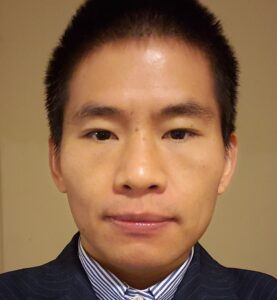 Li Wang合作论文数Developing Brain Computing Lab, University of North Carolina at Chapel Hill;Department of Radiology, School of Medicine, University of North Carolina at Chapel Hill35Gilmore J H合作论文数31
 Gang Li合作论文数Department of Radiology, School of Medicine, University of North Carolina at Chapel Hill30
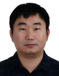 Yaozong Gao (高耀宗)合作论文数United Imaging Intelligence, Shanghai;School of Biomedical Engineering, Shanghai Tech University27
 Pew-Thian Yap合作论文数Image Analysis Core of Biomedical Research Imaging Center, University of North Carolina at Chapel Hill;Department of Radiology, School of Medicine, University of North Carolina at Chapel Hill26Yuwei Xia合作论文数24Qing Zhou (周清)合作论文数22
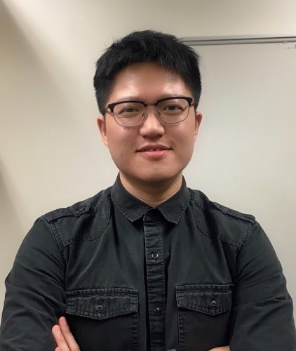 Zehong (Jimmy) Cao合作论文数University of South Australia20
 Xue Zhong (薛忠)合作论文数School of Biomedical Engineering, ShanghaiTech University19
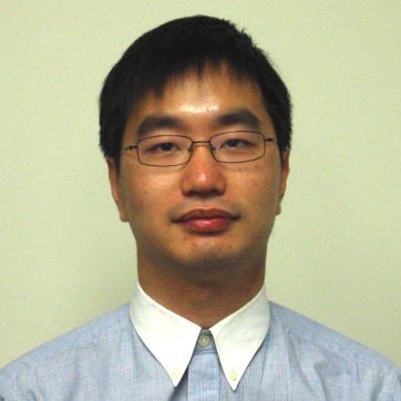 Yiqiang Zhan合作论文数Shanghai Jiao Tong University19
 Zhongxiang Ding (丁忠祥)合作论文数Zhejiang Provincial People's Hospitai19
 Qian Wang (王乾)合作论文数School of Biomedical Engineering, ShanghaiTech University18
 Xiang Zhou (周翔)合作论文数School of Biomedical Engineering, Shanghai Tech University;United Imaging Intelligence, Shanghai18
 Han Zhang (张寒)合作论文数School of Biomedical Engineering, ShanghaiTech University16Jiaojiao Wu合作论文数14
 Yong Fan合作论文数Department of Radiology, Perelman School of Medicine, University of Pennsylvania14
 Chuanming Li (李传明)合作论文数School of Medicine, Chongqing University;Chongqing University Central Hospital13
 Xiaodan Xing合作论文数Shanghai United Imaging Intelligence Co Ltd, Shanghai, Peoples R China13
 Guangming Lu (卢光明)合作论文数State Key Laboratory of Analytical Chemistry for Life Science, Nanjing University;Medical School of Nanjing University13Junbang Feng合作论文数12
 Bin Song (宋彬)合作论文数West China Hospital of Sichuan University12
 Fuhua Yan (严福华)合作论文数Zhoushan Branch, Shanghai Ruijin Hospital12Yichu He合作论文数Shanghai United Imaging Intelligence Co Ltd, Dept Res & Dev, Shanghai, Peoples R China11
 Jianghua Chen (陈江华)合作论文数Kidney Disease Center, The First Affiliated Hospital, Zhejiang University School of Medicine11
 Zi-Wen Peng合作论文数11
 Guorong Wu合作论文数School of Medicine, University of North Carolina at Chapel Hill10
 Tianzi Jiang (蒋田仔)合作论文数University of Chinese Academy of Sciences;Brainnetome Center, Institute of Automation, Chinese Academy of Sciences;Centre for Advanced Imaging, University of Queensland10
 Zhong Xue合作论文数Medical Image Lab,The Methodist Hospital Research Institute10
 Jun Liu (刘军)合作论文数中南大学湘雅第二医院9
 Yuyao Zhang (张玉瑶)合作论文数School of Information Science and Technology, ShanghaiTech University8
 Mianxin Liu合作论文数Shanghai Artificial Intelligence Laboratory, Shanghai, 200232, China.8Rui Hua合作论文数8Fei Shan合作论文数Fudan Univ, Shanghai Publ Hlth Clin Ctr, Dept Radiol, Shanghai, Peoples R China8Yucheng Wang合作论文数8Cuili Wang合作论文数8XiaoQing Cheng合作论文数Department of Medical Imaging, Jinling Hospital, Nanjing University School of Medicine, 305 Zhongshan East Road, Nanjing, 210002, Jiangsu, China.8Zhanhao Mo合作论文数Jilin Univ, China Japan Union Hosp, Changchun, Peoples R China8
 Dijia Wu (吴迪嘉)合作论文数School of Biomedical Engineering, ShanghaiTech University;United Imaging Intelligence8
 An Thai Le合作论文数Intelligent Autonomous Systems Group, Department of Computer Science, Technische Universitat Darmstadt7Tianyang Sun合作论文数7D. Shen合作论文数7
 Zhaohui Su (苏曌慧)合作论文数School of Public Health, Southeast University7
 Hongtu Zhu (朱宏图)合作论文数Department of Biostatistics, Gillings School of Global Public Health, The University of North Carolina at Chapel Hill;Department of Genetics, School of Medicine, The University of North Carolina at Chapel Hill7Chen Lizhou合作论文数7
 Liming Xia (夏黎明)合作论文数Tongji Hospital, Tongji Medical College, Huazhong University of Science and Technology7
 Tao Wang (王涛)合作论文数Shanghai Jiao Tong University School of Medicine Mental Health Center7




