Ultrahigh-field (UHF) magnetic resonance imaging (MRI), i.e., 7T MRI, provides superior anatomical details of internal brain structures owing to its enhanced signal-to-noise ratio and susceptibility-induced contrast. However, the widespread use of 7T MRI is limited by its high cost and lower accessibility compared to low-field (LF) MRI. This study proposes a deep-learning framework that systematically fuses the input LF magnetic resonance feature representations with the inferred 7T-like feature representations for brain image segmentation tasks in a 7T-absent environment. Specifically, our adaptive fusion module aggregates 7T-like features derived from the LF image by a pre-trained network and then refines them to be effectively assimilable UHF guidance into LF image features. Using intensity-guided features obtained from such aggregation and assimilation, segmentation models can recognize subtle structural representations that are usually difficult to recognize when relying only on LF features. Beyond such advantages, this strategy can seamlessly be utilized by modulating the contrast of LF features in alignment with UHF guidance, even when employing arbitrary segmentation models. Exhaustive experiments demonstrated that the proposed method significantly outperformed all baseline models on both brain tissue and whole-brain segmentation tasks; further, it exhibited remarkable adaptability and scalability by successfully integrating diverse segmentation models and tasks. These improvements were not only quantifiable but also visible in the superlative visual quality of segmentation masks.
Medical vision-language models (MVLMs) offer promise in clinical practice but face limitations in generalizability, data quality, and clinically meaningful evaluation. We propose RadiSim-CL, an MVLM trained via curriculum learning by simulating the three-phase pathway of a radiologist: foundational knowledge understanding, anatomical knowledge, and advanced diagnostic reasoning. To support this, we curate RadiSim, a 12-million image-text pair dataset aligned to these phases. We evaluate the model using a five-stage coarse-to-fine validation framework: (1) modality recognition, (2) anatomical recognition, (3) anatomical localization, (4) abnormality and disease diagnosis, and (5) disease differentiation and grading. This framework spans 24 zero-shot subtasks across MR, CT, and DR imaging. RadiSim-CL achieves comparable performance to state-of-the-art baselines in both foundational and anatomical tasks, and demonstrates superior capabilities in complex reasoning (e.g., an AUC of 0.953 for brain tumor diagnosis and an accuracy of 0.764 for meningioma grading). Ablation studies further confirm the curriculum’s effectiveness. RadiSim-CL thus offers a scalable, clinically aligned solution to enhance diagnostic precision.
Tumors display genomic and phenotypic heterogeneity, which holds prognostic significance and may influence therapy response. Radiographic imaging modalities, such as computed tomography, magnetic resonance imaging, nuclear medicine techniques, and ultrasonography, are routinely used to generate parametric maps to identify, measure, and map tumor heterogeneity from different perspectives encompassing anatomy, physiology, and metabolism. This review underscores the potential of artificial intelligence (AI)-based habitat imaging analysis, referred to as Radiomics++, in decoding intratumor heterogeneity compared to conventional radiomics. We highlight the general workflow, underlying principles, detailed methodology, and clinical applications of habitat imaging analysis to guide researchers. Validation advancements are then reviewed to verify the reliability of generated habitats by correlating radiologic phenotypes with biologic underpinnings. Furthermore, we address key challenges and opportunities in clinical translation, including data heterogeneity, model performance, and interpretability. Finally, integrating AI-defined habitats with multi-omics is anticipated to deepen our understanding of tumor evolution and advance precision medicine.
To develop and validate APEX-NET for early diagnosis and severity stratification of acute pancreatitis (AP) using non-contrast CT (NCCT), by leveraging contrast-enhanced CT (CECT) feature learning. This five-center retrospective and prospective study included 3383 patients, comprising AP and Non-AP (abdominal pain patients and healthy individuals) patients. APEX-NET was trained and evaluated to perform pancreas segmentation, AP diagnosis (AP vs Non-AP), and severity prediction (mild, moderately severe, or severe per the revised Atlanta classification) using 3 internal and 2 external cohorts. A feature mapping module was employed to derive simulated CECT features from NCCT based on paired NCCT-CECT feature learning. The model was further evaluated with subgroup analyses, and a reader study was conducted by comparing its performance with six radiologists of varying experiences. Evaluation metrics included the Dice similarity coefficient, area under the receiver operating characteristic curve (AUC), and accuracy. For AP diagnosis, APEX-NET achieved AUCs of 0.949, 0.958, 0.981, and 0.955 in the validation, internal, and two external testing cohorts, respectively. For severity prediction, APEX-NET significantly outperformed the NCCT model (p < 0.05), with macro-average AUCs of 0.873 (validation) and 0.872 (internal testing). The advantage of APEX-NET had been demonstrated in almost all the age, gender, and etiology subgroups. In the reader study, APEX-NET performed comparably to senior radiologists and superior to junior radiologists (p < 0.05). APEX-NET enables accurate NCCT-based diagnosis and early severity stratification of AP, demonstrating strong potential for clinical integration to overcome the inherent delay of CECT-based assessment. Question The absence of an accurate method for predicting AP severity from early NCCT, the initial diagnostic scan, thus forgoing the critical intervention window. Findings Achieve accurate severity prediction for AP by incorporating contrast-enhanced feature learning. Demonstrate robust performance across diverse demographic groups, etiologies, and imaging parameters. Clinical relevance The APEX-NET, an integrated deep learning framework using NCCT, accelerated the diagnosis and severity stratification of AP, demonstrating performance comparable to senior radiologists and direct potential for clinical workflow integration by reducing reliance on delayed contrast-enhanced scans.
Motion artifacts in magnetic resonance imaging (MRI) degrade diagnostic reliability. Existing deep learning methods are typically contrast-specific and fail to generalize across diverse modalities and artifact severities. We propose a unified framework combining parameter-informed contrast disentanglement with severity-aware adaptive correction. ScanCLIP, pretrained on over 30,000 MRI text-image pairs, derives contrast embeddings from acquisition parameters to disentangle contrast style from anatomical content, yielding contrast-free features. A Vision Transformer then estimates motion severity and routes features through a Mixture-of-Experts network, enabling targeted artifact correction. A dual-pathway decoder reconstructs both the clean image and residual artifact map, enforcing image-space consistency. On IXI and HCP benchmarks, our method improves PSNR by 0.75 dB and SSIM by up to 0.0279 over state-of-the-art approaches, with larger gains at higher artifact severities. It further demonstrates robust zero-shot generalization on real-world clinical data acquired with unseen scanning parameters, where existing methods either fail to remove artifacts or introduce additional distortions.
Brain graphs constructed from diverse neuroimaging modalities offer complementary perspectives for characterizing structural and functional connectivity patterns within the human brain. Although multi-modal integration has significantly advanced psychiatric diagnosis, existing fusion methods encounter a critical limitation: they tend to prioritize either modality-specific characteristics or shared complementary information, rather than leverage both synergistically. To address this issue, we propose a Shared-Specific Graph Learning (S2GL) framework, designed to comprehensively explore multi-modal features for improving psychiatric diagnosis. Specifically, we develop two modality-specific graph learning modules to extract multi-level representations from structural and functional brain graphs, respectively. In parallel, a customized structural-functional shared graph learning module captures cross-modal correlations in the feature embedding space, and generates shared representations for high-order feature extraction. The resulting specific and shared representations are then integrated by a shared-specific graph feature aggregation module through multi-modal graph pooling for final diagnosis. Extensive experiments on two psychiatric disorders demonstrate that S2GL consistently outperforms state-of-the-art diagnostic methods. Furthermore, neuroscientific analysis reveals that the identified brain regions align with established clinical findings, highlighting the interpretability and clinical relevance of S2GL for distinguishing complex psychiatric conditions.
Precise brain segmentation is fundamental for quantitative neuroimaging analysis. However, most existing methods lack generalization across the human lifespan and diverse imaging modalities, limiting their utility for Comprehensive Brain Segmentation (CBS) (i.e., tissue segmentation, parcellation, and lesion labeling). To address this, we propose BrainSeg, a novel unified framework, for CBS by using large-scale datasets spanning the entire lifespan, with adaptability to diverse uni- and multimodal input scenarios without the need for retraining or finetuning. Comprehensive experiments are conducted on lifespan data ranging from 14 gestational weeks to 100 years of age, consisting of 45,998 multimodal scans from 26 datasets, which are further augmented by our proposed synthesis strategy. Systematic validation and in-depth analysis demonstrate that our BrainSeg can achieve state-of-the-art performance across all three core CBS tasks, with the averaged Dice ratios reaching up to 96.94% for tissue segmentation, 94.25% for brain parcellation, and 91.06% for lesion labeling in the internal validations. It maintains similarly high accuracy in external validations, with averaged Dice ratios achieving 94.01% for tissue segmentation, and 91.20% for brain parcellation, underscoring its robustness and generalizability across diverse conditions. In summary, BrainSeg serves as a versatile foundation tool, providing flexible and reliable analysis for large-scale neuroimaging studies.
Magnetic resonance imaging (MRI) is an indispensable tool for clinical knee examination, which often scans 2D stacked slices from multiple views. Radiologists typically locate lesion regions in one view, and then refer to other views to formulate a comprehensive diagnosis. However, existing computer-aided diagnosis methods fall short of identifying and fusing local regions in multi-view scans, leading to a decline in diagnostic performance and a heavy reliance on extensively annotated data. This paper introduces a novel framework that represents multi-view MRI scans as a knee graph, and conducts diagnosis using the proposed Knee Graph Network (KGNet). Moreover, KGNet is greatly enhanced by multi-task pre-training, which requires KGNet to reconstruct masked knee local patches and segment unmasked ones working alongside corresponding decoders. Experimental evaluations on public and in-house clinical datasets confirm that our framework outperforms existing approaches in diagnosing cartilage defects, anterior cruciate ligament tears, and knee abnormalities. In conclusion, our framework demonstrates the potential of enhancing knee disease diagnosis by representing multi-view MRI scans as a graph and employing multi-task pre-training in the graph network. The code is publicly available at https://github.com/zixuzhuang/KGNet.
Cervical abnormality screening is pivotal for prevention and treatment. However, the substantial size of whole slide images (WSIs) makes examination labor-intensive and time-consuming. Current deep learning-based approaches struggle with the morphological diversity of cervical cytology and require specialized models for distinct diagnostic tasks, leading to fragmented workflows. Here, we present UniCAS, a cytology foundation model pre-trained on 48,532 cervical WSIs encompassing diverse patient demographics and pathological conditions. UniCAS enables various clinical analysis tasks, achieving state-of-the-art performance in slide-level diagnosis, region-level analysis, and pixel-level image enhancement. In particular, by integrating a multi-task aggregator for slide-level diagnosis, UniCAS achieves area under the curve (AUC) values of 92.60%, 92.58%, and 98.39% for cancer screening, candidiasis testing, and clue cell diagnosis, respectively, while reducing diagnostic time by 70% compared with conventional approaches. This work establishes a paradigm for efficient multi-scale analysis in automated cervical cytology, bridging the gap between computational pathology and clinical diagnostic workflows.
Respiratory diseases cause significant morbidity, yet diagnosis remains labor intensive and dependent on physician expertise. Here, we present LungGPT, a unified multimodal system trained on 147 million tokens of domain-specific electronic health records from 125,917 participants. LungGPT comprises two modules: LungGPT-Dx for respiratory disease diagnosis and early warning of critical illness, and LungGPT-Ex for interpretable diagnostic reasoning and treatment recommendations. In large-scale evaluations, LungGPT-Dx achieves a macro-average area under the curve (AUC) of 0.852 (95% confidence interval [CI]: 0.839-0.865) across 22 respiratory diseases, with disease-specific AUCs exceeding 0.900 for lung cancer and pulmonary tuberculosis. Crucially, the model further improves early warning of critical illness by incorporating chain-of-thought (CoT) reasoning into textual data and integrating computed tomography (CT) imaging features. LungGPT-Ex generates high-quality, interpretable reasoning that outperforms specialized clinical models and matches advanced general-purpose models such as GPT-4o and DeepSeek-R1 in correctness, completeness, and truthfulness. By bridging precision diagnostics and rapid decision-making, LungGPT provides a standardized framework to enhance clinical workflows and improve patient outcomes in respiratory healthcare.
Lung cancer is the leading cause of cancerrelated mortality worldwide. In addition to localizing and segmenting lung nodules, a non-invasive risk assessment system can also help clinicians tailor treatment decisions in a timely manner, ultimately improving patient outcomes. Artificial intelligence (AI) technologies are increasingly being used in medical imaging to assess the risk of lung nodules, especially for malignancy classification. However, little research has been conducted on the assessment of other related risks. This work comprehensively reviews AI applications in lung nodule risk assessment, including malignancy diagnosis, pathological subtype assessment, metastasis risk evaluation, specific receptor expression identification, and disease progression tracking. It details common public databases used and state-of-the-art AI techniques, along with their benefits and challenges like data scarcity, generalizability, and interpretability. We anticipate that future research will tackle these issues, thereby increasing the improved interpretability and generalizability of AI methods in clinical workflows.
Background : In vivo whole-cortex quantification of intracortical signal-defined layering on the routinely acquired structural MRI remains limited. Purpose : To develop and validate an automated framework to reconstruct three intracortical signal-defined layers from 5T three-dimensional (3D) T2-weighted fluid-attenuated inversion recovery (FLAIR) and to characterize whole-cortex morphometrics and regional organization across a prespecified cortical organizational framework. Materials and Methods : In this retrospective study, 5T 3D FLAIR images were acquired between February and July 2024. Brain Multi-Layer Surface Reconstruction (BrainMLSR) reconstructed three intracortical signal-defined layers, and derived intracortical layer thickness and surface area measures and ratios. Performance was evaluated against manual annotations and assessed for test-retest repeatability (n=13) and cross-site feasibility (n=2). Paired two-tailed t-tests and linear mixed-effects models were used. A proof-of-concept analysis compared Heschl's gyrus ratios between 19 patients with temporal lobe epilepsy (TLE) and 19 age-matched healthy controls (HC Results : A total of 270 healthy participants (mean age, 54.4±14.5 years; 146 men) were included. Agreement with manual hypointense-layer annotations was high (Dice, 0.960±0.003), and was similar in the cross-site dataset (Dice, 0.954±0.009). In the test-retest dataset, average symmetric surface distance was less than 0.1 mm. Across prespecified systems, thickness and surface area ratios varied by region; within an auditory-perisylvian hierarchy, banksSTS showed a localized turning point with an increased hyperintense layer thickness ratio and decreased hypointense layer thickness ratio, accompanied by inflections in surface area ratios (P < .001). In bilateral Heschl's gyrus, hypointense (left: 0.619±0.262 vs 0.881 ± 0.102; right: 0.607±0.310 vs 0.907±0.141 mm) and isointense (left: 0.406±0.225 vs 0.678± 0.128; right: 0.478±0.232 vs 0.808 ± 0.176 mm) layer thicknesses were lower in TLE than in HC (all P<.001). Conclusion : BrainMLSR enabled accurate and repeatable in vivo reconstruction of three intracortical signal-defined layers from a single 5T 3D T2-weighted FLAIR acquisition and provided whole-cortex boundary-based morphometry with interpretable regional organization. ### Competing Interest Statement The authors have declared no competing interest. National Natural Science Foundation of China, 82441023, U23A20295, 62131015, 82394432 China Ministry of Science and Technolog, S20240085, STI2030-Major Projects-2022ZD0209000, STI2030-Major Projects-2022ZD0213100 Shanghai Municipal Central Guided Local Science and Technology Development Fund, YDZX20233100001001 The Key R&D Program of Guangdong Province, China, 2023B0303040001
Positron emission tomography (PET) is an advanced nuclear imaging technique and has been widely applied in clinic. However, radiation risks associated with standard-dose PET imaging raise health concerns, whereas the quality of low-dose PET images fails to meet clinical requirements. To reduce the tracer dose while maintaining image quality, it is of great interest to estimate high-quality PET images from low-dose images. However, existing low-dose PET image denoising methods primarily focus on image data, overlooking crucial information in non-image textual data such as patients' clinical tabular and textual descriptions of general image quality. This neglect can lead to subpar denoising quality with inaccurate contexts and poor details. To address these problems, in this paper, we propose Multi-Granularity Textual Prompts, namely MGTP, to denoise low-dose PET images via an adversarial diffusion model. Different from prior methods that rely solely on image conditioning, our MGTP innovatively introduces textual prompts spanning diverse granularities to capture both high-level semantic-related contexts and low-level degradation-related details. To harmonize multi-granularity textual prompts with low-dose PET images, we design a Cross-Modality Selective Conditioning (CMSC) module, which prioritizes semantic- and detail-relevant information while eliminating irrelevant components. The resulting features are fed into diffusion model as conditions, enforcing a more controlled diffusion process. In addition, we develop a Masked Prompt Reconstruction Network (MPR-Net) to enhance the preservation of semantics and details in denoised images, mitigating distortions brought by the random noise in the diffusion process. Experiments on clinical PET data show that our method achieves the state-of-the-art performance.
Automatic sleep staging methods designed on healthy datasets often suffer from performance decline when applied to patients with obstructive sleep apnea (OSA). We attribute We attribute this to two factors: First, OSA sleep exhibits atypical inter-stages dependencies that are not captured by short-context models; Second, many prior approaches rely on one or a few channels and thus ignore the rich multichannel relationships present in clinical Polysomnography (PSG) recordings. To address these issues, we propose TransGATNet, which fuses long-term temporal-frequency features using a Graph-Attentional Transformer. Specifically, each TransGAT layer applies a Transformer encoder to capture global channel context, followed by a top-k sparsified graph attention network to isolate the most informative inter-electrode relationships. On the Sleep-EDF-2018 dataset, our TransGATNet achieves 86.8
Contrast-enhanced multiphase magnetic resonance imaging (MRI), combined with other non-contrast MRI, has become the standard approach for diagnosing focal liver lesions (FLLs). Due to the complex nature of FLLs, it is essential to automatically segment lesions and classify them from multiple MRI sequences. While sequence-specific deep learning (DL) models can be applied, general-purpose segmentation models with a unified encoder and a multitask decoder have shown great effectiveness for multitask multisequence MR analysis, particularly for organ and lesion segmentation through joint learning schemes. The key feature of such foundational integrated models is their ability to process different sequences and to achieve various segmentation tasks using the same model. By fusing the feature vectors encoded from language-based task descriptions, general segmentation models allow specific image features to be used for different segmentation tasks during decoding. Hundreds of segmentation tasks can thus be performed through one general segmentation model, with potential zero-shot capability. Building upon this concept, we propose a multitask deep learning model (MDLM) for segmentation of organs and lesions in multisequence abdominal MRI. Trained on over 15 MRI sequences per subject, our model effectively performs multiple tasks including organ segmentation, hepatic segments segmentation, vessel segmentation, and focal liver lesion segmentation. Lesion classification is also achieved using the same encoder. This approach improves the feasibility of FLL diagnosis and is integrated into our deep learning-assisted FLL diagnosis application. Experimental results demonstrate the model’s effectiveness in image segmentation, providing invaluable clinical decision support in liver imaging.
Attenuation correction is critical for PET imaging to correctly reflect physiological activity. Recent studies perform PET self-attenuation correction, enabling attenuation correction from PET data itself instead of using additional CT or MRI. These methods either predict attenuation-corrected PET (AC-PET) from non-attenuation-corrected PET (NAC-PET) directly or synthesize intermediate CT images to guide PET attenuation correction. However, cross-modality synthesis of CT from NAC-PET is challenging, especially for small yet clinically important lesions. To address these challenges, we propose the Lesion-aware Mutual Guidance Diffusion Model (LMGDM), a coarse-to-fine dual-branch diffusion model that jointly performs PET attenuation correction and CT synthesis, with particular focus on lesion regions. Specifically, we first generate coarse predictions of both AC-PET and CT using individual residual diffusion models. Subsequently, the coarse AC-PET and CT are jointly refined by a proposed dual-branch mutual guidance module to enable feature fusion of the AC-PET and CT branches. Moreover, a lesion-aware refinement module is embedded into the PET branch, encouraging the network to focus on regions with pathologically high uptake rather than physiologically high uptake. In addition, an attenuation prior learned from real CT images is also introduced to further enhance the fidelity of the synthesized AC-PET and CT. Extensive evaluation on eight data centers demonstrates strong superiority of our LMGDM over the state-of-the-art methods.
Accurate vascular structural alignment between 3D computed tomography angiography (CTA) images and 2D digital subtraction angiography (DSA) can significantly enhance visualization during percutaneous coronary intervention (PCI), thereby improving procedural success and reducing surgical risks. Existing methods typically rely on 2D/3D rigid, deformable, or hybrid registration driven by handcrafted features and manually designed matching strategies, which are inefficient and often fail in the challenging clinical scenarios involving vessel overlaps, missing branches, and complex deformations. Although deep learning has demonstrated superior performance in registration, its adoption in this domain is constrained by the lack of algorithms tailored to the unique vascular topology and the scarcity of high-quality paired training data. To address current limitations, we propose a two-stage deep learning registration framework with rigid and deformable stages for robust, efficient vascular structural alignment between 3D CTA and 2D DSA. In the rigid stage, we develop a vascular topology-aware matching network that uses hybrid attention and branch missing prediction to establish correspondences, leveraging a specialized tree attention for robustness in overlapping regions. In the deformable stage, we estimate complex deformations in a coarse-to-fine manner using a pyramid-based hierarchical module, guided by soft correspondence scores from the rigid stage to preserve anatomical consistency. To train and evaluate the framework, we collect 4983 CTA scans and 783 DSA sequences, and further generate 1,050,000 synthetic CTA-DSA pairs via simulations with controlled geometric transformations, anatomical variations such as branch trimming, and diverse imaging artifacts, thereby laying the groundwork for the deep learning-based method in this domain. Extensive experiments on real and simulated datasets demonstrate the effectiveness of our method and highlight its potential to enhance intraoperative PCI visualization. A public repository has been created at Link. The simulation pipeline, pretrained inference weights, and evaluation scripts are being organized for public release to support reproducible testing.
Brain network analysis provides an interpretable framework for characterizing brain organization and has been widely used for neurological disorder identification. Recent advances in self-supervised learning have motivated the development of brain network foundation models. However, existing approaches are often limited by atlas dependency, insufficient exploitation of multiple network views, and weak incorporation of anatomical priors. In this work, we propose MV-BrainFM, a multi-view brain network foundation model designed to learn generalizable and scalable representations from brain networks constructed with arbitrary atlases. MV-BrainFM explicitly incorporates anatomical distance information into Transformer-based modeling to guide inter-regional interactions, and introduces an unsupervised cross-view consistency learning strategy to align representations from multiple atlases of the same subject in a shared latent space. By jointly enforcing within-view robustness and cross-view alignment during pretraining, the model effectively captures complementary information across heterogeneous network views while remaining atlas-aware. In addition, MV-BrainFM adopts a unified multi-view pretraining paradigm that enables simultaneous learning from multiple datasets and atlases, significantly improving computational efficiency compared to conventional sequential training strategies. The proposed framework also demonstrates strong scalability, consistently benefiting from increasing data diversity while maintaining stable performance across unseen atlas configurations. Extensive experiments on more than 20K subjects from 17 fMRI datasets show that MV-BrainFM consistently outperforms 14 existing brain network foundation models and task-specific baselines under both single-atlas and multi-atlas settings.
The substantial size of gigapixel whole slide images (WSIs) presents significant challenges in terms of data storage, transfer, and computational analysis. Existing image compression methods yield suboptimal compression ratios because they (1) overlook redundancy across neighboring/similar patches, and (2) apply uniform compression without considering content differences. To address these issues, we introduce PathoLIC (Pathology Learned Image Compression), a novel learning-based variable-rate compression framework tailored for WSI. Specifically, PathoLIC initially assigns a content score to each non-overlapping patch in the WSI, which reflects its diagnostic relevance. The compression level for each patch is determined based on the content scores, prioritizing detail preservation in diagnostically important regions, e.g., tumor area, while compressing more on less informative regions, e.g., stroma and background. Furthermore, PathoLIC employs attention mechanisms to capture relationships between neighboring or similar patches, which minimize redundancy by compressing shared features. Experimental results demonstrate that PathoLIC achieves over 8 × compression beyond the standard Aperio SVS format while preserving image details. Moreover, it maintains strong performance across various downstream tasks, such as patch-level (WSI-level) cancer subtyping and nuclei segmentation. These results demonstrate its potential for large-scale WSI data management. The source code will be released at https://github.com/wqli498/PathoLIC.
Mild cognitive impairment (MCI) is the prodromal stage of dementia involving complex interactions between the brain and peripheral organs. Emerging evidence indicates that heart dysfunction and gut microbiota dysbiosis contribute to MCI pathogenesis. Here, we present a framework integrating brain-heart-gut interactions using whole-body positron emission tomography (PET) to enhance brain-only diagnostic performance. Our brain-only model achieves diagnostic performance comparable to that of whole-body PET and shows promising generalizability across four datasets comprising 1,543 whole-body PET and 1,721 brain PET images. We identify key brain regions involving the limbic, parietal, frontal, and temporal cortices that engage the default mode, central autonomic, and sensorimotor networks. These regions, along with specific myocardium and distal colon, constitute an integrated brain-heart-gut metabolic network, underscoring multi-organ crosstalk mediated by neural, biochemical, and mechanical pathways. Overall, our generalizable framework not only shows great potential for clinical translation in MCI diagnosis but also provides broad applicability to other systemic diseases beyond MCI.
 Pew-Thian Yap合作论文数Image Analysis Core of Biomedical Research Imaging Center, University of North Carolina at Chapel Hill;Department of Radiology, School of Medicine, University of North Carolina at Chapel Hill255
 Weili Lin合作论文数Biomedical Research Imaging Center, University of North Carolina240
 Feng Shi (石峰)合作论文数School of Biomedical Engineering, ShanghaiTech University;Department of Research and Development, Shanghai United Imaging Intelligence Co194
 Qian Wang (王乾)合作论文数School of Biomedical Engineering, ShanghaiTech University162
 Gang Li合作论文数Department of Radiology, School of Medicine, University of North Carolina at Chapel Hill161
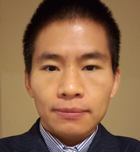 Li Wang合作论文数Developing Brain Computing Lab, University of North Carolina at Chapel Hill;Department of Radiology, School of Medicine, University of North Carolina at Chapel Hill148
 Guorong Wu合作论文数School of Medicine, University of North Carolina at Chapel Hill139
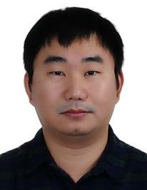 Yaozong Gao (高耀宗)合作论文数United Imaging Intelligence, Shanghai;School of Biomedical Engineering, Shanghai Tech University116
 Gilmore J H合作论文数84
 Christos Davatzikos合作论文数Department of Radiology, Perelman School of Medicine, University of Pennsylvania80
 Mingxia Liu合作论文数Department of Radiology, School of Medicine, University of North Carolina at Chapel Hill78
 Xue Zhong (薛忠)合作论文数School of Biomedical Engineering, ShanghaiTech University74
 Zhiming Cui (崔智铭)合作论文数School of Biomedical Engineering, ShanghaiTech University71
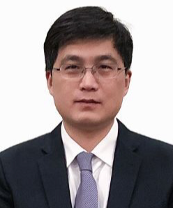 Daoqiang Zhang (张道强)合作论文数Department of Computer Science and Engineering, College of Computer Science and Technology, Nanjing University of Aeronautics and Astronautics66Kaicong Sun合作论文数62
 Tianming Liu合作论文数Department of Computer Science, School of Computing, University of Georgia61
 Chunfeng Lian (练春锋)合作论文数School of Mathematics and Statistics, Xi'an Jiaotong University60
 Geng Chen (陈耿)合作论文数School of Computer Science, Northwestern Polytechnical University57Zhengwang Wu (吴正旺)合作论文数56Dong Nie合作论文数AMap54
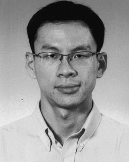 Chong-Yaw Wee合作论文数Department of Radiology and BRIC, University of North Carolina at Chapel Hill52
 Xiaohuan Cao (曹晓欢)合作论文数School of Biomedical Engineering, ShanghaiTech University47
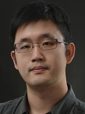 Lichi Zhang (张立箎)合作论文数School of Biomedical Engineering, Shanghai Jiao Tong University47
 Han Zhang合作论文数School of Biomedical Engineering, ShanghaiTech University47
 Islem Rekik合作论文数BASIRA Lab;Faculty of Computer and Informatics, Istanbul Technical University;Imperial College London42
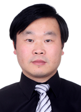 Zhongxiang Ding (丁忠祥)合作论文数Zhejiang Provincial People's Hospitai41
 Ehsan Adeli合作论文数Department of Psychiatry and Behavioral Sciences, School of Medicine, Stanford University41Kim-Han Thung合作论文数39
 Minjeong Kim合作论文数College of Business, Oregon State University38
 Heung Il Suk合作论文数Department of Brain and Cognitive Engineering, Korea University38
 Yongsheng Pan合作论文数Northwestern Polytech Univ, Sch Comp Sci & Engn, Xian 710072, Peoples R China37
 Hongtu Zhu (朱宏图)合作论文数Department of Biostatistics, Gillings School of Global Public Health, The University of North Carolina at Chapel Hill;Department of Genetics, School of Medicine, The University of North Carolina at Chapel Hill37
 Shu Liao (廖术)合作论文数School of Biomedical Engineering, ShanghaiTech University36
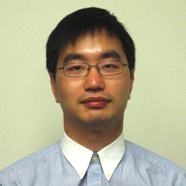 Yiqiang Zhan合作论文数Shanghai Jiao Tong University36
 Luping Zhou (周泸萍)合作论文数School of Electrical and Computer Engineering, Faculty of Engineering, University of Sydney;University of Wollongong36
 Seong-Whan Lee合作论文数Pattern Recognition & Machine Learning Lab, Department of Artificial Intelligence, College of Informatics, Korea University33
 Dajiang Zhu合作论文数Department of Computer Science and Engineering, University of Texas at Arlington32
 Sheng Wang合作论文数Department of Radiology, Perelman School of Medicine, University of Pennsylvania31
 Yong Fan合作论文数Department of Radiology, Perelman School of Medicine, University of Pennsylvania30
 Hongyu An合作论文数30
 Jun Zhang (张军)合作论文数Tencent AI Lab30
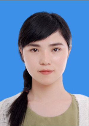 Yan Wang (王艳)合作论文数College of Computer Science, Sichuan University30
 Jun Lian合作论文数Department of Radiation Oncology, School of Medicine, University of North Carolina at Chapel Hill29Feihong Liu (刘飞鸿)合作论文数28
 Xiaofeng Zhu (朱晓峰)合作论文数School of Computer Science and Engineering, University of Electronic Science and Technology of China;School of Computer Science and Technology, Hainan University28
 James J. Xia合作论文数The Methodist Hospital Research Institute, Houston, Texas28
 Xi Jiang (蒋希)合作论文数School of Life Science and Technology, University of Electronic Science and Technology of China27
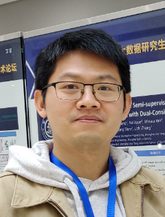 Xi Ouyang (欧阳熹)合作论文数United Imaging Intelligence27




