Palliative care is, according to the World Health Organization (WHO), “a global ethical responsibility” and crucial to person-centered healthcare, for the relief of physical and psychological symptoms and social and spiritual suffering, impacting the improvement of the quality of life not only of pediatric and adult patients dealing with life-threatening illnesses, but also of their families. Despite the growing recognition of palliative care in the hematological setting and the increase in the number of publications on the subject, the available data are still scarce and limited. The objective of this consensus is to establish recommendations for the referral of pediatric and adult hematological patients to specialized palliative care teams and the possible integration of these two specialties.
Background:Sickle cell disease (SCD) is a condition characterized by a prothrombotic state attributed to the simultaneous activation of hemostasis and innate immunity, referred to as thromboinflammation. Previous studies have demonstrated that the podoplanin (PDPN)/C-type lectin-like receptor-2 (CLEC-2) pathway is an emerging and important element of the pathogenesis of conditions in which inflammation and thrombosis coexist, but no data is available regarding its role in SCD. Objectives:To explore the PDPN/CLEC-2 pathway in SCD and correlate it with parameters of disease severity. Methods:Fifty SCD patients (35 with SS genotype; 15 with SC genotype) and 25 healthy individuals were recruited. PDPN and CLEC-2 were assessed for both soluble and surface expression on cells and cell aggregates, along with other classical parameters of hemostasis and platelet activation. An in vitro study was performed to analyze the effect of anti-PDPN antibody on the formation of monocyte-platelet aggregates. Results:Circulating levels and expression of PDPN and CLEC-2 were higher in patients with SCD, particularly in those with genotype SS. The number of CD41+CLEC+ monocytes correlated with hemoglobin, D-dimer, von Willebrand factor, and PDPN+ monocytes. In vitro, PDPN blockade reduced both monocyte-platelet aggregate formation and platelet activation. Finally, patients with a history of vaso-occlusive crises presented a trend toward increased PDPN expression in monocytes (P = .06). Conclusion:Our findings suggest that the PDPN/CLEC-2 pathway may play an important role in the pathogenesis of thromboinflammation in SCD, especially in patients with the SS genotype.
BackgroundMonocytes can interact with erythroid cells and contribute to macrophage pools under anemic stress, thereby supporting the erythropoietic niche. In sickle cell anemia (SCA), extensive hemolysis and ineffective erythropoiesis may expose circulating monocytes to abnormal red blood cells (RBCs), potentially altering their phenotype and function to accommodate the higher erythropoietic response and iron demand.Methods and resultsWe characterized circulating monocytes from SCA patients at steady state using flow cytometry and in vitro phagocytosis assays. Intracellular RBC material was detected in all circulating monocyte subsets from SCA patients, indicating active erythrophagocytosis. Mechanistically, SCA monocytes displayed upregulated VCAM-1 and reduced SIRP-α expression, favoring RBC binding and internalization even when CD47 expression on RBCs was preserved. Following RBC engulfment, monocytes upregulated heme oxygenase-1 (HO-1) and ferroportin (FPN), consistent with enhanced heme degradation and iron export, and expressed higher levels of CD206, suggesting a regulatory phenotype. In vitro assays confirmed that both sickle RBCs and SCA monocytes synergistically promoted erythrophagocytosis.ConclusionCirculating monocytes from SCA patients undergo phenotypic and functional reprogramming upon interaction with sickled RBCs, acquiring features reminiscent of erythroblastic island macrophages. These findings highlight a previously underappreciated role for monocytes in RBC clearance, heme metabolism, and iron recycling in SCA, with potential implications for inflammation and disease progression.
There is no physiological mechanism for the excretion of iron in humans, and excess iron may lead to severe tissue damage if not adequately treated. Iron overload can be caused by genetic factors (hemochromatosis) or acquired conditions (e.g., ineffective erythropoiesis, transfusions, iatrogenic iron treatment, viral hepatitis, alcohol intake, severe liver disease, metabolic dysfunction), and, in many cases, by a conjunction of these factors. Historically, guidelines for the genetic investigation of patients with iron overload have been based on data obtained from Caucasian individuals in Europe and North America. However, due to the genetic heterogeneity of iron overload gene mutations worldwide, these recommendations might not be applicable to other ethnic groups. This study analyzed previously published genetic data obtained from Brazilian patients with iron overload and found a relevant but small prevalence of HFE C282Y/C282Y patients when compared to European populations, while mutations of the TFR2, SCL40A1, HJV, HAMP, BMP6 and SLC11A1 genes seem to be important. This study proposes an adapted algorithm for the investigation and management of iron overload in Brazil.
Priapism is a frequent and debilitating complication in patients with sickle cell disease (SCD), characterized by recurrent ischemic episodes that can culminate in fibrosis of the erectile tissue and irreversible erectile dysfunction. Despite significant advancements in the management of acute episodes, current therapies remain largely ineffective in preventing recurrences, emphasizing the need for novel strategies that target the underlying pathophysiology. This narrative review describes the mechanistic links between oxidative stress and nitric oxide (NO) dysregulation in the pathogenesis of SCD-associated priapism, with a particular focus on the NO–cyclic guanosine monophosphate (cGMP)–phosphodiesterase type 5 (PDE5) signaling axis. We analyze preclinical evidence supporting resveratrol, a natural polyphenolic compound, as well as its NO-donor hybrid derivatives, as emerging therapeutic candidates. Additionally, we discuss the potential of combining resveratrol with current treatment approaches, and address the translational challenges that must be overcome to move from preclinical data to clinical application. Taken together, the evidence presented in this review supports resveratrol-based therapies as a promising approach for oxidative-stress-driven priapism in SCD and delineates critical perspectives for their further investigation.
Continuous vaso-occlusive and inflammatory processes cause extensive end-organ damage in adults with sickle cell disease (SCD), and there is little evidence that longterm hydroxyurea therapy prevents this. In initial trials, P-selectin blockade with crizanlizumab reduced SCD vaso-occlusive crisis frequency, and interleukin (IL)-1β inhibition in SCD patients, using canakinumab, lowered inflammatory markers. We used murine SCD models to examine the effects of acute and chronic blockade of Pselectin and of IL-1β on vaso-occlusive events, their inflammatory profile and organ health. Both approaches improved impaired cutaneous microvascular perfusion in SCD mice by reducing TNF-α-induced vaso-occlusion. Acute P-selectin blockade markedly reduced TNF-α-induced neutrophil-platelet aggregate formation in SCD mice, and decreased leukocyte-rolling movements in the microvasculature, while acute IL-1β inhibition attenuated microvascular leukocyte adhesion. Six weeks of IL-1β-blocking immunotherapy improved the inflammatory profile of SCD mice, considerably reduced hepatic fibrosis and provided some relief from lung injury. In contrast, although Pselectin blockade reduced glomerular congestion, no significant benefit to overall organ pathology was observed. Unexpectedly, while combining the two immunotherapies reduced microvascular occlusion, their prolonged use caused acute liver injury. Notably, inhibition of IL-1β, but not of P-selectin, remarkably decreased hemosiderosis, in association with reduced tissue macrophage infiltration and the correction of biomarkers of dysregulated iron turnover. Our findings suggest that the attenuation of inflammation, as well as of vaso-occlusive processes, may be crucial for mitigating organ damage in SCD. Future trials should explore the ability of cytokine blockade to prevent multiorgan damage in patients with SCD, beyond evaluating vaso-occlusive crisis frequency.
The pathogenesis of priapism in sickle cell disease (SCD) is closely linked to oxidative stress and reduced bioavailability of nitric oxide (NO) in penile tissue. Resveratrol, a potent natural antioxidant, has demonstrated protective effects in various vascular disorders. To evaluate the therapeutic effects of resveratrol on priapism, oxidative stress markers, and NO-cGMP signaling in the penile tissue of transgenic SCD mice. Male wild-type (C57BL/6) and transgenic SCD mice were treated with resveratrol (100 mg/kg/day, gavage) or vehicle for 2 weeks. Functional studies were conducted on CC strips mounted in organ baths to assess relaxation responses to acetylcholine (ACh), sodium nitroprusside (SNP), and nitrergic stimulation (electrical field stimulation, EFS). The oxidative stress markers (NOX-2, 4-HNE, and 3-NT), cGMP levels, and the mRNA expression of endothelial nitric oxide synthase (eNOS) and phosphodiesterase type 5 (PDE5) were evaluated. Resveratrol treatment decreased exaggerated ACh-, SNP-, and EFS-induced relaxation responses in SCD mice. It also reduced oxidative stress markers (NOX-2, 4-HNE, and 3-NT) and normalized eNOS and PDE5 mRNA expression in the CC of SCD mice. Additionally, cGMP levels in the CC were significantly increased by resveratrol treatment. These effects were specific to SCD mice and not observed in wild-type mice. In conclusion, resveratrol reduces oxidative stress and restores NO-cGMP signaling in the penile tissue, reducing the exaggerated cavernosal relaxation characteristic of priapism in SCD. These findings highlight resveratrol as a promising therapeutic candidate for managing priapism in patients with SCD.
Sickle cell disease is a condition characterized by vaso-occlusive episodes and sustained hemolysis, leading to a chronic inflammatory state. Several studies have shown that the release of heme to the extracellular space due to hemolysis contributes to the inflammatory cascade observed in these patients. Hemopexin (HPX), the molecule responsible for removing excess heme from the circulation, is depleted in these patients. We have previously demonstrated that the IV infusion of an adeno-associated virus-based gene transfer vector was capable of inducing the transgenic expression of HPX in a dose-dependent manner in C57Bl6 mice. Here, we explored the effect of this vector in a mouse model of sickle cell anemia. Townes mice were transduced with 2 × 10¹³ vector genomes per kilogram and followed up for up to 48 weeks. HPX expression was confirmed in liver samples by both western blot and quantitative polymerase chain reaction (HPX), but gene transfer did not restore circulating levels of HPX in Townes mice, as shown in models without hemolysis. Indirect surrogate markers of a beneficial effect of delivering HPX were observed, including increased expression of heme-oxygenase 1 upon heme overload, greater weight gain on the long-term follow-up, and a significant decrease in tumor necrosis factor α levels. No signs of liver or hematological toxicity were observed. Our results demonstrate the potential and challenges of therapeutic strategies based on the long-term delivery of HPX in an animal model of sickle cell anemia.
Intravascular hemolysis, a hallmark of sickle cell disease (SCD), leads to elevated plasma heme levels. Although heme is essential for physiological processes, its excess can be deleterious. Heme oxygenase (HO) degrades heme into carbon monoxide (CO), which activates the soluble guanylate cyclase (sGC)–cyclic guanosine monophosphate (cGMP) signaling cascade and can modulate smooth muscle tone. However, the direct effects of heme on bladder function remain unknown. This study investigated whether heme regulates detrusor smooth muscle contractility through the HO–CO–sGC–cGMP pathway. Detrusor strips from C57BL/6 mice were mounted on a myograph for functional analysis. Heme induced a significant, concentration-dependent relaxation of detrusor smooth muscle compared with vehicle-treated tissues. To elucidate the underlying mechanism, tissues were pre-incubated with the sGC inhibitor ODQ (10 µM) or the HO inhibitor 1J (100 µM) before heme exposure. Both inhibitors markedly attenuated heme-induced relaxation, reducing the maximal relaxation response. Moreover, pre-incubation with heme (100 µM) significantly decreased the maximal contractile responses (Emax) to carbachol, KCl, and electrical field stimulation (EFS), effects that were abolished by ODQ or 1J. In parallel, biochemical assays showed that heme markedly increased cGMP levels in detrusor tissue, an effect prevented by both inhibitors, confirming the role of the HO–CO–sGC–cGMP signaling cascade in this response. These findings demonstrate that heme modulates bladder contractility by activating the HO–CO–sGC–cGMP pathway, promoting detrusor relaxation. This mechanism suggests that excessive circulating heme, as occurs in hemolytic disorders such as SCD, may contribute to detrusor hypocontractility and voiding dysfunctions, identifying this pathway as a potential therapeutic target.
Bladder dysfunction, particularly overactive bladder (OAB), is increasingly recognized as a clinical concern in patients with sickle cell disease (SCD), yet its pathophysiological mechanisms remain poorly understood. This study investigated the relationship between oxidative stress, inflammation, and bladder dysfunction in the Townes transgenic SCD mouse model. Cystometric analysis revealed that SCD mice exhibit an OAB phenotype, characterized by increased frequencies of voiding and non-voiding contractions and reduced bladder compliance. In vitro functional assays demonstrated detrusor hypocontractility in SCD mice, associated with a significant reduction in carbachol- and EFS-induced contractions and downregulation of muscarinic M3 receptor expression. Purinergic signaling and calcium-dependent contractility remained preserved. Molecular analyses showed increased mRNA expression of NOX-2 and IL-1β, and elevated protein levels of 3-nitrotyrosine and myeloperoxidase (MPO) activity, indicating redox imbalance and chronic inflammation in bladder tissue. Together, these changes suggest that oxidative and nitrosative stress, combined with inflammation, contribute to bladder remodeling and dysfunction in SCD. This is the first study to characterize bladder alterations in Townes SCD mice, establishing this model as a valuable tool for investigating lower urinary tract complications in SCD. Our findings provide mechanistic insight into the genitourinary manifestations of SCD and identify redox and inflammatory pathways as potential therapeutic targets for bladder dysfunction in affected individuals.
Introduction: Sickle cell disease (SCD) is an inherited hemoglobinopathy characterized by a complex physiopathology. This pathophysiology, which includes hemolysis, inflammation and vaso-occlusion, contributes to a number of different clinical abnormalities. The endothelial damage and activation that is promoted by a cascade of events following red cell sickling is probably directly involved in many of the complications observed in SCD patients, such as acute chest syndrome (ACS), a complication that appears also to be influenced by the hypercoagulability state. Extracellular vesicles (EVs) are small structures released by cells that express and carry several molecules and proteins that are actively involved in cellular communication and have an important role in various diseases. Although several studies suggest the participation of EVs in SCD events, their role in SCD is largely unclear. The possible role of EVs in the pathogenesis of ACS is unknown and we aim, here, to further understand whether EVs could reflect the endothelial damage and coagulation activation in patients who have developed ACS. Aim: Evaluate the profile of endothelial derived EVs and EVs expressing tissue factor (TF) in patients with SCD who have not experienced episodes of ACS during the previous 12 months and those who presented at least one episode of ACS in the 12 months prior to participating in the study. Methods: Patients with at least one episode of ACS in the 12 months before sample collection and patients without ACS were included (n=08 and 70, respectively).Samples were collected and processed as previously described (Olatunya et al., 2019). To quantify and characterize endothelial EVs, we used CD146pos and CD45neg identification. To identify the expression of TF we used the CD142 positivity. The Cytoflex (Beckman Coulter) was used to quantify EVs by flow cytometry and analysis was performed using CytoExpert software. Statistical tests were performed according to the non-parametric distribution for comparative analyzes (Kruskal wallis, Mann Whitney). Values of p<0.05 were considered statistically significant. Results and Discussion: We found higher numbers of EVs expressing the CD142 marker (p=0.019) and of endothelial cell-derived EVs expressing the CD142 marker (p=0.020) in patients with previous ACS, compared to patients that had not experienced ACS during the last 12 months before sample collection. Some functional and observational studies have shown the possible participation of EVs in ACS. A previous study (Jutant et al., 2021) showed that patients present higher plasma levels of EVs from red blood cells, platelets, and pro-coagulant EVs during an ACS episode. In this study, although the sample collection was not carried out at the moment of the event, the EV profile may reflect endothelial activation and the hypercoagulability state in these patients, as augmented levels of endothelial derived-EVs expressing TF were observed in these patients. No difference was observed for endothelial cell-derived EVs with no CD142 marker (p>0.05). As TF is an activator of the coagulation process, we speculate that this could reflect a hypercoagulability state in patients who have had ACS. Moreover, we previously demonstrated (Souza CM, ASH 2023) that EVs from patients with sickle cell anemia in steady state carried proteins that stimulate coagulation. Further analysis should be conducted with samples collected at the time of the complication to better understand the participation of EVs in ACS and how they can be used as a biomarker for ACS. In summary, our data suggest that EVs may provide important insights into the clinical condition of patients with SCD, as they may reflect patterns related to the key events involved in ACS.
Background: Prognosis is the challenging process of making predictions about future health outcomes, generally estimating survival time, in order to help defining the most appropriate therapeutic intervention for each patient. In patients diagnosed with solid tumors undergoing Palliative Care (PC), several prognostic tools have been validated with the aim of predicting prognosis, and are widely used by oncologists and palliativists in order to establish the more proportional therapeutic plan in each phase of the disease. However, in patients with hematological malignancies (HM) at the end of life, there is still no robust validation of these scores, and estimating the prognosis in this context is particularly challenging, resulting from the great heterogeneity of HM natural history, which range from very aggressive and rapidly evolving to pathologies with indolent behavior. Moreover, HM patients experience fluctuations in the trajectory of their disease, leading to delays in identifying and communicating the end-of-life phase, and therefore, in the transition from curative care to exclusive PC. It is known that patients with HM receive more aggressive care at the end of life and are less likely to be referred to PC services when compared to individuals with solid tumors, which may be mainly a consequence from the difficulty of estimating prognosis in HM patients. Aims: To evaluate the capacity of prognostic, performance and symptom assessment scores commonly used for solid tumors patients under PC on predicting prognosis in patients with HM. Methods: Retrospective data were collected from the electronic medical records of patients diagnosed with myeloma, acute leukemia and lymphoma followed at the PC outpatient clinics of the Hematology and Hemotherapy Center of the University of Campinas (Unicamp), Brazil, from 2018 to 2023. Data were evaluated at the date of the first PC consultation. CRP and albumin levels, and ECOG, PPS, CCI, Age-CCI, mGPS and ESAS scores were correlated with overall survival (OS). Statistical analysis was performed on the general population and on each subpopulation according to the underlying pathology. The significance level adopted for the study was 5%. This research was approved by the Institutional Review Board; written informed consent was obtained from all subjects. Results: A total of 181 patients were included in the study (70 multiple myeloma, 49 acute leukemia and 62 lymphoma patients). PPS scores were positively correlated with OS (HR 0.975; 95% CI 0.965-0.986; p<0.0001). Individuals with PPS scores between 80-100% had a survival rate 3.5 times higher than those with PPS<50% (HR 3.501; 95% CI 1.977- 6.199; p=0.0001) and approximately 1.7 times higher than those with PPS between 50-70% (HR 1.695; 95% CI 1.977- 6.199; p=0.0363). Worse performance status assessed by ECOG levels were correlated with OS (HR 1.383; 95% CI 1.154-1.657; p=0.0005). Finally, mGPS scores (HR 2.446; 95% CI 1.103-5.422; p=0.0277) and albumin levels (p<0.0001; HR 0.391; 95% CI 0.289-0.528) also predicted survival in these patients. CCI, Age-CCI, number of comorbidities, CRP and ESAS scores did not predict survival in this cohort. When analyzing separately the subpopulations with myeloma, acute leukemias and lymphomas, PPS scores (HR 0.963 [0.942-0.985], HR 0.974 [0.954-0.995] and HR 0.979 [0.964-0.993], respectively, all p<0.05) and albumin levels (HR 0.272 [0.164-0.450], HR 0.497 [0.255-0.97] and HR 0.541 [0.339-0.864], respectively, all p<0.05) remained statistically related to OS. In patients with myeloma and lymphomas, ECOG was also related to OS (HR 1.816; 95% CI 1.211-2.724; p=0.0039, and HR 1.323; 95% CI 1.043-1.678; p=0.0213, respectively). CCI scores correlated with survival only in the myeloma subgroup (HR 1.306; 95% CI 1.064-1.602; p=0.0105). Conclusions: PPS and albumin levels predicted survival in our cohort of HM patients undergoing PC. Also, ECOG and CCI predicted survival in individuals with multiple myeloma, and ECOG in lymphoma patients. These results indicate that these prognostic assessment tools could be helpful for OS prediction in patients with HM undergoing PC. Also, the routine application of prognostic tools in patients with HM could help hematology-oncologists to find the better time-points for referral to the PC team. Given the limitations of the study design, these results need to be validated in further studies.
Introduction and aims: Chronic pain is a frequent and debilitating complication of Sickle Cell Disease (SCD). The aim of this study was to evaluate the impact of chronic pain on the mental health and quality of life of patients, in addition to seeking to identify molecules associated with its occurrence, in prospecting for new therapeutic targets. Methods: Adult patients with SCD followed at the UNICAMP Blood Center completed PROMIS (Patient-Reported Outcomes Measurement Information System) questionnaires validated for the Portuguese language, in the domains of depression, anxiety and pain interference in daily life. The results were reported as T scores in relation to a reference population, and used to search for correlations with clinical and laboratory data. In parallel, plasma samples from the same patients and healthy controls were subjected to metabolomic profiling analysis by untargeted Ultra Performance Liquid Chromatography-High Resolution Mass Spectrometry (UHPLC-HRMS) in the search for possible molecular markers differentiating patients with or without chronic pain. Results: Forty-three patients were included, with a median age of 43 years (15-78), of whom 27 (63%) were female, 25 had HbSS, 08 had HbSC, 7 had Sbeta-thalassemia and 3 had HbSD. Comparing individuals according to the presence or absence of chronic pain: individuals with chronic pain had a higher median age (39.3 vs. 48.7, p = 0.026), higher Hgb levels (9.9 vs. 8.7 g/dL, p=0.038), lower total leukocyte counts (6.35 vs. 7.68 x 10³/mm3, p=0.04), though no significant differences in the PROMIS test results. However, the occurrence of at least one vasoocclusive crisis (VOC) in the previous 6 months or the diagnosis of avascular bone necrosis had statistically significant relationships with worse results in the 3 PROMIS domains tested. Furthermore, the lower the patients' income, the worse the impact of pain on daily life (r=-0.46, p<0.001). Regarding the UHPLC-MS analyses, 107 molecules with significant differences in expression were identified between healthy controls, patients with chronic pain or without chronic pain. Among them, a higher expression of molecules of the endocannabinoid system (2-Arachidonoylglycerol and 2-Eicosapentaenoyl-glycerol) was observed in patients without chronic pain, and a higher expression of lysoplasmenylcholine and adrenic acid in the group affected by chronic pain. Conclusions: In this cohort of Brazilian patients, the occurrence of VOCs, the diagnosis of bone necrosis and low income were demonstrated to be related to worse rates of anxiety, depression and pain interference in daily life. The increased expression of molecules of the endocannabinoid system may indicate a protective factor in patients who are not affected by chronic pain. In patients with chronic pain, the increase in adrenic acid may be associated with a higher occurrence of ferroptosis, a mechanism previously described as being involved in the development of neuropathic pain; and lysoplasmenylcholine is released in response to hypoxia and thrombin generation, and increases cellular adhesion to the endothelium, revealing possible microvascular mechanisms associated with a higher occurrence of pain. This is the first study using untargeted UHPLC-HRMS for exploratory analysis of metabolites involved in chronic pain in Sickle Cell Disease, identifying potential therapeutic targets to be explored in future clinical trials.
Patients with sickle cell disease (SCD) display priapism, a prolonged penile erection in the absence of sexual arousal. The current pharmacological treatments for SCD-associated priapism are limited and focused on acute interventions rather than prevention. Thus, there is an urgent need for new drug targets and preventive pharmacological therapies for this condition. This review focuses on the molecular mechanisms linked to the dysfunction of the NO-cyclic guanosine monophosphate (cGMP)-phosphodiesterase type 5 (PDE5) pathway implicated in SCD-associated priapism. In murine models of SCD, reduced nitric oxide (NO)-cGMP bioavailability in the corpus cavernosum is associated with elevated plasma hemoglobin levels, increased reactive oxygen species levels that inactive NO, and testosterone deficiency that leads to endothelial nitric oxide synthase downregulation. We discuss the consequences of the reduced cGMP-dependent PDE5 activity in response to these molecular changes, highlighting it as the primary pathophysiological mechanism leading to excessive corpus cavernosum relaxation, culminating in priapism. We also further discuss the impact of intravascular hemolysis on therapeutic approaches, present current pharmacological strategies targeting the NO-cGMP-PDE5 pathway in the penis, and identify potential pharmacological targets for future priapism therapies. In men with SCD and priapism, PDE5 inhibitor therapy and testosterone replacement have shown promising results. Recent preclinical research reported the beneficial effect of treatment with haptoglobin and NO donors. SIGNIFICANCE STATEMENT: This review discusses the molecular changes that reduce NO-cGMP bioavailability in the penis in SCD and highlights pharmacological targets and therapeutic strategies for the treatment of priapism, including PDE5 inhibitors, hormonal modulators, NO donors, hydroxyurea, soluble guanylate cyclase stimulators, haptoglobin, hemopexin, and antioxidants.
Introduction: Vaso-occlusive crises (VOC) are the most common complication of sickle cell disease (SCD) and can be elicited by different physiological events, including infections, dehydration, cold temperature and psychological stress. Currently, there exists no specific treatment to resolve VOC, or limit its intensity, other than pain management and rehydration. Agents targeting platelet (plt) aggregation, hemoglobin polymerization and selectin molecule activity can prevent vaso-occlusion (VO) in standardized models of VOC in SCD mice. However, all have, so far, failed to bring relief to patients in real-life conditions, where VOC triggers are polymorphic. Hypothesizing that VOCs may differ in their molecular mechanisms, depending on their triggers, we compared the effects of physiological triggering events and inflammatory signaling molecules on in vivo and in vitro models of SCD VO. Methods: Townes HbSS mice (20 wks) were submitted to molecular triggers, consisting of tumor necrosis factor (TNF)α, hemin, lipopolysaccharide (LPS), or interferon (IFN)α. Other mice were submitted to hypoxia/reoxygenation, cold temperature or dehydration. Cutaneous VO was then evaluated by laser Doppler perfusion monitoring (LDPM), cremaster microvascular leukocyte recruitment was observed by intravital microscopy, and neutrophil-plt aggregate (NPA) formation in peripheral blood was quantified by flow cytometry. For in vitro protocols, peripheral blood from sickle cell anemia (SCA) patients was incubated with TNFα, LPS, IFNα or RBC lysis products. We then investigated blood cell adhesion to endothelial cells using microfluidic adhesion protocols, the architecture and composition of the VO aggregate using confocal imaging, and the phenotype of neutrophils and their aggregation with RBC/ plts using flow cytometry. Results: In the SCD mouse model, LDPM indicated that all of the molecular and physiological triggers promoted VO, significantly decreasing both blood flow velocity and perfusion in the cutaneous microcirculation. The physiological triggers all augmented microvascular leukocyte rolling and moderately increased adhesion, while hypoxia/ reperfusion and dehydration, but not cold, induced circulating NPA formation. Notably, the molecular triggers differed more distinctly in their mechanistic responses. In SCD mice, the bacterial component, LPS, caused extensive microvascular leukocyte adhesion without NPA formation, while TNFα cytokine amplified leukocyte rolling, adhesion and caused significant NPA formation. Consistently, stimulation of blood from SCA patients with LPS led to global activation of neutrophils, with increased expressions of the MAC-1 integrin (CD18/CD11b), CD66b, CD64 and CD33, and RBC recruitment at the heart of the VO aggregate, indicating enhanced RBC adhesion to other blood cells, rather than to endothelial cells. TNFα stimulation of SCA patient blood, however, caused a specific increase in neutrophil MAC-1 expression, but not of other adhesion molecules, and an overrepresentation of the plts inside the VO cluster. In contrast, the major effect of hemin in SCD mice was an acceleration of leukocyte rolling, but without significant leukocyte adhesion. Once again, this was consistent with the lack of overexpression of adhesion or activation molecules on neutrophils after the addition of RBC lysis content to SCA whole blood. Finally, in SCD mice, the viral response cytokine, IFNα, induced leukocyte rolling, in association with moderate adhesion, but with a very significant increase in NPA formation. Significantly, IFNα also drastically increased the adhesion of plts from SCA blood to endothelial cell layers during microfluidic flow assays, together with increased RBC adhesion both to endothelial cells and to other cell types, but in the absence neutrophil phenotype alterations. Conclusions: Our study is the first to demonstrate that VO is not a uniform phenomenon, and probably depends on the specific trigger of the crisis. Molecular triggers amplified inflammatory responses more than physiological events, revealing specific VOC mechanistics. For instance, phenotypic leukocyte changes were crucial to bacterial component-induced VO, while plt activity was central to the IFNα response. Findings represent an important step towards defining targeted strategies for personalized VOC reversal in patients, according to the triggering event.
Introduction: Intravascular hemolysis (IVH) results in the release of damage-associated molecular patterns (DAMPs) into the circulation, particularly hemoglobin (Hb) and heme, which can trigger NLRP3 inflammasome activation and a sterile inflammatory response. Complications of hemolytic diseases include thrombosis, inflammation, and organ damage. In sickle cell disease (SCD), IVH can lead to vaso-occlusive crises and ischemic complications such as cutaneous leg ulcers, acute chest syndrome, and multiorgan infarction. Aim: To investigate the in vivo effects of inflammasome formation on microvascular function and its role in the pathophysiology of hemolytic conditions. Methods: We utilized two murine models to simulate intravascular hemolysis: an acute model where C57BL6 mice received osmotic stress (150 μl sterile water, i.v. -HEM); and a chronic model induced by repeated low doses of phenylhydrazine (10 mg/kg, i.p. -CHEM). Control mice received saline under similar conditions. Microvascular dysfunction was assessed by laser Doppler fluxometry (LDF) in the skin of the pelvis and by intravital microscopy of the cremaster muscle. Inflammasome activation was evaluated via flow cytometry measurement of caspase-1 (casp-1) activity, measurement of interleukin (IL)-1β and IL-18 release by ELISA, and NLRP3 protein expression in the liver using western blot. Results and discussion: Our findings show that acute hemolysis immediately increased plasma cell-free Hb and heme levels in HEM mice, along with decreased Hb scavenger haptoglobin (Hp). CHEM mice also displayed a significant depletion of hemopexin (Hx), indicating the persistent removal of this heme-neutralizing protein. These results confirm that these mouse models effectively mimic acute and chronic hemolytic stress. Acute IVH resulted in microvascular dysfunction characterized by a vaso-occlusive-like process, as demonstrated by an increase in rolling, adherent, and extravasated leukocytes. The blood flow velocity of HEM mice was reduced in the microcirculation, resulting in impaired tissue blood perfusion. These findings were accompanied by increased casp-1 activity in peripheral monocytes and elevated levels of IL-1β and IL-18, all hallmarks of inflammasome formation, suggesting that inflammasome assembly during hemolytic stress contributes to tissue microcirculation dysfunction. While acute hemolysis caused microvascular defects and impaired blood perfusion in mice, chronic hemolysis significantly affected the liver, leading to an increase in the macrophage population, which displayed elevated active casp-1 and increased NLRP3 protein expression. Next, we investigated whether the NLRP3 inflammasome contributes to hemolysis-induced microvascular pathology. As expected, casp-1 activation was reduced by the NLRP3 inhibitor, MCC950, in both the neutrophils and monocytes of HEM mice. Moreover, NLRP3 inflammasome inhibition reduced leukocyte recruitment to the venule walls of HEM mice, indicating an improvement in hemolysis-induced microvascular dysfunction. This was further corroborated by LDF, which showed improved blood perfusion in the skin of MCC950-treated HEM mice. Conclusion: These results demonstrate that NLRP3 inflammasome assembly, in response to hemolysis, compromises microvascular function by modulating leukocyte adhesion to the endothelium and impairing blood flow to tissues, which could contribute to clinical complications in hemolytic disorders such as SCD.
 Sara Teresinha Olalla Saad合作论文数Departamento de Clinica Medica, Faculdade de Ciencias Medicas, Universidade Estadual de Campinas215
 Dulcinéia Martins de Albuquerque合作论文数Faculdade de Ciências Médicas, Universidade Estadual de Campinas154
 Nicola Conran合作论文数Hemotherapy Center;State University of Campinas;Hemotherapy Center, State University of Campinas145
 Sto Saad合作论文数130Fabiola Traina合作论文数101
 Carolina Lanaro合作论文数Faculty of Medical Sciences, UNICAMP Hemocenter82
 Kleber Yotsumoto Fertrin合作论文数Universidade de Campinas - UNICAMP73
 Irene Gyongyver Heidemarie Lorand Metze合作论文数Departamento de Clínica Médica, Faculdade de Ciencias Medicas, Universidade Estadual de Campinas73Kimura Elza合作论文数71
 Aderson Araujo合作论文数Fundação de Hematologia e Hemoterapia de Pernambuco63
 Marcos André Cavalcanti Bezerra合作论文数Departamento de Biofisica e Radiobiologia, Universidade Federal de Pernambuco;Núcleo de Hematologia Clínica e Laboratorial62Carla Fernanda Franco Penteado合作论文数Faculdade de Ciências Médicas, Universidade Estadual de Campinas62Maria De Fatima Sonati合作论文数Department of Clinical Pathology, Universidade Estadual de Campinas62MF Sonati合作论文数58
 Valder R. Arruda合作论文数Department of Pediatrics, Perelman School of Medicine, University of Pennsylvania;Children's Hospital Research Institute, The Children's Hospital of Philadelphia56
 Mônica Barbosa De Melo合作论文数Haematology and Haemotherapy Centre, UNICAMP Cidade Universitária "Zeferino Vaz"53
 João Agostinho Machado-Neto合作论文数Department of Pharmacology, Institute of Biomedical Sciences, University of São Paulo52
 Flavia Costa Leonardo合作论文数43S. T. O. Saad合作论文数38de Melo Campos Paula合作论文数38A. S. S. Duarte合作论文数Faculdade de Ciências Médicas, Universidade Estadual de Campinas;Biochemistry and Molecular Biology Laboratory, Blood Center, State University of Campinas35
 Lilian Maria de Castilho合作论文数Centro de Hematologia e Hemoterapia, State University of Campinas33
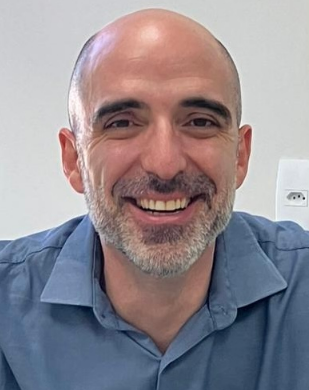 Erich Vinicius De Paula合作论文数Department of Clinical Medicine, School of Medical Sciences, University of Campinas;Faculdade de Ciências Médicas, Universidade Estadual de Campinas32
 Fabio Henrique da Silva合作论文数Universidade São Francisco;Universidade Estadual de Campinas;FCM - UNICAMP31
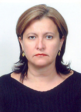 Joyce Maria Annichino Bizzacchi合作论文数Departamento De Clinica Medica, Faculdade De Ciencias Medicas, Universidade Estadual de Campinas31
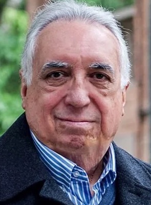 Carmino Antonio de Souza合作论文数Centro de Inovação Teranóstica em Câncer;Centro de Hematologia e Hemoterapia, Universidade Estadual de Campinas31Favaro Patricia合作论文数Department of Biological Sciences, Federal University of Sao Paulo29
 Daniela Sanchez Bassères合作论文数Laboratório de Pesquisas Integradas em Sinalização Celular e Câncer, Departamento de Bioquímica, Instituto de Química, Universidade de São Paulo28
 Anderson Ferreira da Cunha合作论文数Departamento de Genética e Evolução, Universidade Federal de São Carlos;Centro de Ciências Biológicas e da Saúde, Universidade Federal de São Carlos26
 Margareth Castro Ozelo合作论文数Hematology and Hemotherapy Center, State University of Campinas25C. Lanaro合作论文数24
 Manoela M Ortega合作论文数24
 Venina Marcela合作论文数Triangulo Mineiro Federal University24
 Camila B. Almeida合作论文数Hematology and Hemotherapy Center - Instituto Nacional de Ciência e Tecnologia do Sangue (INCTS),, University of Campinas - UNICAMP24Susan Elisabeth Jorge合作论文数24S. Saad合作论文数23
 Antonio R Lucena-Araujo合作论文数22Ana Flávia Brugnerotto合作论文数21Igor De Farias Domingos合作论文数Department of Genetics, Federal University of Pernambuco21
 Gotardo Érica Martins Ferreira合作论文数Clinical Pharmacology and Gastroenterology Unit, São Francisco University Medical School21
 Maria Fernanda Rios Manrique Rios合作论文数and the Science and Technology Laboratory, New York Blood Center21Csp Lima合作论文数20
 Calmasini Fabiano B合作论文数Faculty of Medical Sciences, State University of Campinas20Carmen S.P. Lima合作论文数19
 Vanessa Tonin Garrido合作论文数19
 Mariana Lazarini合作论文数Instituto Nacional de Ciência e Tecnologia do Sangue, University of Campinas19C BOTTURA合作论文数18Pamela L. Brito合作论文数18
 Andre Fattori合作论文数Faculty of Medical Sciences, State University of Campinas18




