We investigate how vibrotactile wrist feedback can enhance spatial guidance for handheld tool movement in optical see-through augmented reality (AR). While AR overlays are widely used to support surgical tasks, visual occlusion, lighting conditions, and interface ambiguity can compromise precision and confidence. To address these challenges, we designed a multimodal system combining AR visuals with a custom wrist-worn haptic device delivering directional and state-based cues. A formative study with experienced surgeons and residents identified key tool maneuvers and preferences for reference mappings, guiding our cue design. In a cue identification experiment (N = 21), participants accurately recognized five vibration patterns under visual load, with higher recognition for full-actuator states than spatial direction cues. In a guidance task (N = 27), participants using both AR and haptics achieved significantly higher spatial precision (5.8 mm) and usability (SUS = 88.1) than those using either modality alone, albeit with modest increases in task time. Participants reported that haptic cues provided reassuring confirmation and reduced cognitive effort during alignment. Our results highlight the promise of integrating wrist-based haptics into AR systems for high-precision, visually complex tasks such as surgical guidance. We discuss design implications for multimodal interfaces supporting confident, efficient tool manipulation.
Cancers evade anti-tumor immunity by the infiltration of immunosuppressive myeloid cells such as macrophages and neutrophils into the tumor microenvironment (TME). The limited efficacy of current immunotherapy is attributed to the presence of these immunosuppressive cells. Here, we describe a novel immunotherapy approach, using a single-cycle alphavirus replicon particle (VRP) that carries a self-amplifying RNA encoding interleukin-12 (IL-12). Leveraging the natural tropism of alphavirus to myeloid cells, single cell transcriptomic analyses of mouse tumor tissues demonstrated that intratumoral (i.t.) injection of the vector effectively reprogrammed tumor-associated macrophages and neutrophils toward a pro-inflammatory, anti-tumor phenotype via interferon signaling, further enhanced by IL-12 expression. The treatment also induced cytotoxic NK and T cell responses, resulting in a significant reduction of tumor growth and effectively preventing nodal and distant metastases in solid tumor mouse models. By targeting tumor-supporting myeloid cells to transform the TME into an immune-active state, our myeloid modulation approach has the potential to enhance the efficacy of current immunotherapies. ### Competing Interest Statement Declaration of interests M.I, J.S., F.B., K.I., F.T., K.T., D.L., K.M. are employees of VLP Therapeutics, Inc.; W.A. is a board member of, an employee of, and holds stocks in VLP Therapeutics, Inc. and is a management board member of VLP Therapeutics Japan, Inc.; J.F.S. are employees of and hold stocks in VLP Therapeutics, Inc.; and W.A. and J.F.S. are inventors on a related patent. J.B.S. is scientific co-founder and member of the scientific advisory board of Indapta Therapeutics.
BACKGROUND:Psychosocial well-being remains a significant unmet need among head and neck cancer (HNC) survivors in the United States, with limited understanding of the role of faith-based communities and religious coping in addressing social isolation. METHODS:HNC patients not actively undergoing treatment were recruited to complete validated surveys assessing physical and psychological symptoms, religious participation, and coping. RESULTS:52 patients completed surveys, of which most had early-stage cancer (61.5%). The majority of participants reported clinically significant pain (52.0%) or fatigue (53.1%). Nearly half of participants reported regular religious or spiritual involvement, with 60.5% attending services at least a few times per year and 48.9% engaging in private practices like prayer or meditation weekly. Negative religious coping was not significantly associated with distress (odds ratio: 1.17, 95% confidence interval: 1.00-1.37, p = 0.06), though this association approached significance. CONCLUSIONS:These findings suggest a need for further research on integrating spiritual support into HNC survivorship care.
Importance:Oral tongue is the most common subsite of oral cavity cancers, and incidence is increasing. Tongue resection is associated with morbidity, affecting speech, swallowing, and quality of life. Objective:To assess the association of tumor volume and extent of tongue resection with functional outcomes. Design, Setting, and Participants:Patients with squamous cell carcinoma who underwent surgical resection of the oral tongue at a tertiary-level academic institution between 2014 and 2025 were included in this retrospective cohort study. Demographic, tumor, treatment, and postoperative outcomes measured with validated instruments were identified. Exposure:Partial or hemiglossectomy (PG) vs total or subtotal glossectomy (TG). Main Outcomes and Measures:Functional Oral Intake Scale (FOIS) score of 3 or lower, indicating gastrostomy tube dependency. Multivariable analysis assessed risk factors for the primary outcome. Radiographic volumetric analysis of tumor, tongue, and flap volumes was performed. Linear and nonlinear regression modeling assessed the association between measured volumes and functional outcomes. Results:Of 357 patients (median age 62.3 [range, 18-95] years; 51.0% female), 88.2% [n=315] had PG and 11.8% [n=42] had TG. Tumors were classified as T1 or T2 in 71.2% of patients (254 of 357) and as T3 or T4 in 28.8% (103 of 357). Free-flap reconstruction was performed in 150 (42.0%) patients, and 186 (52.1%) received adjuvant radiation. Median follow-up was 36.1 (IQR, 18.2-65.6) months. The TG cohort had worse median FOIS score (2 [IQR, 1-5] vs 5 [IQR, 4-6]), aspiration (3 [IQR, 2-5] vs 1 [IQR, 1-2]), tongue strength (17 [IQR, 15-20] vs 36 [IQR, 25-42]), tongue range of motion (16 [IQR, 0-25] vs 88 [IQR, 63-100]), speech understandability (50 [IQR, 50-75] vs 100 [IQR, 100-100]), and patient-reported quality of life (67 [IQR, 60-71] vs 77 [IQR, 65-86]). On multivariable analysis, older age, current or former smoking status, use of free flap, TG, and greater resection volume were associated with FOIS score of 3 or lower. Preoperative tumor fraction greater than 31% and resection volume fraction greater than 67% were associated with the outcome of FOIS score of 3 or lower, indicating gastrostomy tube dependence. Preoperative tumor fraction greater than 26% and resection volume fraction greater than 58% were associated with understandability of speech score lower than 75, indicating poorly intelligible speech. On nonlinear regression analysis, tongue volume restoration to 100% of baseline value was associated with improved oral intake. Conclusions and Relevance:In this study, volumetric analysis demonstrated that tumor, resection, flap, and total tongue volumes were associated with speech and swallow outcomes. These findings suggest that quantitative knowledge of incremental changes in tongue resection and restoration may contribute to improved preoperative counseling and functional outcomes.
In this paper, we develop and clinically evaluate a depth-only, markerless augmented reality (AR) registration pipeline on a head-mounted display, and assess accuracy across small, or low-curvature anatomies in real-life operative settings. In HoloLens 2, we align Articulated HAnd Tracking (AHAT) depth to Computed Tomography (CT)-derived skin meshes via (i) depth-bias correction, (ii) brief human-in-the-loop initialization, (iii) global and local registration. We validated the surface-tracing error metric by comparing “skin-to-bone” relative distances to CT ground truth on leg and foot models, using an AR tracked tool. We then performed seven intraoperative target trials (feet × 2, ear × 3, leg × 2) during the initial stage of fibula free-flap harvest and mandibular reconstruction surgery, and collected 500+ data per trial. Preclinical validation showed tight agreement between AR-traced and CT distances (leg: median |Δ d| 0.78 mm, RMSE 0.97 mm; feet: 0.80 mm, 1.20 mm). Clinically, per-point error had a median 3.9 mm. Median errors by anatomy were 3.2 mm (feet), 4.3 mm (ear), and 5.3 mm (lower leg), with 5 mm coverage 92–95 Δ median ≈ 1.1 mm; p<0.001 ) A depth-only, markerless AR pipeline on HMDs achieved ∼ 3–4 mm median error across feet, ear, and lower leg in live surgical settings without fiducials, approaching typical clinical error thresholds for moderate-risk tasks. Human-guided initialization plus global-to-local registration enabled accurate alignment on small or low-curvature targets , improving the clinical readiness of markerless AR guidance.
BACKGROUND:Elective neck dissection (END) is the standard treatment for early-stage oral cavity squamous cell carcinoma (OCSCC). This study compared costs of sentinel lymph node biopsy (SLNB) and END from the Medicare payer perspective. METHODS:Thirty-two patients with T1-2N0M0 OCSCC (16 SLNB and 16 END) treated at an academic institution from 2017 to 2025 were included. RESULTS:32 patients were included: median age 62, 46.9% male, 38% had adjuvant RT. The SLNB cohort had shorter operative time and length-of-stay. Four patients had reoperations: 2 after SLNB (completion neck dissection, tumor re-resection) and 2 after END (neck hematomas). The SLNB cohort had lower costs for postoperative admission ($9567 vs. $12 920, p = 0.015). Total costs were not statistically different ($19 777 vs. $22 126, p = 0.45). On break-even analysis, SLNB with a completion ND rate ≤ 35% had lower costs than END. CONCLUSIONS:Costs of SLNB were 10.6% less than END, and remained lower with completion ND rates ≤ 35%.
Reflectance confocal microscopy offers a noninvasive approach for diagnosing skin lesions at the point of care, but it remains underutilized owing to the specialized skill required for interpretation. Artificial intelligence provides an opportunity to automate this process. We developed deep learning models to automate the analysis of reflectance confocal microscopy block images. Reflectance confocal microscopy images acquired from 3rd and 4th generation VivaScope 1500 devices were preprocessed and split for training and testing. Two models were developed: a modified convolutional neural network ResNet-18, for skin layer detection, and a ResNet-34 integrated with a gated recurrent unit for lesion classification. The models were pretrained on 3rd generation images and fine tuned on 4th generation data, utilizing 5-fold cross-validation. Our cohort included 845 patients, 1147 lesions, and 4391 VivaBlock images. The layer detection model identified the dermis, epidermis, and dermoepidermal junction, achieving an area under the curve of 0.70, 0.71, and 0.57, respectively. The lesion classification model distinguished malignant from benign lesions with an area under the curve of 0.80 and specificity of 0.91. Our convolutional neural network gated recurrent unit approach effectively distinguished benign from malignant lesions, showing impressive diagnostic accuracy mimicking expert dermatological assessments. This highlights artificial intelligence's potential in improving reflectance confocal microscopy image interpretation, reducing unnecessary biopsies, and paves the way for future research.
Background For patients with cutaneous melanoma, sentinel lymph node biopsy (SLNB) is used to stage regional lymph nodes pathologically and inform prognosis, treatment, and surveillance. To reduce unnecessary surgeries, predictive tools aim to identify those at lowest risk for node-positive disease. The Melanoma Institute of Australia (MIA)’s Prediction Tool for Sentinel Node Metastasis Risk estimates risk of a positive SLNB using patient age and primary melanoma Breslow depth, histologic subtype, ulceration, mitotic rate, and lymphovascular invasion. Methods A single-institution validation was performed of the MIA Calculator with 982 cutaneous melanoma patients that included all relevant clinicopathologic factors and SLNB pathology outcomes. The study evaluated discrimination via receiver operating characteristic (ROC) curves, calibration via calibration plots, and clinical utility via decision curve analysis of the MIA model in various subgroups. The data were fit to MIA model parameters via a generalized linear model to assess the odds ratio of parameters in our dataset. Results The Calculator demonstrated limited discrimination based on ROC curves (C-statistic, 0.709) and consistently underestimated risk of SLN positivity. It did not provide a net benefit over SLNB performed on all patients or reduce unnecessary procedures in the risk domain of 0% to 16%. Compared with the original development and validation cohorts, the current study cohort had thinner tumors and a larger proportion of acral melanomas. Conclusions The Calculator generally underestimated SLN positivity risk, including assessment in patients who would be counseled to forego SLNB based on a predicted risk lower than 5%. Recognition of the tool’s current limitations emphasizes the need to refine it further for use in medical decision-making.
Nerve damage during surgery is a common and serious complication, often leading to chronic pain, functional impairments, and diminished quality of life. However, existing methods for intraoperative nerve identification remain insufficient, especially for detecting small or hidden nerve branches. Here we present a new application of a clinically approved fluorescent agent, sodium fluorescein, to enhance nerve visualization during surgery. Utilizing both clinical and customized imaging systems, fluorescein remarkably improved nerve contrast, revealing structures undetectable with white light, including small branches embedded within tissues. With its established safety profile, low cost, and immediate clinical applicability, sodium fluorescein offers the potential to revolutionize surgical practice by minimizing nerve injuries and improving patient outcomes. Clinical Trial Registration: NCT06054178 . Graphical abstract:
Background: Sentinel lymph node biopsy (SLNB) is integral to management of head and neck melanomas (HNM). Here, we evaluate how radiotracer selection (Tc-99m tilmanocept or Tc-99m sulfur colloid) and imaging modality (planar imaging or single positron emission computed tomography (SPECT)) may influence SLNB and its outcomes. Methods: We performed a retrospective chart review of patients who received SLNB for HNM at our institution between January 2000 and July 2022. Results: A total of 211 patients received Tc-99m tilmanocept (Tc-tilmanocept) and 113 received Tc-99m sulfur colloid (Tc-SC). There was no difference between Tc-tilmanocept and Tc-SC in terms of number of nodal levels to which the radiotracers localized (1.48 +/- 0.93 SD vs 1.38 +/- 0.93, respectively). Tc-tilmanocept had a lower rate of failed sentinel node identification as reported on radiology reports compared to Tc-SC (1.9% vs 11.5%, P < .001). There was a lower 5-year recurrence rate with Tc-tilmanocept versus Tc-SC (20.9% vs 28.3%, respectively P = .07), but this difference was not significant. When analyzing only cases in which SPECT was obtained, this there was no observed difference in 5-year recurrence rate. With SPECT, fewer patients had non-specific description of their node localization as "cervical" without further detailing of nodal basin (0% SPECT vs 9.7% non-SPECT, P < .001). Multivariable linear regression analysis for predictors of HNM 5-year recurrence found the odds ratio for SPECT imaging was 0.43, but not statistically significant (P = .06). Conclusions: Tc-tilmanocept may be associated with greater rate of successful radiotracer migration along lymphatics compared to Tc-SC. SPECT imaging may confer more precision of the nodal basin to which radiotracer localizes as fewer patients receiving SPECT had radiology findings showing nonspecific localization of nodes to "cervical" basins without further specification. More patients are necessary to determine if Tc-tilmanocept and SPECT imaging are associated with lower melanoma recurrence rate.
OBJECTIVE:Injection laryngoplasty relies on accurate placement of filler, which can be difficult to visualize in the visible light spectrum (400-700 nm). Shortwave infrared (SWIR, 1000-2000 nm) wavelengths offer advantages due to increased penetration depth and reduced scattering. The study aims to perform a multispectral characterization of the larynx in the SWIR and to provide accurate visualization of injection laryngoplasty via SWIR imaging techniques. STUDY DESIGN:Basic science. SETTING:Laboratory. METHODS:Larynges were prepared from fresh porcine samples and preserved human cadaveric specimens. Using a custom SWIR imaging system, multispectral imaging was conducted on porcine and human larynges at 1050, 1200, 1300, 1450, 1550, and 1650 nm. Following injection laryngoplasty with common injection agents (saline, carboxymethylcellulose gel, and calcium hydroxyapatite sphere suspension), larynges were imaged with different SWIR imaging modalities: reflection imaging and fluorescence imaging (798 nm excitation). RESULTS:Multispectral imaging of the larynx revealed a significant increase in contrast between the vocal fold stratified squamous epithelium and adjacent respiratory mucosa at 1550 nm compared to visible light. With injection laryngoplasty, saline and superficial carboxymethylcellulose gel injections were successfully visualized and localized at 1550 nm. Calcium hydroxyapatite sphere suspension had SWIR fluorescence properties due to hydroxyapatite, allowing visualization and material localization during injection. CONCLUSION:Multimodal SWIR imaging can successfully provide accurate visualization of filler materials during injection laryngoplasty. This novel visualization technique opens the path toward precision-guided surgical techniques in laryngology.
Sentinel lymph node biopsy (SLNB) helps stage melanoma. Pre-surgical single-photon emission computed tomography/computed tomography (SPECT/CT) visualizes draining lymph nodes, but intraoperative gamma probe detection only estimates SLN location. This study evaluates augmented reality (AR) for projecting pre-surgical SLN imaging onto patients to aid precise localization and extraction. Molecular sieves (8 mm) incubated in fluorine-18 simulated lymph nodes and were implanted in the head and neck region of cadavers. Positron emission tomography/magnetic resonance imaging (PET/MRI) replaced SPECT/CT due to institutional restriction on cadavers. Virtual PET/MRI renderings were projected using the HoloLens 2 and custom software. Five cadavers underwent surgeries with standard, AR, and AR with head movement compensation methods. AR achieved a mean surface localization error of 2.5±2.0 mm (range, 0–8 mm) and a depth error of 2.3±1.7 mm (range, 1–7 mm), both within PET voxel resolution. For more challenging level V nodes, the mean surface error slightly increased to 2.9 mm. Compared to manual surface marking, which had an average error of 18.6±13.0 mm (range, 6–62 mm), the AR system significantly reduced errors both in the head-straight and rotated positions (p <.001). Additionally, the AR system reduced the task completion time by 74
PURPOSE To evaluate the feasibility of administering three doses of neoadjuvant atezolizumab before curative surgical resection in patients with advanced cutaneous squamous cell carcinoma (cSCC). PATIENTS AND METHODS This single-arm phase II trial included patients with cSCC of the head and neck with stage III or IV disease, or stage II lesions for which standard therapy would incur an unacceptable morbidity. Patients received up to three doses of atezolizumab at a fixed dose of 1,200 mg every 21 days before undergoing surgical resection. The primary end point was the percentage of patients able to complete three doses of therapy and be eligible for surgical resection without discontinuation due to toxicity or progression. Secondary end points included unconfirmed RECIST 1.1 response rate, pathological response rate (major and complete pathological response), and safety and tolerability of atezolizumab. RESULTS Twenty patients were enrolled and treated. Sixteen (80%) of 20 patients completed three doses of atezolizumab, and all patients were eligible for surgical resection. A pathological complete response was seen in seven (35%; 95% CI, 15.4 to 59.2) patients, and a major pathological response (<10% viable tumor) was seen in four (20%; 95% CI, 5.7 to 43.7) patients. RECIST responses were seen in eight (40%; 95% CI, 19.1 to 64.0) patients, and one (5%) patient progressed. Grade 3 or higher adverse events were seen in one (5%) patient who developed pneumonitis after the first dose of atezolizumab. CONCLUSION Neoadjuvant atezolizumab is feasible and well tolerated in patients with advanced cSCC and results in a high rate of major and complete pathological responses.
In patients with head and neck cancer (HNC), surgical removal of cancerous tissue presents the best overall survival rate. However, failure to obtain negative margins during resection has remained a steady concern over the past 3 decades. The need for improved tumor removal and margin assessment presents an ongoing concern for the field. While near-infrared agents have long been used in imaging, investigation of these agents for use in HNC imaging has dramatically expanded in the past decade. Targeted tracers for use in primary and metastatic lymph node detection are of particular interest, with panitumumab-IRDye800 as a major candidate in current studies. This review aims to provide an overview of intraoperative near-infrared fluorescence-guided surgery techniques used in the clinical detection of malignant tissue and sentinel lymph nodes in HNC, highlighting current applications, limitations, and future directions for use of this technology within the field.
Background:Three-dimensional (3D) printing has emerged as a promising new technology for the development of surgical prosthetics. Research in orthopedic surgery has demonstrated that using 3D printed customized prosthetics results in more precise implant placements and better patient outcomes. However, there has been little research on implementing customized 3D printed prosthetics in otolaryngology. The program sought to determine whether computed tomography (CT) serves as feasible templates to construct 3D printed palatal obturator prosthetics for defects in patients who have been treated for head and neck cancers.Observations:A retrospective review of patients with palatal defects was conducted and identified 1 patient with high quality CTs compatible with 3D modeling. CTs of the patient's craniofacial anatomy were used to develop a 3D model and a Formlabs 3B+ printer printed the palatal prosthetic. We successfully developed and produced an individualized prosthetic using CTs from a veteran with head and neck deformities caused by cancer treatment who was previously treated at the Veterans Affairs Palo Alto Health Care System. This project was successful in printing patient-specific implants using CT reproductions of the patient's craniofacial anatomy, particularly of the palate. The program was a proof of concept and the implant we created was not used on the patient.Conclusions:Customized 3D printed implants may allow otolaryngologists to enhance the performance and efficiency of surgeries and better rehabilitate and reconstruct craniofacial deformities to restore appearance and function to patients. Additional research will strive to enhance the therapeutic potential of these prosthetics to serve as low-cost, patient-specific implants.
BACKGROUND:Positive surgical margin rates remain high in head and neck cancer surgery. Relocation is challenging given the complex, three-dimensional (3D) anatomy. METHODS:Prospective, multi-institutional study to determine accuracy of head and neck surgeons and pathologists relocating margins on virtual 3D specimen models using written descriptions from pathology reports. Using 3D models of 10 head and neck surgical specimens, each participant relocated 20 mucosal margins (10 perpendicular, 10 shave). RESULTS:A total of 32 participants, 23 surgeons and 9 pathologists, marked 640 margins. Of the 320 marked perpendicular margins, 49.7% were greater than 1 centimeter from the true margin with a mean relocation error of 10.2 mm. Marked shave margins overlapped with the true margin a mean 54% of the time, with no overlap in 44 of 320 (13.8%) shave margins. CONCLUSIONS:Surgical margin relocation is imprecise and challenging even for experienced surgeons and pathologists. New communication technologies are needed.
Additional Cover: The cover image is based on the article How far are we off? Analyzing the accuracy of surgical margin relocation in the head and neck by Alexis Miller BS et al., https://doi.org/10.1002/hed.27793 .
Importance Depression is more prevalent among individuals with cancer than in the general population and is correlated with increased mortality in patients with head and neck cancer (HNC) in particular. Objective To compare the prevalence of depression between patients with HNC and patients with other cancers. Design, Setting, and Participants This retrospective cohort study used population-level data on patients aged 18 years or older with cancer who participated in the 2019 National Health Interview Survey and had completed the Personal Health Questionnaire-8 (PHQ-8). The analysis was performed between August 7, 2023, and April 5, 2024. Exposure Any cancer diagnosis. Main Outcomes and Measures The main outcome was prevalence and severity of depression based on the PHQ-8. The magnitude of the difference in baseline characteristics was measured between patients with HNC and those with other cancer types, and 95% CIs were used to measure the precision of these estimates. Multivariable logistic regressions were used to evaluate the association of demographic, socioeconomic, anxiety, and clinical variables with depression. Results From a weighted cohort of 23 496 725 adult patients with cancer, 377 080 were diagnosed with HNC (87.5% aged 51-84 years; 77.9% male). The prevalence of any depression on the PHQ-8 (mild, moderate, or severe) was 40.1% in patients with HNC vs 22.3% in patients with other cancers. Compared with patients with other cancers, patients with HNC were equally likely to screen positive for anxiety (23.6% vs 16.0%; difference, 7.6%; 95% CI, -5.9% to 21.1%), take medication for depression (10.1% vs 13.9%; difference, -3.8%; 95% CI, -11.9% to 4.4%), and state that they never feel depressed (59.7% vs 53.7%; difference, 6.0%; 95% CI, -9.1% to 21.0%). On multivariable logistic regression analysis, having HNC was associated with an increased likelihood of depression (odds ratio [OR], 2.94; 95% CI, 1.39-6.22). Other factors associated with depression were being unmarried or not living with a partner (OR, 1.94; 95% CI, 1.55-2.43) and having anxiety (OR, 23.14; 95% CI, 17.62-30.37). Conclusions and Relevance This cohort study found that patients with HNC were twice as likely to screen positive for depression on a validated survey than those with other cancers, despite having similar rates of self-reported depression and depression medication use. These findings suggest that self-reporting of depression may result in underreporting and undertreatment in this population and, thus, a need for further work in developing interventions to improve identification of and optimize treatment for patients with HNC and comorbid depression.
Head and neck surgeons often have difficulty in relocating sites of positive margins due to the complex 3-dimensional (3D) anatomy of the head and neck. We introduce a new technique where resection specimens are 3D scanned with a smartphone, annotated in computer-assisted design software, and immediately visualized on augmented reality (AR) glasses. The 3D virtual specimen can be accurately superimposed onto surgical sites for orientation and sizing applications. During an operative workshop, a surgeon using AR glasses projected virtual, annotated specimen models back into the resection bed onto a cadaver within approximately 10 minutes. Colored annotations can correspond with pathologic annotations and guide the orientation of the virtual 3D specimen. The model was also overlayed onto a flap harvest site to aid in reconstructive planning. We present a new technique allowing interactive, sterile inspection of tissue specimens in AR that could facilitate communication among surgeons and pathologists and assist with reconstructive surgery.
Introduction: Oral squamous cell carcinoma (OSCC) is peculiarly increasing in non-smokers, many of whom are younger than traditional, smoking related OSCC. The cause of this trend is unknown, and it is unclear whether these cancers represent a distinct disease, as compared to smoking-related OSCC. We hypothesize that young non-smokers with OSCC have immunological differences that increase susceptibility to infectious and immunological disease (including cancer) as compared to smokers with OSCC. Therefore, we investigated the incidence of bacterial, viral, and autoimmune illness in young OSCC patients. Methods: We performed a retrospective cohort study at a single tertiary care institution of patients under age 55 diagnosed with OSCC from 2003-2021. Multiple encounters for the same ICD-10 code in the same year were counted as one. Bacterial infections included bacterial pneumonia (ICD-10 J13-J15), cellulitis and lymphangitis (L03), urinary tract infection (N39.0), and post-surgical infection (T81.4). Viral infections included viral pneumonia (J10-J12), skin herpesvirus (A60, A63, B00, B07), infectious mononucleosis (B27), herpes zoster-related infections (B02), HIV (B20), viral hepatitis (B15-19), CMV disease (B25), viral conjunctivitis (B30), and other unclassified viral diseases (B33-34). Autoimmune disease included rheumatoid arthritis (M05), systemic Lupus (M32, L93), psoriasis (L40), inflammatory bowel disease (K50-52), Sjogren disease (M35.0), type 1 diabetes (E10), multiple sclerosis (G35), myasthenia gravis (G70), and autoimmune thyroiditis (E06.3). When patients were known to be deceased, but date of death was unavailable, they were presumed deceased at date of last encounter. Kaplan Meier analysis was used to analyze survival. Poisson regression was used to model the incidence of illness. Results: In total, 749 patients were analyzed; 467 were never smokers (NS) and 282 were current or former smokers (S). In NS, median age (interquartile range) was 47.9 (40.4-51.9) and 267 (57.2%) were male. In S, median age was 49.3 (44.6-52.7) and 208 (73.8%) were male. Five-year OS was 89% (95% confidence interval 86-93%) in NS vs. 89% (85%-93%) in S (p=0.66). The incidence of bacterial infection was 25.9 per 1000 person-years (py) in NS vs. 37.0/1000py in S (p<0.01). The incidence of viral infection was 37.8/1000py in NS vs. 31.4/1000py in S (p=0.14). The incidence of autoimmune disease was 26.7/1000py in NS and 36.0/1000py in S (p=0.02). Conclusions: NS were more likely to be female vs. S. OS was similar between NS and S. The incidence of bacterial infections and autoimmune diseases were significantly higher in S vs. NS, in line with pre-existing evidence that smoking is associated with increased susceptibility to bacterial infection and increased incidence and severity of autoimmune disease. Interestingly, there was a trend towards increased viral infections in NS vs. S, although not statistically significant. In the context of pre-existing evidence showing that smoking increases risk of viral infection, this finding warrants further study. Citation Format: Maxwell Y. Lee, Fred M. Baik, John B. Sunwoo. Patterns of immune susceptibility in young, non-smoking, oral cancer patients [abstract]. In: Proceedings of the AACR-AHNS Head and Neck Cancer Conference: Innovating through Basic, Clinical, and Translational Research; 2023 Jul 7-8; Montreal, QC, Canada. Philadelphia (PA): AACR; Clin Cancer Res 2023;29(18_Suppl):Abstract nr PO-081.
 Mark L. Urken合作论文数Division of Head and Neck Surgical Oncology, Department of Otolaryngology, Mount Sinai Beth Israel;Institute for Head and Neck and Thyroid Cancer, Mount Sinai Beth Israel12
 Eben Lloyd Rosenthal合作论文数Department of Otolaryngology - Head & Neck Surgery, School of Medicine, Vanderbilt University11
 Lauren E Yue合作论文数THANC (Thyroid, Head and Neck Cancer) Foundation, Icahn School of Medicine at Mount Sinai, Mount Sinai Beth Israel, New York, New York.10Kayvon F Sharif合作论文数10
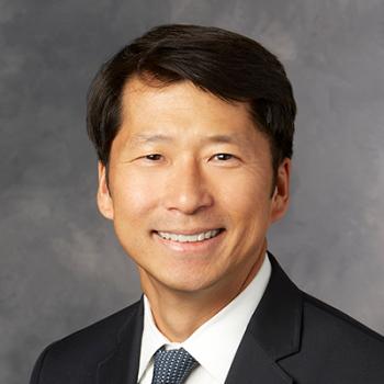 John B. Sunwoo合作论文数Department of Dermatology, School of Medicine, Stanford University;Stanford Cancer Institute, School of Medicine, Stanford University10
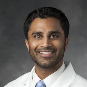 Vasu Divi合作论文数Otolaryngology (Head and Neck Surgery) Department, Stanford University8Margaret Brandwein-Weber合作论文数7Finegersh Andrey合作论文数Departments of Anesthesiology and Pharmacology &Chemical Biology, University of Pittsburgh;Departments of Anesthesiology and Pharmacology & Chemical Biology, University of Pittsburgh7
 Azita Khorsandi合作论文数6
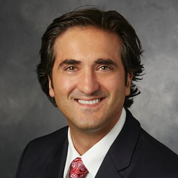 Davud Sirjani合作论文数Otolaryngology (Head and Neck Surgery) Department, Stanford University6Fabian Necker合作论文数From the Institute of Functional and Clinical Anatomy-Digital Anatomy Laboratory, Friedrich-Alexander University Erlangen-Nürnberg;Graduate School of Medicine, Osaka Metropolitan University;Friedrich-Alexander University Erlangen-Nürnberg, Osaka Metropolitan University5
 Quyen Nguyen合作论文数Department of Surgery, School of Medicine, University of California, San Diego5
 Michael Topf合作论文数Med Ctr, Vanderbilt Univ5
 Chris Holsinger合作论文数Otolaryngology (Head and Neck Surgery) Department, Stanford University5van den Berg Nynke S合作论文数5
 Quan Zhou合作论文数4Sedef Everest合作论文数4
 Giri Krishnan合作论文数Dept Otolaryngol Head & Neck Surg, Univ Adelaide4John R Sims合作论文数4
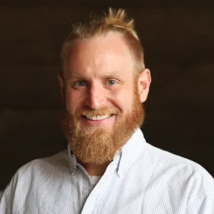 Brock A. Martin合作论文数University Of Louisville Physicians, Inc.4
 Bruce Daniel合作论文数Bioengineering Department, Stanford University;Radiology Department, Stanford University4
 Alexander Dimitrios Colevas合作论文数Medicine Department, Stanford University4
 Christoph Leuze合作论文数4
 Michael J. Kaplan合作论文数Department of Otolaryngology–Head and Neck Surgery, School of Medicine, Stanford University4Michelle M. Chen合作论文数3Pooja Dhorajiya合作论文数3Mobeen Rahman合作论文数3Alexis Miller合作论文数3Marisa E Hom合作论文数Dept Otolaryngol Head & Neck Surg, Vanderbilt Univ3Xulei Liu合作论文数3Muhammad Qazi合作论文数3Martha J Griffin合作论文数3
 Naoki Nishio合作论文数Department of Otorhinolaryngology, Nagoya University Graduate School of Medicine3
 Guolan Lu合作论文数3Stevens Todd合作论文数Department of Pathology, Creighton University Medical Center3
 Yu-Jin Lee合作论文数3
 Eduardo Mendez合作论文数3Maxwell Y. Lee合作论文数2Stacey Hansen合作论文数2Nikita Bedi合作论文数2Tulika Singh合作论文数2Disha Sahetya合作论文数2
 Sainiteesh Maddineni合作论文数2Sue E Knoblaugh合作论文数2Julia Parrish-Novak合作论文数2Jeffrey Harris合作论文数2
 Jacqueline Pei合作论文数Department of Otolaryngology, Stanford University School of Medicine2
 Heying Duan合作论文数Department of Radiology, Stanford University2




