Three experiments investigating the patient M.S.'s semantic memory are reported. Experiments 1 and 2 involved a category-membership decision task, in which M.S. was asked to determine whether a noun was a member of a specified semantic category. His performance in Experiment 1 was impaired for nouns from living categories in comparison with nouns from nonliving categories, and this impairment was especially marked for nouns of low typicality. Experiment 2 demonstrated an equivalent pattern of very poor performance to nouns of low familiarity from living categories. In Experiment 3 the effect of a category label on lexical decision was examined, using category labels as primes preceding nouns or pronounceable nonwords. Facilitation from related category label primes was found for typical and untypical members of living and nonliving semantic categories. These findings demonstrate that M.S. has impaired knowledge of the structure of living semantic categories when explicit access to this information is required (Experiments 1 and 2), but that some form of preserved category structure can be demonstrated in tasks which assess this implicitly (Experiment 3).
Current theoretical models of face perception postulate separate routes for processing information needed in the recognition of a familiar face, for matching photographs of unfamiliar faces, and for the analysis of facial expressions. This study investigated this claim in a group of ex-servicemen who had sustained unilateral brain injuries affecting posterior areas of the left or right cerebral hemisphere. Care was taken to confirm the nature of impairment by using two different tasks to assess each of the three theoretically defined abilities (leading to a total of six tasks). The results showed selective impairments of all three abilities on accuracy scores. Response latency data confirmed the finding of a selective deficit in the processing of facial expressions, but produced evidence suggesting that impairments affecting familiar face recognition and unfamiliar face matching were not completely independent from each other in this group of ex-servicemen.
AbstractA previous study of the performance of men with chronic unilateral focal brain lesions (due to wartime missile injury) on a standard test of line orientation suggested a left hemisphere (LH) as well as a right hemisphere (RH) contribution to visuospatial processing. The present study was designed to fractionate the variables that could underlie this unexpected finding and thereby to tease out the mechanisms involved in LH as compared with RH processing. A simpler (“purer”) version of the standard line orientation task was used, as were two other versions in which matching in an array and matching with distractors were measured. The findings confirmed the hypothesis of RH involvement in the purer task of metric measurement and suggested that the LH has an important role in keeping track of decisions and updating decisions in more complex aspects of line orientation judgment. (JINS, 1996, 2, 335–339.)
This single case study of the ability to generate verbal and non-verbal imagery in a woman who sustained a gunshot wound to the brain reports a significant difficulty in generating images of word shapes but not a significant problem in generating object images. Further dissociation, however, was observed in her ability to generate images of living vs non-living material. She made more errors in imagery and factual information tasks for non-living items than for living items. This pattern contrasts with our previous report of the agnosic patient, M.S., who had severe difficulty in generating images of living material, whereas his ability to image the shape of words was comparable to that of normal control subjects. Furthermore, with regard to the generation of images of living compared with non-living material, M.S. shows more errors with living than non-living items. In contrast, the present patient, S.M., made significantly more errors with non-living relative to living items. There appear to be two types of double dissociation which reinforce the growing evidence of dissociable impairments in the ability to generate images for different types of verbal and non-verbal material. Such dissociations, presumably related to sensory and cognitive processing demands, address the problem of the neural basis of imagery.
This paper reviews outcome studies of wartime head-injury in Europe and North America. These include Goldstein's major but neglected contribution: intensive clinical and experimental studies of World War I veterans, in which he emphasizes the need for rehabilitation and community care. The present paper reports a follow-up, in Oxford U.K., of a cohort of World War II veterans drawn from over a thousand head-injured servicemen admitted to St Hugh's Military Hospital in Oxford, usually within one to two days of wounding. This cohort was initially examined in 1963 and thereafter until the present day. Behavioural outcome is remarkably good. Cognitive data show a striking preservation of ability in the group as a whole, despite previously reported selective impairments related to specific loci of lesion. Psychiatric illness is rare; and there is little difference in self-report on rating scales between the veterans and a carefully recruited, age-matched control panel. This positive outcome is attributed to a number of factors, including the psychosocial context of World War II for British servicemen, the expert and systematic medical care that commenced within hours of wounding, and the positive attitude of the civilian population. It reinforces the crucial importance of after-care.
A short account is given of a case of right occipito-parietal abscess first studied by Oldfield in 1939 and followed up over a period of 30 years. The main disabilities lay in the sphere of spatial orientation and visual memory and underwent little substantial change over this period. None the less, appreciable re-adaptation was achieved by compensatory measures. Some theoretical issues are briefly touched upon.
The concept of agnosia as a higher order functional impairment, which can occur in the absence of low level visual perceptual deficits, continues to provoke debate. This controversy is complicated by the fact that, on close examination, agnosic patients do tend to have some perceptual difficulties. Thus the issue centres around the question as to whether these deficits play a causal part in the aetiology of agnosia or whether they are functionally independent, with both impairments resulting from the substantial cerebral lesions involved in agnosia. In 1956, Ettlinger published a study in which he compared the performance of patients with visual recognition deficits and patients with posterior brain lesions whose recognition abilities were intact. He argued that visual perceptual problems could not explain the recognition deficit in agnosia as he saw far worse perceptual impairments in patients who did not experience any problems in visual recognition. Although the logic of Ettlinger's argument is not disputed, some criticisms have arisen concerning the study, such as the fact that his experimental group did not include a truly object agnosic patient. In addition, Ettlinger's visual-sensory assessment can no longer be considered comprehensive in the light of present day knowledge of the cerebral visual apparatus. This study therefore investigated three (prosop)agnosic patients and five patients with unilateral brain lesions without recognition deficits on an extensive battery of visual sensory tests. The results support Ettlinger's original claim that (in some cases) agnosia cannot be explained as resulting from lower level visual impairments.
A sample of the literature on acquired dyslexia is reviewed with special reference to the nature of paralexic errors. Linguistic studies of six cases of dyslexia are presented, with appropriate neurological and psychological details; there are two cases of each of three hypothesized types of impairment, visual dyslyxia, “surface” (grapheme-phoneme) dyslexia, and “deep” (syntactico-semantic) dyslexia. Finally, a scheme for acquired dyslexia is proposed and related to the error patterns observed in “normal” adult readers and in children learning to read; our approach to lexical look-up is one that the diagram makers may have found sympathique.
This study addresses the possibility that cognitive sequelae--albeit of a transient or minor character--can be associated with mild head injury. Twenty men (aged 16-30 years of age), whose post-traumatic amnesia did not exceed eight hours, were examined within 48 hours of their accident and again one month later. This unselected sample had no previous history of head injury. A control group of 20 men of similar socioeconomic background, was selected from medical wards (where they had been admitted for orthopaedic treatment or a minor operation). They were also retested one month after the first examination. Neuropsychological tests were selected to measure abilities often compromised after significant head injury, namely memory and attention. The experimental component consisted of the fractionation of a complex skill (paced addition) to probe for deficits at different stages of information processing: perception and input into storage; search for and retrieval of information from working memory; and paced and unpaced addition. In general, no significant difference was found between the experimental and control groups, with the possible exception of an initial decrement on two working memory tasks: probe digits and a keeping track task (where the subject has to keep in mind and update a number of variables at the same time). The keeping track paradigm, ostensibly of ecological relevance, may well be worth further exploration in memory research, and in studies of more severely head-injured patients. It is further suggested that the appropriate management and counselling of mildly head-injured patients may help to avert symptoms that are of psychological rather than pathophysiological origin.
We assessed the ability of an achromatopsic patient to detect and discriminate colour and form concealed in a static or dynamic checkerboard display where the luminance differences among adjacent squares were randomly assigned. There were no conditions under which he could discriminate two very different saturated colours from each other. Nevertheless, he could discriminate chromatic from luminance boundaries in static displays when the colour defining the boundary was saturated and the achromatic boundaries all had similar luminance contrast, i.e. varied over a narrow range. However, he could not readily detect chromatic boundaries from among many achromatic boundaries that differed widely in luminance contrast. In addition, he was able to detect chromatic boundaries even when they were concealed by dynamic random luminance masking. His ability to pick out chromatic borders was abolished when desaturated colours were used. However, he was singularly proficient at detecting coloured forms in static or dynamic displays even when the saturation of the colours of which the form was composed were such that they were rendered invisible when concealed as a single square in a checkerboard. This implies that signals about chroma are still available in extracting shape. The patient performed flawlessly when asked to indicate the direction of motion of a horizontal red/green isoluminant grating which was phase shifted by 90 degrees in either direction, demonstrating unequivocally that he has access to the sign of colours that he nevertheless does not perceive.
Current theoretical models of face perception postulate separate routes for processing information needed in the recognition of a familiar face, for matching photographs of unfamiliar faces and for the analysis of facial expressions. The present study investigated this claim in a group of ex-servicemen who had sustained unilateral brain injuries affecting posterior areas of the left or right cerebral hemisphere. Care was taken to confirm the nature of impairment by using two different tasks to assess each of the three theoretically defined abilities (leading to a total of six tasks). We adopted a stringent application of the double dissociation methodology to investigate the pattern of performance across tasks of individual ex-servicemen. A selective impairment was defined as a significantly impoverished performance on both tests of a specific ability, while all other tasks were performed within normal limits. In addition, we used both accuracy and response latency measures to substantiate evidence for spared or defective abilities. The results showed selective impairments of all three abilities on accuracy scores. Response latency data confirmed the finding of a selective deficit in the processing of facial expressions, but produced evidence suggesting that impairments affecting familiar face recognition and unfamiliar face matching were not completely independent from each other in this group of ex-servicemen.
A selection of the literature on vision published in the last 2 years from various neuroscience disciplines is reviewed. It is organized around the notion of independent processing mechanisms for sensory cues such as wavelength, intensity and orientation; these are subsequently integrated for the perception of colour, brightness and contours, for example, which then feed into higher order systems for visual recognition (words and objects) and visuospatial cognition. A strict hierarchical system is, however, not plausible. Apart from the physiological evidence against such a concept, the widespread observations of 'knowledge without awareness' suggest direct links between different levels, which by-pass conscious experience.
Despite the absence of "conscious", overt identification, some patients with face recognition impairments continue covertly to process information regarding face familiarity. The fact that by no means all patients show these covert effects has led to the suggestion that indirect recognition tasks may help in identifying different types of face recognition impairment. The present report describes a number of experiments with the patient NR, who, after a closed head injury, has been severely impaired at recognizing familiar faces. Investigations mostly failed to show overt or covert face recognition, but NR performed at an above-chance level in selecting the familiar face on a task requiring a forced-choice between a familiar and an unfamiliar face. This discrepancy between a degree of rudimentary overt recognition and absence of covert effects on most indirect tests was addressed using a cross-domain identity priming paradigm. This examined separately the possibility of preserved recognition for faces that NR consistently chose correctly in a forced-choice familiarity decision and those on which he performed at chance level. Priming effects were apparent only for the faces that were consistently chosen as "familiar" in forced-choice. We suggest that NR's stored representations of familiar faces are degraded, so that face recognition is possible only via a limited set of relatively preserved representations able to support a rudimentary form of overt recognition and to facilitate performance in matching and priming tasks.
Experiments were designed to examine the imagery abilities of an agnosic patient, M.S., who has consistently shown more severe deficits in recognizing visually, and in retrieving knowledge of living as compared with non-living items. Judgements of visual similarity were required for named objects and for object-pictures, as well as for the factual properties of these stimuli. The same disproportionate difficulty in processing living ('natural') objects was found in these tasks as well as in forced-choice recognition. In contrast, no deficit was found on analogous tasks concerned with word-shape similarities. These findings have a bearing on concepts of semantic memory.
 John C Marshall合作论文数University of Oxford28
 Andy Young合作论文数Department of Psychology, University of York14
 EHF DEHAAN合作论文数13Graham Ratcliff合作论文数11Ziyah Mehta合作论文数10Robert Hiorns合作论文数5
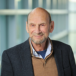 Arthur Wingfield合作论文数Department of Psychology, Brandeis University/School of Life Sciences, Brandeis University4R. C. Oldfield合作论文数4Dc Hay合作论文数3Charles a Heywood合作论文数3
 Edward Haan合作论文数Faculty of Social and Behavioural Sciences, Universiteit Van Amsterdam3
 Alan Baddeley合作论文数Department of Psychology, University of York2Young Alan合作论文数2Hanna Damasio合作论文数2
 Paula A. Tallal合作论文数Scientific Learning Corporation;Center for Molecular and Behavioral Neuroscience, Rutgers University2Alan Cowey合作论文数2C.W.M. Whitty合作论文数2A. J. Mistlin合作论文数1Wr Russell合作论文数1B Steinberg合作论文数1M. Small合作论文数1AW YOUNG合作论文数1E.H.F. de Hann合作论文数1Pj Carrivick合作论文数1cfe heeschen合作论文数1Rc Tallis合作论文数1Ma Jeeves合作论文数1Ls Illis合作论文数1Oppenheimer D合作论文数1
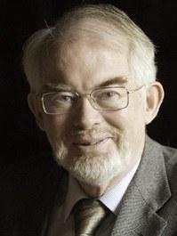 John Mollon合作论文数Department of Psychology, University of Cambridge1Hellawell D合作论文数1Adams C B合作论文数1
 David Perrett合作论文数School of Psychology and Neuroscience, University of St Andrews;Institute of Behavioural and Neural Sciences, University of St Andrews;Centre for Social Learning & Cognitive Evolution, University of St Andrews1W. Ritchie Russell合作论文数1Neil Brooks合作论文数1Jane M. Holmes合作论文数1Dennis C. Hay合作论文数1Christopher Ounsted合作论文数1A.J. Chitty合作论文数1Marian Small合作论文数1F. X. Plooij合作论文数1Janet Ross合作论文数1Victoria A. Fromkin合作论文数1
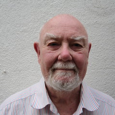 Patrick Rabbitt合作论文数Department of Experimental Psychology, University of Oxford;Department of Psychology, University of West Australia;University of Manchester1E.H.F. De Haan合作论文数1




