AIM To study if T-cell activation related to portasystemic shunting causes osteoclast-mediated bone loss through RANKL-dependent pathways. We also investigated if T-cell inhibition using rapamycin would protect against bone loss in rats. METHODS Portasystemic shunting was performed in male Sprague-Dawley rats and rapamycin 0.1 mg/kg was administered for 15 wk by gavage. Rats received powderized chow and supplemental feeds to prevent the effects of malnutrition on bone composition. Weight gain and growth was restored after surgery in shunted animals. At termination, biochemical parameters of bone turnover and quantitative bone histology were assessed. Markers of T-cell activation, inflammatory cytokine production, and RANKL-dependent pathways were measured. In addition, the roles of IGF-1 and hypogonadism were investigated. RESULTS Portasystemic shunting caused low turnover osteoporosis that was RANKL independent. Bone resorbing cytokine levels, including IL-1, IL-6 and TNFalpha, were not increased in serum and TNFalpha and RANKL expression were not upregulated in PBMC. Portasystemic shunting increased the circulating CD8+ T-cell population. Rapamycin decreased the circulating CD8+ T-cell population, increased CD8+ CD25+ T-regulatory cell population and improved all parameters of bone turnover. CONCLUSION Osteoporosis caused by portasystemic shunting may be partially ameliorated by rapamycin in the rat model of hepatic osteodystrophy.
P190 Aims: The immunosuppressive properties of rapamycin are well documented and it is used routinely in renal transplant recipients. Rapamycin also inhibits fibroblasts which may be important for proper healing of the ureteric anastomosis after renal transplantation. In this study we investigated the effect of rapamycin on the healing of the ureteric anastomosis. Methods: Large White X Landrace pigs (n=12) weighing 22-30 kg were subjected to laparotomy and mobilization of both urethers. Each ureter was divided, spatulated and re-anastomosed using continuous 6/0 PDS. The animals were randomly allocated to receive either rapamycin 2mg orally per day, or no treatment. On the fifth postoperative day the anastomoses were excised and examined histologically (Haematoxylin-eosin and Chromotrop Aninline Blue) and biochemically (hydroxyproline levels). Thin strips of ureter (±5mm) were used to measure the breaking strength of the anastomosis. Results: The ureteric anastomoses were all healed and patent, although the proximal ureters were markedly dilated. The serum creatinine levels were similar in the two groups (1.57±0.06 and 1.55±0.09mg/ml). The breaking strength of the ureteric anastomosis was lower in the rapamycin treated animals compared to the control group (221±24g versus 261±16g). The hydroxyproline levels in the ureter were lower in the rapamycin treated animals (12.8±2.7 versus 22.4±5.3μg/ml). Histological examination showed no difference in the morphology or collagen staining between the two groups. Conclusions: Thus in summary, healing of the ureteric anastomosis appears to be impaired in animals treated with rapamycin.
Rapamycin is a potent immunosuppressive agent that also inhibits fibroblastic activity and therefore may affect the healing of various tissues. The aim of this study was to investigate the effect of rapamycin on wound healing and the healing of the ureteric anastomosis. Large White/Landrace pigs were subjected to a laparotomy and division and immediate anastomosis of the ureter. The animals were randomly allocated to receive either rapamycin or placebo. The animals were sacrificed on postoperative day 5, and strips of the skin and fascia closure and the ureteric anastomosis excised and used to determine the tensile strength, hydroxyproline levels, and histological changes. The tensile strength and the hydroxyproline levels in the ureter and fascia were lower in the rapamycin-treated animals. There was no difference in the tensile strength in the skin, although the hydroxyproline levels were lower. This study shows that healing of the ureteric anastomosis and fascia and skin closure may be impaired by rapamycin.
Besides its potent immunosuppressive properties, rapamycin also has antitumor and antifungal effects. Rapamycin also inhibits the proliferation of fibroblasts and therefore may impair the healing of various tissues. We investigated the effect of rapamycin on the healing of the bile duct anastomosis. The study was undertaken in pigs that were subjected to a laparotomy under general anesthesia. The bile duct was mobilized and divided and immediately reanastomosed. The animals were randomly allocated to receive either rapamycin or placebo. The animals were sacrificed on the postoperative day 5, then the biliary anastomosis was excised and used to determine the tensile strength, hydroxyproline levels, and the histological changes. The tensile strength and the hydroxyproline levels in the biliary anastomosis were lower in the animals treated with rapamycin. The liver function tests were normal. These studies show that rapamycin may impair the healing of the biliary anastomosis.
A302 Aims: The use of rapamycin to prevent graft rejection in renal transplantation is well documented. However the inhibitory effect of rapamycin on fibroblasts has raised concerns about its effect on wound healing. Anecdotal reports have suggested an increased incidence of post transplant wound problems. In this study in pigs we investigated the effect of rapamycin on wound healing. Methods: Large White X Landrace pigs (n=12) weighing 22-30kg were subjected to a midline laparotomy. The sheath was closed with a continuous 0 Nylon suture and the skin with a continuous subcuticular 2/0 Nylon suture. The animals were randomized to receive either rapamycin 2mg orally daily or no treatment. The animals were sacrificed on the fifth postoperative day and the skin and fascia closure examined histologically (Haematoxylin-eosin and Chromotrop Aninline Blue [CAB]) and biochemically (hydroxyproline levels). The breaking strength of strips of skin and fascia were also measured. Results: One animal in the rapamycin treated group developed a full-thickness wound dehiscence on the 4th postoperative day and had to be sacrificed. The wounds of the remaining animals were intact at the time of sacrifice. The breaking strength of the fascia was lower in the rapamycin treated animals compared to the control animals (351±62g versus 444±54g). The breaking strength of the skin was the same in the two groups (239±34g versus 237±32g). The animals treated with rapamycin had lower hydroxyproline levels in both the fascia (28.7±11.8 versus 34.1±9.9 μg/ml) and the skin (12.3±3.7 versus 19.4±5.7 μg/ml) compared to control animals. On histological examination there were no differences in morphology or collagen staining between the two groups. Conclusions: These findings suggest that rapamycin impairs wound healing. Special attention should be given to wound closure and wound management in patients treated with rapamycin.
Background and aims: In chronic liver disease, bone disease frequently develops. The contributions of the different features of liver disease such as parenchymal inflammation, portal hypertension, and portasystemic shunting on bone metabolism have not been systematically studied. The aim of this study was to identify the features of liver disease contributing to bone disease using rat models. Methods: Parenchymal liver disease was induced by carbon tetrachloride administration, portal hypertension by partial portal vein ligation, and portasystemic shunting by end to side anastomosis of the portal vein to the inferior vena cava. Normal and sham operated surgical animals served as controls. Serum calcium, 25-hydroxy vitamin D (25-OH vit D), and osteocalcin levels, and urinary deoxypyridinoline excretion were analysed. Testosterone and oestradiol levels were determined in male and female rats, respectively. Interleukin 1, interleukin 6, and tumour necrosis factor α (TNF-α) were determined in serum. Bone density was measured in all groups and in addition, in the surgical groups, histomorphometry was performed on undecalcified specimens of the proximal tibia. The calcium content of the femurs, removed at termination and ashed, was determined. Results: Early parenchymal disease and portal hypertension did not affect bone metabolism or body mass. Portasystemic shunting increased bone resorption, decreased bone formation, bone density, and trabecular bone volume which were commensurate with a reduction in body mass. TNF-α levels were elevated and testosterone levels were low in male portasystemic shunted rats. Conclusions: Portasystemic shunting in the rat adversely affects bone metabolism as part of a generalised catabolic state where high TNF-α and low testosterone and 25-OH vit D levels may play a role.
BACKGROUND:The portal venous system plays a major role in a variety of disease processes. Although the anatomy, histology and physiology of the portal vein have been extensively studied over the last century, very little attention has been given to its unique helical structure. Previous documentation of the helical structure has been based on microscopic, macroscopic and radiological appearances.MATERIALS AND METHODS:A more refined method than the above was used to image the portal vein helix. Resin casts of the portal venous system were performed in 6 rats, 1 baboon and 1 human.RESULTS:All species had a clearly demonstrated helical structure of the portal vein situated above the splenic vein.CONCLUSION:The unique structure of the portal vein and its subsequent helical flow has been neglected in medical research. The significance of the activated portal helix and its effect on the blood flow into the liver needs further investigation.
Free-flap failure is in the order of 4 to 10 percent. Heparin is more effective at preventing venous thrombosis than arterial thrombosis. This study was undertaken to investigate the efficacy of delivering heparin at a high dose locally but low dose systemically (heparin infusion via a catheter placed proximal to the venous anastomosis) to prevent venous thrombosis in microsurgery. A model of venous thrombosis was first established by a venous inversion graft in the rat femoral vein (this was performed in seven animals and resulted in 100 percent thrombosis). Saline and heparin were delivered proximal to the inverted vein graft to assess the effect of each in preventing venous thrombosis. Flow/patency distal to the inverted vein graft was assessed by observation under the microscope, the milk test, and rate of flow (flowmeter). Saline infused via a catheter proximal to the venous inversion graft resulted in 100 percent thrombosis in 10 animals. Heparin (100 U/ml at 2 to 3 ml/hour) infused through a catheter for 2 hours proximal to the anastomosis resulted in flow in all 10 animals during the infusion. Blood was also taken before beginning the procedure (control) and after the heparin infusion distal to the anastomosis (local partial thromboplastin time) as well as in the contralateral femoral vein (systemic). The control for all animals that received heparin was <3 minutes. The systemic partial thromboplastin time after heparin infusion was <3 minutes in seven animals, 3.3 minutes in two animals, and >7 minutes in one animal. The local partial thromboplastin time distal to the inverted vein graft was >10 minutes in nine animals and 3.7 minutes in one animal. The study also had a clinical component, in which a catheter was placed in a vein of the free flap, and heparin was infused over 5 days. This technique has been used in 83 consecutive free flaps. In three recent free flaps performed on the limbs, the local partial thromboplastin time (close to the anastomosis) was raised but the systemic time was normal. This technique offers a method in preventing venous thrombosis in microsurgery. It is simple to implement and is not associated with the systemic complications of heparin.
We have evaluated the link between haemostatic abnormalities and immune dysfunction in liver disease by evaluating parameters of cellular and humoral immunity in conjunction with coagulation profiles in rats following portacaval anastomosis, induction of portal hypertension by portal vein stenosis or by sham surgical procedures. Twelve weeks following surgery, portacaval shunted rats were markedly anaemic (8.9 +/- 0.6 g/dl; controls 12.3 +/- 1.4 g/dl, p < 0.05), had low plasma fibrinogen levels (0.6 +/- 0.3 g/l, controls 2.5 +/- 0.2 g/l p < 0.05) and markedly elevated fibrin(ogen) degradation products (FDP) titres (1/40-1/80; controls < 1/10. p < 0.05). Portal vein stenosed rats were less anaemic (11.5 +/- 0.8 g/dl), had near normal fibrinogen levels (2.1 +/- 0.3 g/l) but elevated FDP levels (1/40-1/80). Both portacaval shunted and portal vein stenosed rats had elevated serum IgG levels (35.1 +/- 14.1 g/l; 29.2 +/- 13.9 g/l respectively; control values 20 +/- 5.9 g/l p < 0.05 for comparison with both experimental groups). Intrinsic lymphocyte proliferation to T and B cell mitogens was markedly depressed in the portacaval anastamosed rats when compared to controls. Serum factors inhibitory to control lymphocyte proliferation were noted in the shunted rats. Phagocytosis of complement and immunoglobulin sensitised sheep RBC by Kupffer cells purified from rats that had undergone portacaval shunting was markedly reduced (p < 0.05). The increased degree of phagocytosis following exposure to LPS-endotoxin (50 micrograms/ml) was proportionate in degree to the control group. Spontaneous release of bioactive lymphocyte activating factors (IL-1 and IL-6) by purified rat sinusoidal cell populations was decreased in the portacaval shunted group, and decreased still further following stimulation with LPS (50 micrograms/ml) in vitro. The observation that many of the haemostatic and immunological abnormalities associated with chronic liver disease are present in rats with surgically created portacaval shunts or with induced portal hypertension, lends credence to the hypothesis that shunting of portal blood is, at least in part, responsible for many of the systemic manifestations associated with chronic liver disease.
The effect of various components of the transplant procedure upon the regenerative process in reduced liver grafts is not known. In this study, partially hepatectomised rat liver remnants were flushed with 5 ml of either Ringer's lactate, Euro Collins solution or University of Wisconsin solution at 4 degrees C and then 5 ml Ringer's lactate at 4 degrees C. After partial hepatectomy alone, the peak increase in thymidine kinase was measured at 24 hours (36,021 +/- 8,060 disintegrations per minute per milligram protein; the mitotic index was 25 +/- 7). In all the groups in which the remnant was flushed, peak thymidine kinase and mitotic index were measured at 48 hours. The pattern of ornithine decarboxylase activity was disorganised in all groups. Flushing of the liver remnant therefore delays the regenerative response by 24 hours. In large animals, including humans, regeneration appears to commence within the first 5 days after resection. A comparable delay doubling this time might coincide with the onset of rejection and further compromise liver function.
This study was conducted to determine the pattern of early regenerative response to orthotopic intact liver transplantation in the rat and to investigate whether the response differed in grafts with or without revascularisation of the arterial bed. Outbred male Long Evans (LE-LE allogeneic, non rejector) rats weighing 300-350g were subjected to orthotopic intact liver allograft using a "sleeve" anastomosis for the hepatic artery. Total warm ischaemia ranged from 19 to 34 minutes and no storage was employed. Comparison was made with a group of control rats which were subjected to 25 minutes total inflow occlusion and regeneration was measured with tissue thymidine kinase (TK) and mitotic figures. Samples were taken at 1, 2, 4, 7, 10 and 20 days post-operatively. Plasma aspartate aminotransferase (AAT) and light microscopy were used to evaluate hepatocyte necrosis. There was a brief sharp increase in TK and AAT in the first 24 hours after sham operation but no appearance of mitotic figures. A similar but more prolonged increase in TK occurred in the arterialized transplant group with the highest levels recorded on day 4. The level remained significantly elevated above pre-operative until 10 days and declined within 20 days. Mitotic figures appeared at 2 days, reached significance at 7 and 10 days and had disappeared by 20 days. The pattern of changes was accentuated in animals in which the artery was not reanastomosed and the increases in TK and AAT were still significant at 20 days. Whilst similar degrees of peri-portal cellular infiltrate occurred in both groups of rats, bile duct proliferation was most obvious in non-arterialized animals.(ABSTRACT TRUNCATED AT 250 WORDS)
It has been shown previously that liver regeneration after partial hepatectomy in rats is delayed if the liver is subjected to either concurrent ischaemia, flushing with cold solution, or grafting. We have shown recently that treatment with CsA preoperatively overcomes the suppressive effect of flushing and returns the regenerative response to a normal time scale. The present study was designed to investigate whether administration of FK506 would also return the observed delayed regenerative response to normal. Long-Evans rats weighing 250-350 g were subjected to standard 68% partial hepatectomy. Group 1 had no further treatment; in group 2, the liver remnant was flushed with 10 ml cold (4 degrees C) Ringers lactate solution, and in group 3, FK506 (1 mg/kg/day) was administered by intramuscular injection for 3 days before the partial hepatectomy and flushing as in group 2; a final dose was given after completion of the procedures. Animals were killed in sets of 6 per group at 4, 24, 48, 72, and 96 hr after surgery and blood samples were taken for measurement of plasma aspartate amino-transferase. Liver biopsies were analyzed for measurement of thymidine kinase and ornithine decarboxylase activity and for counting of mitotic figures. While the highest recorded thymidine kinase activity occurred in group 1 at 24 hr, this was delayed to 48 hr in both group 2 and 3 and counts remained high up to 96 hr in group 3. Mitotic indices were only significantly elevated (compared with group 1 at 96 hr), while ornithine decarboxylase activity did not correlate with these changes being significantly lower than in groups 2 and 3 at 4 hr and in group 3 also at 24 hr. Plasma aspartate aminotransferase was also significantly higher in group 3. It is concluded that the administration of FK506 preoperatively to rats subjected to partial hepatectomy and flushing did not restore the delayed regenerative response to normal but enhanced the response (as measured by thymidine kinase but not by mitotic indices) which commenced at 48 hr and was still present at 96 hr.
Current knowledge of liver regeneration after reduced liver transplantation is limited. Warm ischemia is one component of the reduced liver transplantation procedure that could have an impact on the regenerative response. To study this effect, we performed partial hepatectomy on male Long-Evans rats, with animals divided into four groups: group 1 underwent partial hepatectomy only; group 2 underwent partial hepatectomy and 40 min of ischemia; group 3 underwent partial hepatectomy, 40 min of ischemia and portocaval shunt surgery; and group 4 underwent partial hepatectomy and orthotopic autograft surgery. Group 5 consisted of sham-operated animals. Animals were killed 4, 24, 48, 72 and 96 hr after surgery. Thymidine kinase activity, mitotic index, a liver mass index and ornithine decarboxylase levels were used as parameters of liver regeneration. Aspartate transaminase was recorded. Maximal thymidine kinase and mitotic index were observed in group 1 animals at 24 hr. In groups 2, 3 and 4 maximal thymidine kinase activity and mitotic activity were observed 24 hr later at 48 hr. The magnitude of the peak response in these groups appeared to correlate with the duration of portal venous occlusion, with greatest increases occurring in those groups where portal stasis was most prolonged. The increase in liver mass for these groups was also delayed with respect to group 1 animals. The anticipated peak in ornithine decarboxylase levels was seen at 4 hr in group 1. The ornithine decarboxylase response in the other groups was disorganized, with delay of the recorded peaks.(ABSTRACT TRUNCATED AT 250 WORDS)
Present techniques for renal transplantation in the rat include a period of 20-25 minutes warm ischemia. Our method combines a recently described sleeve anastomotic technique for the renal artery, conventional end-to-end anastomosis of the renal vein, and implantation of the ureter into the bladder. This has resulted in a reproducible ischemic interval of 12-14 minutes. Plasma creatinine and histological features in animals sacrificed from 10 to 30 days after transplantation were within normal limits with no evidence of ischemic damage. A further advantage of the technique is that kidneys can be exchanged between the donor and recipient. It is recommended that this procedure, which reduces the ischemic interval by up to 50%, should be learned and employed in studies of renal transplantation in the rat, especially if such studies include the prior administration of cyclosporine, which may aggravate the effects of ischemia.
The precise cause of allograft dysfunction after renal transplantation often cannot be established by non-invasive means. In clinical practice, radionuclide scans form an integral part of the clinician's armamentarium in the assessment of these patients [1, 2]. Unfortunately, in the clinical setting more than one pathological process may be responsible for the impaired function, making it difficult to correlate the scan appearances with the pathology. In this study in rats we compared the renal DTPA scan appearances of the various pathological processes which may cause renal allograft dysfunction in the immediate post-transplant period.
 R Hickman合作论文数16Marilyn Tyler合作论文数9Zoe Lotz合作论文数8Delawir Kahn合作论文数8OL MEYERS合作论文数5John Terblanche合作论文数5M. Keraan合作论文数5F. Duminy合作论文数Groote Schuur Hospital4
 Heather McLeod合作论文数4Sj Saunders合作论文数3RE KIRSCH合作论文数3
 Anwar Suleman Mall合作论文数Department of Surgery and The Medical Research Council Liver Research Centre, University of Cape Town3SW van der Merwe合作论文数2K Jaskiewicz合作论文数2E Shepherd合作论文数2
 Wendy Spearman合作论文数Division of Hepatology, Department of Medicine, Faculty of Health Sciences, University of Cape Town2J. Fevery合作论文数2Del Kahn合作论文数2Ralph Kirsch合作论文数2J.B. Van Den Bogaerde合作论文数2N Matsiliza合作论文数2Don A. Hudson合作论文数2R VANHOORNHICKMAN合作论文数1Schalk W. van der Merwe合作论文数1P CHILD合作论文数1G BOLITHO合作论文数1L O Frith合作论文数1Robert Bond合作论文数1Brenda J Olivier合作论文数1LP Adams合作论文数1
 Jakobus Ernst Van Zyl合作论文数1J Bendahan合作论文数1D Kahn合作论文数1
 M J Kotze合作论文数Div Chem Pathol, Stellenbosch Univ1Bolitho D G合作论文数1Govender Dhirendra合作论文数Div Anat Pathol, Univ Cape Town1
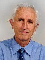 Eric Bateman合作论文数Lung Institute, University of Cape Town;Department of Medicine, University of Cape Town;Department of Global Health, University of Washington1
 Stephen Hough合作论文数1
 Yohannes T Ghebremariam合作论文数1
 Tomas Slavik合作论文数Ampath Pathology Laboratories and Department of Anatomical Pathology, University of Pretoria1C Goosen合作论文数1
 Simon Robson合作论文数Division of Gastroenterology, Department of Medicine, Beth Israel Deaconess Medical Center;Department of Anesthesia, Beth Israel Deaconess Medical Center;Division of Medical Sciences, Harvard Griffin Graduate School of Arts and Sciences, Harvard University;Liver Center, University of Cape Town;Harvard Medical School, Harvard University;Inner Mongolian Medical College;Department of Medicine, New England Deaconess Hospital1Girish J. Kotwal合作论文数National Institute of Allergy and Infectious Diseases, National Institutes of Health1
 Rhena Delport合作论文数Department of Chemical Pathology, University of Pretoria1O. L. Meyers合作论文数1J.H. Crosier合作论文数1
 M M Conradie合作论文数Division of Endocrinology;Department of Medicine;Faculty of Health Sciences;University of Stellenbosch;Faculty of Health Sciences, University of Stellenbosch1




