The reduction of blood damage is still a big challenge in blood-carrying medical devices. In vitro experiments are performed to investigate the damage-causing effects, but due to the opaqueness of blood cells, only near-wall flows can be observed. Thus, several transparent blood models to visualize the rheologic behavior of blood have been proposed and examined. Nevertheless, two-phase blood models with added particles still represent the properties of blood inadequately or are very expensive and complex to produce. In this in vitro study, the viscosity, the flow behavior and the cell deformation of human red blood cells have been compared to a novel, easy-to-produce, two-phase blood model fluid with deformable alginate microspheres. The comparison has been performed in a cone-plate rheometer, a straight and a hyperbolic converging microchannel. The viscosity of the blood model fluid with a particle fraction of 30 2000 s^-1 . The alginate microspheres were deformable in an extensional flow and formed a cell free layer comparable to that of blood in a straight microchannel. The experiments showed a good optical accessibility of the two-phase flow with traceable movements of individual microspheres in the center of the microchannel. It could be shown that our proposed blood model fluid is a promising tool for the analysis of two-phase flows in complex flow geometries.
Artificial OrgansVolume 44, Issue 10 p. 1019-1020 IN MEMORIAM Obituary: Klaus Affeld Ulrich Kertzscher, Corresponding Author Ulrich Kertzscher ulrich.kertzscher@charite.de Biofluid Mechanics Laboratory, Institute for Imaging Science and Computational Modelling in Cardiovascular Medicine, Charité - Universitätsmedizin Berlin, Campus Virchow-Klinikum, Berlin, Germany Correspondence Ulrich Kertzscher, Biofluid Mechanics Laboratory, Institute for Imaging Science and Computational Modelling in Cardiovascular Medicine, Charité - Universitätsmedizin Berlin, Campus Virchow-Klinikum, Augustenburger Platz, 13353 Berlin, Germany. Email: ulrich.kertzscher@charite.deSearch for more papers by this authorMichael Lommel, Michael Lommel Biofluid Mechanics Laboratory, Institute for Imaging Science and Computational Modelling in Cardiovascular Medicine, Charité - Universitätsmedizin Berlin, Campus Virchow-Klinikum, Berlin, GermanySearch for more papers by this authorGrischa Gabel, Grischa Gabel Biofluid Mechanics Laboratory, Institute for Imaging Science and Computational Modelling in Cardiovascular Medicine, Charité - Universitätsmedizin Berlin, Campus Virchow-Klinikum, Berlin, GermanySearch for more papers by this authorSetsuo Takatani, Setsuo Takatani Department of Artificial Organs, Institute of Biomaterials and Bioengineering, Tokyo Medical and Dental University, Tokyo, JapanSearch for more papers by this authorUlrich Steinseifer, Ulrich Steinseifer orcid.org/0000-0002-8065-9803 Department of Cardiovascular Engineering, Institute of Applied Medical Engineering, Helmholtz Institute, Medical Faculty, RWTH Aachen University, Aachen, GermanySearch for more papers by this authorHorst Klinkmann, Horst Klinkmann Honorary President ESAO, Rostock, GermanySearch for more papers by this authorPiotr Ładyżyński, Piotr Ładyżyński orcid.org/0000-0003-3268-9896 Department for Modelling and Support of the Internal Organs Function, Nałęcz Institute of Biocybernetics and Biomedical Engineering, Polish Academy of Sciences, Warsaw, PolandSearch for more papers by this authorHeinrich Schima, Heinrich Schima orcid.org/0000-0002-8003-7617 Center for Medical Physics and Biomedical Engineering, Medical University of Vienna, Vienna, AustriaSearch for more papers by this author Ulrich Kertzscher, Corresponding Author Ulrich Kertzscher ulrich.kertzscher@charite.de Biofluid Mechanics Laboratory, Institute for Imaging Science and Computational Modelling in Cardiovascular Medicine, Charité - Universitätsmedizin Berlin, Campus Virchow-Klinikum, Berlin, Germany Correspondence Ulrich Kertzscher, Biofluid Mechanics Laboratory, Institute for Imaging Science and Computational Modelling in Cardiovascular Medicine, Charité - Universitätsmedizin Berlin, Campus Virchow-Klinikum, Augustenburger Platz, 13353 Berlin, Germany. Email: ulrich.kertzscher@charite.deSearch for more papers by this authorMichael Lommel, Michael Lommel Biofluid Mechanics Laboratory, Institute for Imaging Science and Computational Modelling in Cardiovascular Medicine, Charité - Universitätsmedizin Berlin, Campus Virchow-Klinikum, Berlin, GermanySearch for more papers by this authorGrischa Gabel, Grischa Gabel Biofluid Mechanics Laboratory, Institute for Imaging Science and Computational Modelling in Cardiovascular Medicine, Charité - Universitätsmedizin Berlin, Campus Virchow-Klinikum, Berlin, GermanySearch for more papers by this authorSetsuo Takatani, Setsuo Takatani Department of Artificial Organs, Institute of Biomaterials and Bioengineering, Tokyo Medical and Dental University, Tokyo, JapanSearch for more papers by this authorUlrich Steinseifer, Ulrich Steinseifer orcid.org/0000-0002-8065-9803 Department of Cardiovascular Engineering, Institute of Applied Medical Engineering, Helmholtz Institute, Medical Faculty, RWTH Aachen University, Aachen, GermanySearch for more papers by this authorHorst Klinkmann, Horst Klinkmann Honorary President ESAO, Rostock, GermanySearch for more papers by this authorPiotr Ładyżyński, Piotr Ładyżyński orcid.org/0000-0003-3268-9896 Department for Modelling and Support of the Internal Organs Function, Nałęcz Institute of Biocybernetics and Biomedical Engineering, Polish Academy of Sciences, Warsaw, PolandSearch for more papers by this authorHeinrich Schima, Heinrich Schima orcid.org/0000-0002-8003-7617 Center for Medical Physics and Biomedical Engineering, Medical University of Vienna, Vienna, AustriaSearch for more papers by this author First published: 10 September 2020 https://doi.org/10.1111/aor.13803Read the full textAboutPDF ToolsRequest permissionExport citationAdd to favoritesTrack citation ShareShare Give accessShare full text accessShare full-text accessPlease review our Terms and Conditions of Use and check box below to share full-text version of article.I have read and accept the Wiley Online Library Terms and Conditions of UseShareable LinkUse the link below to share a full-text version of this article with your friends and colleagues. Learn more.Copy URL Share a linkShare onFacebookTwitterLinkedInRedditWechat No abstract is available for this article. Volume44, Issue10October 2020Pages 1019-1020 RelatedInformation
Prof. Dr Klaus Affeld, aerial and medical engineer, forerunner in the field of heart support and mentor of young scientists, passed away in Berlin, Germany, on 19 November 2019. Born on 22 July 1935 in Oberschöneweide, Berlin, he went through many historic turning points. To enable him to study and start an academic career, his parents moved from the former East Berlin to West Berlin. Parallel to the beginning of his scientific career, Klaus pursued a creative activity as a sculptor. Following his studies in aeronautics and his diploma in 1962 at the Technical University of Berlin (TU Berlin), he was a research assistant at the Institute of Aeronautics at the TU Berlin from 1963 to 1969. There, he carried out studies on the swimming behaviour of fish, dolphins and whales, which were also partly conducted on trawlers in Iceland, and began to work in the field of biomechanics. This was followed by his summa cum laude doctorate at the TU Berlin in 1969 at the Institute of Aeronautics. Klaus habilitated in 1974 in the subject ‘Fluid Mechanics of Biological Systems’. In 1977, Klaus became a full professor at the TU Berlin and an external member of the Hermann-Föttinger Institute for Thermo-Fluid Dynamics at the TU Berlin. From 1969 to 1980, he was technical director of the working group for the development of an artificial heart within Prof. Emil Bücherl’s Department of Experimental Surgery at the Surgical Clinic at Charlottenburg Hospital (Westend), Free University of Berlin. During his time there, a series of novel blood pumps and corresponding drive units were designed. The first portable artificial heart drive was developed under his supervision. Moreover, various methods for measuring venous pressure in animals over a longer period of time were developed followed by extensive work on the flow of artificial heart valves. The first measurements of such a flow with laser Doppler velocimetry were carried out. The artificial hearts developed during this time were later produced by the company Berlin Heart, which still exists and manufactures heart support systems today. The resignation of the head of department – Prof. Bücherl – in 1985 led to Klaus’ foundation of the Biofluid Mechanics Laboratory at the former University Hospital Charlottenburg. A large number of innovative ideas have since been developed in the field of medical technology, which has resulted in a series of innovation awards and corresponding patent applications. In 1986, for example, he did the first synopsis of different rotary blood pump concepts with an analytical estimation of mechanically induced blood trauma. Later on, a number of different concepts to evaluate the flow and shear in cardiovascular prostheses, such as studies on magnified models, special test fluids and coatings, and new concepts for driving units originated from these activities. For 34 years, the laboratory has been one of the leading research centres in the field of artificial organs. Several successful spin-offs could be founded in this time. In addition, Klaus was one of the first and most active members of the European Society for Artificial Organs (ESAO), which was always a unique place for him to exchange new ideas and discuss his endeavours. He was a member of the Board of Governors of ESAO for 4 years, President of ESAO from 1998 until 2000 and Congress President of ESAO in 1995. His commitment was honoured by the Society with the EmilBücherl-Award for life-time achievement in 2015. Klaus has never limited his activity in the Society. Still in 2019, he co-authored eight presentations at the Annual ESAO Congress; he was a reviewer in the ESAO-PhD Award contest and a lecturer during the ESAO Winter School. Many researchers and lab leaders have to thank him also for background advice and highly valuable comments. He will be remembered as a great inspiration. What has contributed to this great success as a researcher? Certainly his alert and lively spirit; his curiosity and passion to search for new and unusual solutions for In Memoriam: Klaus Affeld
Colonization of in-dwelling catheters by microbial biofilms is a major concern in patient health eventually leading to catheter-related blood stream infections. Biofilms are less susceptible to standard antibiotic therapies that are effective against planktonic bacteria. Standard procedure for the detection of microorganisms on the catheter tip is culture. However, viable but non-culturable cells (VBNCs) may be missed. The aim of this study was to evaluate the use of fluorescence in situ hybridization (FISH) as an indicator to visualize and quantify the effect of the antibiotics daptomycin and vancomycin on biofilms in situ. We established an in vitro catheter biofilm model of Staphylococcus epidermidis biofilms on polyurethane catheters. Biofilm activity was measured by FISH and correlated to colony forming units (CFU) data. Digital image analysis was used for quantification of total biofilm mass and the area of the FISH positive biofilm cells. FISH showed a pronounced effect of both antibiotics on the biofilms, with daptomycin having a significantly stronger effect in terms of both reduction of biofilm mass and number of FISH-positive cells. This supports the anti-biofilm capacity of daptomycin. Interestingly, neither antibiotic was able to eradicate all of the FISH-positive cells. In summary, FISH succeeded in visualization, quantification, and localization of antibiotic activity on biofilms. This technique adds a new tool to the arsenal of test systems for anti-biofilm compounds. FISH is a valuable complementary technique to CFU since it can be highly standardized and provides information on biofilm architecture and quantity and localization of survivor cells.
Continuous non-invasive blood pressure measurement for long-term application remains an unsolved technical challenge. Determining blood pressure by measuring pulse transit times is a promising technique; however, it needs repeated recalibration. The correlation between blood pressure and pulse transit time changes with the elastic properties of the arteries. Experimental data are required to develop a longer calibration interval with a model-based time series analysis. A polymeric vascular model of three artery sections connected by an arterial bifurcation was set up with physiological flow and pressure curves along with physiological pulse transit times. The elastic properties of the three modelled arteries can be changed separately within a physiological range during the experiments. The vascular model provides the pressure signal and pulse wave signal upstream and downstream of the bifurcation; the flow is determined in the inlet. Physiologic pulse transit time changes in the model are mainly realized through changes in the elasticity and not variations in fluid pressure.
Fish are able to extract oxygen from water effectively and with low energy consumption. In order to understand this trait, we investigated the fl ow in a fi sh gill model. We performed particle image velocimetry measurements on an enlarged simplifi ed model of a fi sh gill. Two fl uid models were used in the experiments: a one-phase and a two-phase fl ow, the latter of which models blood fl ow. The local hematocrit value distribution was investigated for the two-phase fl ow. In addition, the fl ow was visualized by dye injection for both fl uid models. It was concluded from the results that the effective gas exchange is due to an enhanced transversal exchange within the gill with a two-phase fl ow, a deformation of the erythrocytes, and a local increase in hematocrit.
 Ulrich Kertzscher合作论文数9
 Klaus Affeld合作论文数Biofluid Mechanics Laboratory, Institut für kardiovaskuläre Computer-assistierte Medizin, Charité – Universitätsmedizin Berlin6
 Michael Lommel合作论文数3
 Piotr Ładyzynski合作论文数Nalecz Institute of Biocybernetics and Biomedical Engineering, Polish Academy of Sciences2
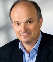 Heinrich Schima合作论文数Institute of Biomed Engineering and Physics, Medical University of Vienna;Department of Cardiac Surgery, Medical University of Vienna2
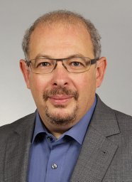 Leonid Goubergrits合作论文数Institute of Computer-assisted Cardiovascular Medicine, Charité – Universitätsmedizin Berlin2Ch. Große-Siestrup合作论文数2A. Wiessner合作论文数1
 Johannes Grosshauser合作论文数1Julia Parnell合作论文数1Vera Froese合作论文数1Laura Kursawe合作论文数1A. Petrich合作论文数1K. Reiter合作论文数1Sarah Schneider合作论文数1Albert Prause合作论文数FG Phys Chem Mol Mat Sci, Tech Univ Berlin1
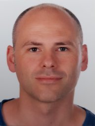 Ulrich Doll合作论文数Paul Scherrer Institut1
 Judith Kikhney合作论文数1Setsuo Takatani合作论文数1
 Ulrich Steinseifer合作论文数Institute of Applied Medical Engineering, Department of Cardiovascular Engineering, RWTH Aachen University1Tobias Timmel合作论文数1
 Annette Moter合作论文数1




