Introduction: There is limited literature to guide prognostication after resection of AVMs located in eloquent locations. The existing literature suffers from an over-reliance on gross measures of disability such as mRS which are less sensitive to non-motor deficits such as speech, sensory or visual deficits. In addition, all eloquent locations are frequently grouped together in common grading scales which reduces the clinical utility of such scales. This study is the first to provide granular outcomes data relating to each eloquence subtype in a large sample of this rare disease. Methods: Retrospective review of a prospectively maintained institutional database of AVMs was undertaken. Medical records and imaging were reviewed. Eloquence subtype was defined according to the potential neurological deficit corresponding to cortical or subcortical structures adjacent to the AVM (motor, sensory, visual, coordination, speech) Primary outcomes were focal neurological deficits and mRS at discharge, 6 months and 2 years postoperatively. Neurological deficits were defined as major, minor or intact at each timepoint (Table 1). Results: Of >1500 patients with AVMs in the database 815 patients have undergone one or more surgical resections. From 2000-2025, 274 patients underwent resection of AVMs in eloquent locations. 171 (62%) were ruptured. 40 patients (15%) were ≤18 years of age. 141 (51%) underwent preoperative embolization. 122 (45%) were neurologically intact preoperatively. Mean followup was 7.0±6.4 years. In patients who were intact preoperatively, 104 (85%) had a mRS of 0-2 at last followup. At last followup postoperatively 9 (7%) had a major deficit and 27 (22%) a minor deficit. Any deficits present at 2 years persisted. Eloquence subtype predicted persistent neurological deficits and mRS downgrade with speech, sensory and coordination deficits more likely to experience complete recovery of postoperative deficits (12/16, 64%) compared to visual (5/22, 23%), or brainstem deficits (1/6, 16%) (p<0.001, Table 2). Among patients with an unruptured AVM and a preoperative focal deficit, 4/9 (44%) experienced improvement or resolution of their preoperative deficit after surgical resection (1 speech, 3 visual). Conclusion: Functional preservation after AVM surgery is influenced by eloquence subtype. Preoperative focal deficit may be a novel indication for surgery.
Background: Post-stroke brain stimulation is a promising neurorestorative strategy, yet the underlying molecular mechanisms driving recovery remain unclear. Our prior work demonstrated that post-stroke optogenetic stimulation of the ipsilesional motor cortex (iM1) enhances functional recovery, and our RNA sequencing suggested cholesterol metabolism as a key pathway modulated by stimulation. Here, we examined the temporal dynamics of 3-hydroxy-3-methylglutaryl-CoA synthase 1 (HMGCS1), a key cholesterol enzyme, after stroke and assessed how optogenetic iM1 stimulation influences this expression. Methods: Male C57BL/6 mice (6-7 weeks) underwent stereotaxic surgery to express Channelrhodopsin in iM1 excitatory neurons and optical fiber implantation. After 5-6 weeks, mice received transient middle cerebral artery occlusion (30 min). Optogenetic stimulations were delivered from post-stroke days (PD) 5–14. Motor performance was assessed using the rotating beam test at pre-stroke baseline, PD4, 7 and 14. Brains were collected from stroke (PD1, 7&15) and sham control mice (n=4-5/group), and processed for immunohistochemistry using antibodies against HMGCS1, NeuN and CD68. Results: iM1 stimulation enhanced recovery at PD14, improving beam performance in both travel distance and speed (p<0.05). Previous RNA sequencing revealed involvement of multiple cholesterol biosynthesis and metabolism pathways at PD15 in iM1-stimulated mice. Immunostaining demonstrated that HMGCS1 was primarily expressed in primary motor cortex neurons and in peri-infarct glia. Compared with sham, iM1 showed a transient HMGCS1 increase at PD1, followed by reduced levels at PD7 and PD14, while contralateral M1 displayed reduced expression at all time points. At PD15, neuronal HMGCS1 in both stimulated and non-stimulated iM1 was significantly lower than in their respective contralateral M1 (p<0.05). In both regions, stimulated mice showed a trend toward higher HMGCS1 than non-stimulated mice. Stimulated mice also showed increased glial HMGCS1 in the peri-infarct region. Conclusions: Our findings link optogenetic stimulation-induced recovery to modulation of cholesterol metabolism, highlighting HMGCS1 as a potential molecular contributor. Future studies will validate cholesterol-related gene changes and clarify cell-type-specific contributions, with implications for targeting metabolic pathways to enhance neurorestoration.
Introduction: Moyamoya disease (MMD) is a rare cerebrovascular disease causing nonatherosclerotic intracranial arterial stenosis in children and young adults. Recent investigations have found that some genetic variants, such as RNF213 p.R4810K variant among East Asian populations, play an important role in MMD. Previously, we have identified a subset of patients with unique imaging features characterized by stenosis of the internal carotid artery (ICA) localized proximal to the terminal portion of ICA (non-terminal ICA), differing from the typical stenosis in MMD. This non-terminal stenosis was more common in Caucasian than in Asian patients; thus, investigating this unique feature may elucidate novel pathophysiology of MMD, especially in non-Asian population. Herein, we investigated the genetic background of non-terminal ICA stenosis in diverse ethnicities. Methods: We selected 46 sporadic nonhemorrhagic bilateral MMD patients aged 18-50 years old with diverse ethnic backgrounds and no significant comorbidities (Fig. 1). MRAs of each patient were visually assessed to evaluate the location of stenosis in ICAs and were categorized as “terminal stenosis” or “non-terminal stenosis”. Blood samples were analyzed for known single-nucleotide polymorphisms with risk of MMD in RNF213, ZXDC, DIAPH1, ACTA2, and GUCY1A1. Results: Representative images of patients with terminal ICA stenosis and non-terminal ICA stenosis were shown in Fig. 2. Non-terminal ICA stenosis was more frequent in Caucasians (5/15, 33.3%) compared to Asians (2/13, 15.4%) and other ethnicities (2/19, 10.5%). Genetic analysis revealed RNF213 p.R4810K variant only in Asian patients with terminal stenosis (Table 1). Among patients with non-terminal stenosis, ZXDC p.P562L variant was detected in two Caucasian patients; however, this variant was also detected in two Hispanic patients with terminal ICA stenosis. Two Black patients with terminal stenosis exhibited DIAPH1 c.534-2A>G variant, but no variants of DIAPH1 , ACTA2, and GUCY1A1 were detected in patients with non-terminal ICA stenosis Conclusion: Previously established variants of disease-causing genes, particularly RNF213 p.R4810K mutation, are insufficient to explain non-terminal ICA stenosis, an atypical feature of MMD. Future genomic analysis focusing on other rare genetic variants and epigenetic analysis may clarify the factors inducing this unique phenotype.
Background: Moyamoya disease (MMD) is a rare progressive cerebrovascular disease that can lead to ischemic and hemorrhagic strokes. The pathology of MMD arteries includes thickening of the innermost layer (intima), leading to the narrowing of arteries that supply oxygen to the brain. However, the mechanism underlying this process is largely unknown. We investigated the transcriptome differences of arterial samples from MMD patients and other vascular diseases. Specifically we analyzed the principal cell types - endothelial cells (EC) and vascular smooth muscle cells (VSMC) in MMD superior temporal artery (STA) and middle cerebral artery (MCA). Methods: STA and/or MCA were collected during surgeries in MMD, aneurysm, or cavernous malformation patients. MCA and STA samples were sectioned into 10µm thick slices and RNA transcripts stained through Advance Cell Diagnostics’ multiplex protocol for endothelial cells (PECAM1), vascular smooth muscle cells (ACTA2), proliferation marker (ki67), extracellular matrix (ECM), and DAPI for cell nuclei. Sections were imaged with Zeiss laser scanning microscope (LSM800) and analyzed with ImageJ. For spatial transcriptomic analysis, samples were formalin fixed, paraffin embedded and then processed through the Visium HD pipeline. Results: RNA transcriptome detection through RNAscope noted the presence of ki67, PECAM, ECM, and ACTA2. This aligns with previous immunohistochemistry staining for endothelial cells (CD31) and vascular smooth muscle cells (α-SMA). In addition, the morphology of VSMCs in MMD STA differs significantly; control STAs display a contractile phenotype with elongated, spindle-shaped cells, whereas MMD STAs exhibit synthetic phenotype with shorter, cobblestone appearance. Quantification of the intima layer thickness also show increased intima thickness in MMD STA. Spatial transcriptome analysis of these vessels have been processed to identify gene expression differences in these vessel layers in MMD. Conclusions: Our findings found RNA evidence of proliferation in the media layer, predominantly VSMCs, indicating a shift towards synthetic phenotype associated with increased cellular proliferation and reduced contractility, which can contribute to the MMD pathology. Ongoing studies include analysis of Visium HD transcriptome changes across different layers in MMD, aneurysm, and cavernous malformation to identify key molecular changes driving these pathological MMD features.
Stem cell transplantation holds promise for promoting recovery in the sub-acute and chronic phases of stroke. With this promise comes a mystery of the molecular mechanisms underlying stem cell actions, yet illumination of these mechanisms is crucial to enhance stem cell efficacy. In this study, we utilize Translating Ribosome Affinity Purification followed by RNA sequencing (TRAP-seq) in combination with secretome profiling of cell media to identify stem cell-secreted factors that could act as molecular targets for enhancing stem cell-mediated stroke recovery. Our clinically tested human neural stem cells (NR1), modified to express a ribosomal tag, were transplanted into the cortex of adult male Sprague Dawley rats (n=5), one week following stroke induced by distal middle cerebral artery occlusion. Two days following transplantation, brains were harvested and cortical tissue, surrounding the transplantation, excised. TRAP-seq was performed on the excised tissue (i.e. Translating Ribosome Affinity Purification, followed by RNAseq) to identify gene transcripts unique to NR1 transplantation. We identified 175 different NR1 genes encoding for known secreted factors. In parallel, to characterize the NR1 secretome in vitro at the protein level, we conducted a 63-plex Luminex Assays on NR1-conditioned cell media. Integrated transcriptomic and proteomic analysis reveals a subset of stem cell-secreted proteins known to be involved in pathways of synaptic plasticity, axonal remodeling, and neuroinflammation. These are all pathways that have been previously characterized in stroke brain recovery. Four novel gene candidates[GKS1] LGALS1, SPARC, VEGFA, and CXCL12,were prioritized for further characterization and functional screening using in vitro CRISPR-based knockout strategies. Overall, our findings identify key stem cell-secreted factors with potential roles in stem cell-based neural repair enhancing stroke recovery. Utilizing gene manipulation we will further characterize stem cell-based repair.
Background: Critically eloquent brain regions such as the brainstem, thalamus, internal capsule, basal ganglia and hypothalamus pose a challenge for treatment of vascular malformations such as cavernous malformations. This is due to their deep location, with significant mortality and morbidity associated with surgical treatment. Surgery remains the treatment of choice, although the core dilemma of how—and how much—to traverse functional brain remains an unaddressed. In principle, reducing the size of pial entry should limit disruption to eloquent structures and improve functional outcomes. Aims: We sought to determine whether the size of pial entry relative to lesion size predicts and/or contributes to determining long-term functional outcomes related to microsurgical treatment of vascular malformations in critically eloquent brain regions. We evaluated whether the use of the flexible ominiguide microtip laser achieves smaller entry geometries and better outcomes as compared to conventional approaches. Methods: We retrospectively analyzed 234 patients (140 laser, 94 conventional) lesion and entry size. A subgroup of 188 patients (101 laser, 87 conventional) had preoperative and long-term follow-up modified Rankin Scale (mRS) scores. Entry-to-lesion size ratios were compared across groups. Functional improvement was defined as a reduction in mRS from baseline to last follow-up. Logistic regression, spline modeling, and a causal mediation analysis were used to evaluate the predictive and mechanistic role of entry geometries. Results: Laser-assisted resection resulted in significantly lower entry-to-lesion ratios than conventional surgery (mean 0.47 vs. 0.66; p < 0.001), with lower average entry size as compared to traditional microsurgery (4.9 mm vs 8 mm). In the outcome-assessable subgroup, lower entry-to-lesion ratios independently predicted functional improvement ( p = 0.045; OR = 0.981, 95% CI: 0.964–0.998, p = 0.029). Spline modeling identified thresholds corresponding to ‘ideal’, ‘safe,’ and ‘risk’ zones for recovery probability. Mediation analysis demonstrated approximately 51% of the benefit of laser surgery was mediated through a reduction in entry-to-lesion ratio ( p = 0.034). Conclusion: Entry-to-lesion ratio is a significant, quantifiable predictor of neurological recovery after resection of deeply placed vascular malformations. Laser microsurgery facilitates smaller, more precise pial and ependymal entry, which may mediate its functional advantage.
Introduction: Acute hyperglycemia affects ~40% of stroke patients and worsens outcomes despite standard glucose control, yet no targeted therapy exists. We identify a previously unrecognized Metabolic–Complement–Vascular (MCV) axis, where hyperglycemia rapidly disrupts the endothelial glycocalyx and activates vascular complement within hours of stroke onset, defining a new, time-sensitive therapeutic target. Method: Male C57BL/6 mice (10-11 weeks) underwent 30 min transient MCAO with reperfusion to mimic thrombectomy. Hyperglycemia was induced by intraperitoneal glucose injection 10 min before occlusion. BBB disruption was assessed at multiple time points by Evans blue or IgM/IgG staining; neurological deficits, motor function (open field), and mortality were recorded. Glycocalyx disruption was measured by electron microscopy and IB4 staining; complement activation assessed by C3d immunostaining. The role of complement C3 was tested using C3 knockout mice and targeted inhibitor CR2-Crry. Human post-mortem ischemic stroke and control brain tissues (n = 5/group) were analyzed for glycocalyx integrity (UEA I lectin) and vascular immune injury (C3d/IgG). Pre-thrombectomy plasma complement markers (n=66) were analyzed via elastic net regression to predict outcomes. Result: Hyperglycemia caused rapid and severe luminal vascular injury within 4.5 h of stroke, with glycocalyx loss, luminal IgM/IgG deposition, vascular C3 activation, and BBB leakage (n=5, p<0.0001); not observed in normoglycemic stroke. Complement activation persisted after glucose normalization, was exacerbated by reperfusion, and propagated into the brain. This early vascular damage increased mortality (100% vs. 25%, p=0.0008) and worsened neurological deficits (p<0.001). C3 knockout mice had reduced BBB leakage (n=6, p<0.01) and improved function (p<0.01). Targeted C3 inhibition with CR2-Crry at 30 min post-reperfusion preserved BBB integrity and improved function (n=4, p<0.01), providing proof-of-concept for adjunct complement-targeted therapy. In human stroke brain, C3 activation colocalized with luminal glycocalyx loss. In human pre-thrombectomy plasma circulating complement activation markers (Ba, Bb, C4a, C3a) independently predicted modified Rankin Scale outcomes at discharge. Conclusion: These findings reframe acute hyperglycemic stroke as a rapid luminal vascular disorder and identify complement inhibition as a promising adjunct to reperfusion therapy.
Introduction: Moyamoya disease is a progressive cerebrovascular disorder. Revascularization is an effective procedures to reduce the risk for stroke. While the goal is to restore normal cerebral blood flow, predicting the outcome of this procedure is challenging. AI has shown great promise in predicting and synthesizing imaging results. Here, we a present a novel strategy using AI to predict post-operative CBF from pre-operative MRI scans in moyamoya. Methods: A conditional latent diffusion model based on the MONAI framework was used to train the prediction model. The model included variational autoencoders with 2D spatial dimensions and a diffusion process operating in the latent space. A total of 124 retrospective paired pre- and post-operative CBF images derived from ASL MRI were included for training the diffusion model. Preoperative CBF images were used as conditional inputs to predict 6-month postoperative outcomes as model outputs. A total of 25 cases were reserved for validation. Training utilized a multi-component loss function: perceptual loss to preserve anatomical features, adversarial loss to ensure realistic image quality, and mean squared error to ensure pixel-wise accuracy. We assessed prediction accuracy using structural similarity index (SSIM), mean squared error (MSE), and voxel-wise correlation coefficients. Results: Figure 1 shows the prediction of an example patient. Overall, the model successfully predicted the improved CBF. The model generated spatially coherent predictions that preserved anatomical structure and maintained reasonable intensity distributions. In terms of quantitative results, validation on the independent test set demonstrated mean SSIM scores of 0.77 and correlation coefficients of 0.63 between predicted and actual postoperative images. MSE values were low and averaged 0.14. Discussion: This novel application of conditional latent diffusion models demonstrates clinically relevant accuracy in predicting postoperative CBF changes in Moyamoya disease. The model offers potentially patient-specific predictions that could enhance patient selection and procedural planning. The approach advances precision medicine in cerebrovascular surgery by leveraging generative AI for medical imaging applications. While the approach demonstrates the feasibility of applying generative AI to CBF prediction, further optimization is needed to improve voxel-wise correspondence with postoperative changes, which is an area of our ongoing investigation.
Introduction: Except for vagal nerve stimulation, no treatment exists to restore function in chronic stroke patients. Several prior intracerebral stem cell trials were promising, but are not being further developed. Objective: NR1 is a human embryonic derived neural stem cell that improved motor-sensory function in rodent stroke models, and was expanded to produce GMP cryopreserved cell lots. The safety&efficacy of NR1 intracerebral transplantation in chronic stroke patients was assessed over 12 months. Methods: Inclusion Criteria: 18-75 yo; 6-60 mos post-ischemic subcortical MCA stroke; mRS 3-4. Subjects were transplanted with 2.5M, 5M, 10M or 20M. Primary Outcomes: Adverse events 0-12 mos; Change in total Fugl-Meyer motor score (FMMS, max 100) compared to baseline at 12 months (≥10 points improvement considered “clinically meaningful”). Other outcomes: UE FMMS, LE FMMS, Gait Speed test, Barthel Index (BI), NIHSS, MR FLAIR, Resting State fMRI and [18F]FDG PET. Results: 18 patients were transplanted. Adverse events included headache, worsened baseline expressive aphasia and asymptomatic chronic subdural hygroma, all resolving spontaneously. All 17 pts with f/u ≥3 mos demonstrated improved total FMMS and 11 of these 17 subjects showed clinically meaningful recovery in total FMMS. At 12 mos subjects increased 12.1 (+/- 1.8) points for total FMMS (p=0.00002), 7.4 (+/-1.6) points for UE FMMS (p=0.00057), 4.7 (+/-0.5) points for LE FMMS (p =0.0000009), 7.7 (+/-2.5) points for BI, while NIHSS improved by 1.77 (+/-0.47) and gait speed improved substantially. 14/18 pts demonstrated new transient FLAIR signal in premotor cortex at d7, that resolved by 2 mos, which was highly correlated with sustained neurologic recovery. Resting state fMRI showed improved functional brain connectivity in sensorimotor network, both ipsilesionally&contralesionally. FDG PET showed increased activity in the ipsilesional motor cortex&contralesional cerebellum. Conclusions: Intraparenchymal transplantation of NR1 cells in chronic stroke patients appears safe and well tolerated. Results suggest improved motor function starting at 1 mos and increasing to clinically meaningful recovery in most patients at 12 mos post-implant. UE FMMS improvement surpassed vagal nerve stimulation outcomes.
Introduction: The cross-talk between the brain and peripheral immune organs plays a crucial role in the response to stroke injury. Spleen responses are implicated in stroke pathology and inflammatory responses. Hyperglycemia is known to worsen stroke outcomes, with increased immune cell infiltration into the brain. However, it remains unclear whether hyperglycemia exacerbates spleen immune cell responses and whether this contributes to the heightened immune response in hyperglycemic stroke. Method: Male C57/BL6 mice (10-12 weeks) were subjected to transient middle cerebral artery occlusion (MCAO) for 30 minutes, followed by reperfusion to mimic ischemic stroke. Acute hyperglycemia was induced by glucose injection 10 minutes before MCAO. The study included three groups: sham, MCAO only, and MCAO with hyperglycemia (n=6-7 per group). At 24 hours post-stroke, spleen and brain immune cell populations were analyzed using our established 12-color flow cytometry technique, which simultaneously analyzes myeloid and lymphoid subpopulations. Results: Compared with sham controls, stroke significantly decreased the numbers of dendritic cells (2.33 ± 0.48 vs. 1.50 ± 0.42 million; p<0.05), NK cells (2.30 ± 0.37 vs. 1.21 ± 0.37 million; p<0.01), Ly6C+ macrophages (0.29 ± 0.12 vs. 0.11 ± 0.06 million; p<0.01), and total lymphocytes (56.9 ± 16.6 vs. 37.0 ± 5.1 million; p<0.05) at 24 hours post-stroke in the spleen, whereas there were no significant changes in neutrophils and Ly6C+ monocytes cell counts. Hyperglycemia during stroke did not alter spleen immune cell numbers compared to stroke alone. However, hyperglycemia significantly increased brain-infiltrating immune cells at 24 hours after stroke, including neutrophils, B cells, and CD8+ T cells compared to normoglycemic mice. Conclusion: This study provides insights into spleen immune cell responses to acute ischemic stroke and hyperglycemia. Significant changes in spleen immune cell populations after stroke confirm their potential roles in stroke pathology and recovery, warranting further investigation. Acute hyperglycemia, however, does not significantly affect spleen immune cell numbers after stroke, implying that the spleen is not the primary target of hyperglycemia-exacerbated immune cell infiltration into ischemic stroke brains. These results pave the way for exploring other mechanisms driving immune cell infiltration under hyperglycemic conditions.
The current spatial resolution of PET images is 3-4 mm for whole body PET/MR. Anatomical MR images with higher resolution and superior image quality have been used in PET reconstruction to improve the image quality and spatial resolution; however, mismatches between MR priors and actual tracer distribution can hinder accuracy. A novel PET reconstruction with MR priors, Magnetic Resonance-guided Block Sequential Regularized Expectation Maximum (MRgBSREM), that is robust to mismatches between anatomical priors and true activity distribution is proposed. This method is evaluated in diverse clinical settings using various tracers: 18F-florbetaben (FBB) in 373 subjects from a dementia study, 18F-FDG in a patient with chronic ischemic stroke, 18F-NaF in a knee study, and 15Owater in a patient with Moyamoya disease. Reconstruction using MRgBSREM visually improved both spatial resolution and image quality in all studies. In the 18F-florbetaben study, it mitigated white-matter spill-in into gray-matter as well as gray-matter spill over to the adjacent tissues, potentially leading to more accurate measurement of FBB uptake in the gray-matter. Visual assessment suggests that the proposed PET reconstruction enhances spatial resolution, which may contribute to improved diagnostic accuracy, while it displays robustness to mismatches between MR priors and true activity distribution.
Arterial spin labeling (ASL) MRI is a non-invasive perfusion imaging technique with potential for assessing hemodynamics in children. However, understanding hemodynamic changes in developing brains remains challenging. This study investigates the impact of normal brain development on ASL-derived cerebral blood flow (CBF) and arterial transit time (ATT) in MR-negative children. Thirty-two pediatric subjects (ages 0.7–17.9 years, mean 9.3 ± 5.3y, 19 male) with MR-negative findings were retrospectively included. Four pseudo-continuous ASL (PCASL) scans were acquired: single-delay (PLD 1525 or 2025 ms) and multi-delay (3 or 7 delays). CBF and ATT in supratentorial gray matter (GM), white matter (WM), and total-brain (TB) regions were analyzed using paired t-tests, Cohen’s d, Spearman correlation, and mixed linear regression models. Single-delay CBF was significantly higher than 3-delay CBF in GM and WM (p < 0.001 PLD 2025 ms; p = 0.02 PLD 1525 ms). WM and TB CBF correlated negatively with age (rho=-0.56, p < 0.001), whereas GM CBF showed no significant correlation (rho=-0.03, p = 0.87); the trends differed significantly (p = 0.01). GM and TB ATT increased with age (r2 > 0.11, p < 0.021). WM and TB CBF correlated with WM and combined WM/GM volumes (rho=-0.42, p = 0.02; rho=-0.46, p = 0.008). GM and WM exhibit distinct age-related hemodynamic patterns. WM perfusion declines with age and correlates with WM volume, while GM perfusion remains stable. The progressive increase in GM ATT highlights the need for cautious interpretation of single-delay ASL data in pediatric studies.
Background and ObjectivesFamilial cavernous malformations (FCMs) are vascular lesions that pose a lifelong risk of symptomatic hemorrhage (SH) and seizures, yet their natural history remains unclear. This study aims to determine the cumulative lifetime risk of a first SH and/or seizure and assess whether genetic variations influence these risks.MethodsThis international, multicenter retrospective cohort study included data from 16 tertiary referral centers and 1 patient advocacy group. Eligible patients had confirmed or suspected FCM, available magnetic resonance imaging (MRI) data, documented baseline clinical features, and longitudinal follow-up (FU). Functional outcomes were assessed using the modified Rankin Scale (mRS) at last FU. Direct adjusted survival curves and mixed-effects Cox regression analyses were performed to estimate cumulative lifetime risk. The association between genetic variations and SH/seizure rates was evaluated, and mixed-effects logistic regression assessed the effect of SH/seizures on mRS outcomes.ResultsA total of 1,592 patients with FCM were included, with a mean age of 37.6 years (SD 17.1) and 55.7% female. The median FU was 42 years (IQR: 27-55), totaling 64,146 person-years. Of these, 869 (54.6%) had confirmed FCM, 775 (48.7%) experienced at least 1 hemorrhage, and 447 (28.1%) had at least 1 seizure. Genetic testing was performed in 47.7%, identifying CCM1 (31.0%), CCM2 (4.8%), and CCM3 (1.9%) variations. The lifetime risk of a first SH was similar to 80%, with an event rate that remained constant beyond age 20. The lifetime risk of a first seizure was similar to 45%. Patients with CCM3 variations exhibited a more aggressive hemorrhagic course than those with CCM1 (hazard ratio 1.799, 95% CI 1.008-3.208). SH and seizures were independently associated with worse mRS outcomes at last FU.DiscussionThe event rate of SH and seizures remained stable over time, leading to high cumulative lifetime risks. Patients with CCM3 variations exhibited a more aggressive disease course. Limitations include the non-population-based design, selection bias from tertiary centers, retrospective data collection, and variability in data extraction across centers. However, this study represents the largest international FCM cohort to date, improving the precision of risk estimates and providing valuable insights into disease progression.
 Michael P. Marks合作论文数Radiology Department, Stanford University111
 Tonya Bliss合作论文数80
 Ma Yenari合作论文数63
 Michelle Y. Cheng合作论文数Department of Biology, Stanford University62
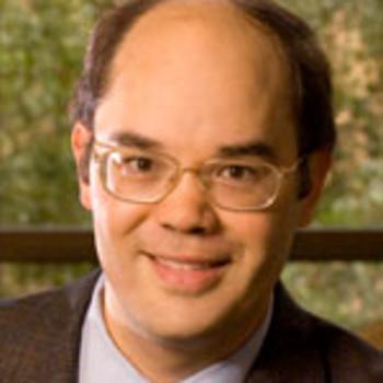 Steven D. Chang合作论文数Department of Neurosurgery, School of Medicine, Stanford University60
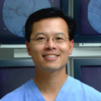 Huy M. Do合作论文数Center for Academic Medicine, Stanford University Medical Center54
 Heng Zhao合作论文数Department of Neurosurgery, Stanford University53Guohua Sun合作论文数53
 Robert Sapolsky合作论文数Department of Biological, Stanford University52GH Sun合作论文数47
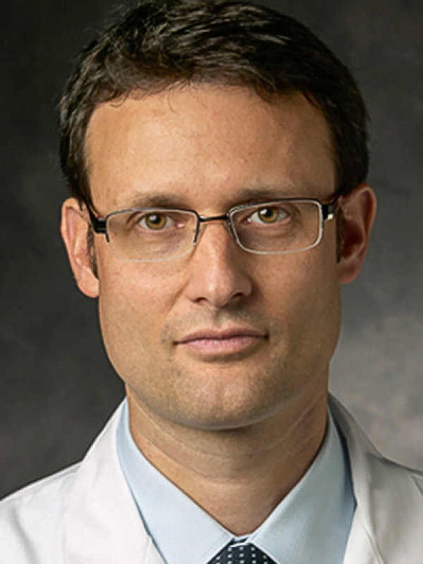 Raphael Guzman合作论文数Department of Neurosurgery, University Hospital Basel;Department of Biomedicine, Medizinische Fakultät, University of Basel41Terrance Chiang合作论文数39
 Robert Dodd合作论文数Neurosurgery Department, Stanford University/Radiology Department, Stanford University39
 Teresa Bell-Stephens合作论文数36
 Arjun Pendharkar合作论文数School of Medicine, Stanford University30
 Greg Zaharchuk合作论文数Center for Advanced Functional Neuroimaging, School of Medicine, Stanford University;Wu Tsai Neurosciences Institute, Stanford University24
 Ahmet Arac合作论文数Brain Research Institute, University of California, Los Angeles23Marcel Daadi合作论文数22
 Mario Teo合作论文数22
 Theo D. Palmer合作论文数Neurosurgery Department, Stanford University22
 Achal Singh Achrol合作论文数20
 Jeremy J. Heit合作论文数Stanford Neuroscience Health Center, Stanford University20
 Michael Moseley合作论文数Radiological Sciences Laboratory, Department of Radiology, Stanford Medicine;Richard M. Lucas Center for Imaging, Stanford Medicine20
 Chen Hansen合作论文数School of Chinese Medicine, The University of Hong Kong19
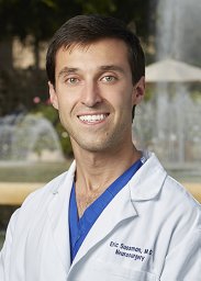 Sussman Eric S合作论文数19Alexander Lee合作论文数17Uchino Haruto合作论文数Sapporo Asabu Neurosurgical Hospital17David M. Kunis合作论文数17Marco Lee合作论文数17K. Frankel合作论文数16Anika Kim合作论文数16Ji Fabrikant合作论文数16
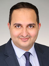 Omar A Choudhri合作论文数Department of Neurosurgery, University of Pennsylvania16
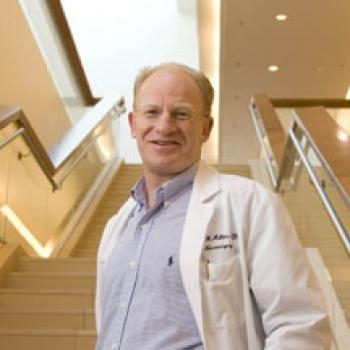 John R. Adler合作论文数Department of Neurosurgery, School of Medicine, Stanford University16
 Gerald Arthur Grant合作论文数Department of Neurosurgery, School of Medicine, Stanford University;Department of Neurosurgery, Duke University16
 Robert H Andres合作论文数16Nathan Manley合作论文数15
 Jaime R. Lopez合作论文数Neurosurgery Department, Stanford University15Robert Delapaz合作论文数Department of Radiology, Memorial Sloan-Kettering Cancer Center15
 Summer Han合作论文数Department of Adult Neurosurgery, School of Medicine, Stanford University15Mary L. Marcellus合作论文数14
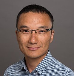 Moss Y Zhao合作论文数Dept Radiol, Stanford Univ14
 Kristen Yeom合作论文数Department of Pediatric Neurosurgery, Stanford University;Bio-X, Stanford University14
 Gregory W. Albers合作论文数Department of Neurosurgery, School of Medicine, Stanford University;Stanford Stroke Center, Stanford Medical Center, Stanford University14Cm Maier合作论文数14IG Fleetwood合作论文数13Eric Wang合作论文数13Cao Zhijuan合作论文数13
 Hannes Vogel合作论文数Department of Neurosurgery, School of Medicine, Stanford University;Department of Pathology, School of Medicine, Stanford University13




