Objective: Platform studies are an innovative clinical trial design to evaluate multiple potential therapies simultaneously using a master protocol framework for more rapid discovery of critical new disease-modifying therapies. Initially pioneered in cancer clinical trials, platform studies have more recently been adopted for new vaccine trials and other disease states. This approach has not been used for biomarker(s) validation.Approach: We present the development and successful implementation of a master protocol for a platform study in the National Institute of Diabetes and Digestive and Kidney Diseases Diabetic Foot Consortium (DFC) that concomitantly tests multiple biomarkers that could be used to predict diabetic foot ulcer (DFU) healing outcomes in people with DFUs.Results: This flexible master protocol, allowing the study of multiple biomarkers in a perpetual manner, designed to be inclusive of a broad spectrum of people with DFUs operating under the mantra "No DFU Goes Unstudied" successfully collects participant data and biospecimens for biomarker development and validation in a standardized manner across multiple DFC sites.Innovation: Integrating multiple biomarkers with a rich database of participants' clinical characteristics, patient-reported outcomes, and standard of care practices at various sites collected longitudinally over 52 weeks following a pragmatic approach and thus relevant to people with DFU as seen in today's daily practice, enable their subsequent implementation at the point of care to guide timely personalized DFU management decisions.Conclusion: Leveraging this robust platform will help to identify new therapeutic targets and design and conduct effective interventional clinical trials for DFU wound healing, which could inform the standards of care.
Wound healing is a complex biological process involving hemostasis, inflammation, proliferation, and remodeling. While this orchestrated response is essential for tissue repair, dysregulated healing often culminates in scar formation, characterized by fibrosis and loss of physiological tissue architecture. Scar formation impairs both aesthetic and functional outcomes, posing significant clinical challenges. We previously identified mechanical signaling, specifically through the Focal Adhesion Kinase (FAK) pathway, as a critical regulator of wound healing and fibrosis across various disease states. Elevated mechanical signaling drives fibrosis and scarring, whereas targeted manipulation of this pathway either by device or pharmacologic approaches blocks these adverse effects. We have performed extensive preclinical and clinical studies to demonstrate the potential of these interventions to minimize scarring, promote regenerative healing, and restore dermal architecture. This review highlights the importance of mechanical signaling modulation to achieve scarless healing in humans.
The inflammatory process resulting in the fibrotic encapsulation of implants has been well studied. However, how acellular dermal matrix (ADM) used in breast reconstruction elicits an attenuated foreign-body response (FBR) remains unclear. Here, by leveraging single-cell RNA-sequencing and proteomic data from pairs of fibrotically encapsulated specimens (bare silicone and silicone wrapped with ADM) collected from individuals undergoing breast reconstruction, we show that high levels of the extracellular-matrix protein osteopontin are associated with the use of ADM as a silicone wrapping. In mice with osteopontin knocked out, FBR attenuation by ADM-coated implants was abrogated. In wild-type mice, the sustained release of recombinant osteopontin from a hydrogel placed adjacent to a silicone implant attenuated the FBR in the absence of ADM. Our findings suggest strategies for the further minimization of the FBR. Transcriptomic and proteomic analyses show that high levels of the extracellular-matrix protein osteopontin are associated with the attenuated foreign-body response elicited by breast silicone implants wrapped with acellular dermal matrix.
SIGNIFICANCE:The Wound Healing Foundation recognized the need for consensus-based unbiased recommendations for the treatment of wounds. Consensus statements on the treatment of chronic wounds and acute wounds have been developed and published previously. The current publication on diabetic wounds represents the next step in this process. Diabetic wounds constitute a major problem. Population-based and meta-analytic studies indicate that the presence of foot wounds in patients with diabetes increases their mortality risk by more than twofold. The management of diabetic wounds requires consistent and evidence-driven intervention to achieve optimal clinical outcomes. This consensus statement provides the clinician with the necessary foundational approaches to the causes, diagnosis, and therapeutic management of diabetic wounds. Presented in a structured format, this is a useful guide for clinicians and learners in all patient care settings. RECENT ADVANCES:Continuous glucose monitoring and other new tools have facilitated better diabetes management and the management of associated wounds. Diabetic limb salvage should focus on achieving and optimizing function for the patient with diabetes rather than preserving limb tissue at all costs. CRITICAL ISSUES:Successful management of diabetic wounds requires a multidisciplinary approach encompassing comprehensive assessment, timely intervention, and collaborative care by the wound clinician with providers who can address critical aspects to achieve healing, including careful management of blood glucose levels, optimization of off-loading and physical therapy, assessment and treatment of limb ischemia, control and prevention of wound infection, and optimal pain management. FUTURE DIRECTIONS:Emerging treatments offer hope and promise, but the heterogenicity of diabetic wounds poses a challenge to performing good studies, which will be necessary to advance new treatments for diabetic wounds.
Trogocytosis is the process by which a recipient cell siphons small membrane fragments and proteins from a donor cell and can be utilized by cancer cells to avoid immune detection. We observed lymphocyte specific protein expressed by triple negative breast cancer (TNBC) cells via immunofluorescence imaging of patient samples. Image analysis of Cluster of Differentiation 45RA (CD45RA) expression, a naïve T cell specific protein, revealed that all stages of TNBCs express CD45RA. Flow cytometry revealed TNBC cells trogocytose CD45 protein from T cells. We also showed that the acquisition of these lymphoid markers is contact dependent. Confocal and super-resolution imaging further revealed CD45+ spherical structures containing T cell genomic DNA inside TNBC cells after co-culture. Trogocytosis between T cells and TNBC cells altered tumor cell expression of PTPRC, the gene that encodes for CD45. Our results revealed that CD45 is obtained by TNBC cells from T cells via trogocytosis and that TNBC cells express CD45 intracellularly and on the membrane.
OBJECTIVE:The National Institute of Diabetes and Digestive and Kidney Diseases Diabetic Foot Consortium tested the hypothesis that compromised restoration of the skin barrier function of closed diabetic foot ulcers (DFUs), as measured by high transepidermal water loss (TEWL), is associated with an increased risk of DFU recurrence. RESEARCH DESIGN AND METHODS:This was a multicenter noninterventional study measuring TEWL in 418 adult participants with diabetes and a recently healed DFU. TEWL was measured at the center of the closed wound and at an anatomically similar reference area on the contralateral foot within 2 weeks of wound closure (visit 1); measurements were repeated at a wound closure confirmation visit 2 weeks later (visit 2). Participants were observed for up to 16 weeks to assess for wound recurrence. Participant self-reported and clinician assessments of DFU wound recurrence were recorded. RESULTS:DFU recurrence by week 16 occurred in 21.5% of participants. Mean TEWL at the center of the healed DFU at visit 1 was higher for those with recurrence compared with those without (P = 0.006). Among participants with high TEWL (>30.05 g · m-2 · h-1), 35% reported wound recurrence by 16 weeks versus 17% of those with low TEWL. The odds ratio for recurrence for participants with high TEWL was 2.66 (P < 0.001). Self-reported wound recurrence was highly concordant with clinician assessment of wound recurrence. CONCLUSIONS:Compromised wound healing mechanisms culminating in wound closure associated with defective skin barrier function is associated with increased risk of DFU recurrence. Measurement of TEWL has value as a predictor of functional wound healing and could affect clinical practice, leading to better outcomes.
In response to injury, a variety of different cells are recruited to sites of injury to facilitate healing. Recent studies have examined the importance of the heterogeneity of tissue resident fibroblasts and mechanical signalling pathways in healing and fibrosis. However, tissue repair and the inflammatory response also involves blood cells that are recruited from the circulation. Here we identify mechanoresponsive myeloid subpopulations present in scar and unwounded skin. We then modulate these subpopulations by manipulating mechanical strain in vivo and in vitro and find that specifically targeting myeloid mechanical signalling is sufficient to reduce the pro-fibrotic myeloid subpopulations and restore the native, anti-inflammatory subpopulations. In addition, myeloid-specific mechanotransduction ablation also downregulates downstream pro-fibrotic fibroblast transcriptional profiles, reducing scar formation. As inflammatory cells circulate and home to injury sites during the initial healing phases in all organs, focusing on mechanoresponsive myeloid subpopulations may generate additional directions for systemic immunomodulatory therapies to target fibrosis and other diseases across other internal organ systems. Myeloid-specific mechanotransduction ablation downregulates pro-fibrotic fibroblast transcriptional profiles to reduce scar formation in human cells
Modulating mechanotransduction by inhibiting yes-associated protein (YAP) in mice yields wound regeneration without scarring. However, rodents are loose-skinned and fail to recapitulate key aspects of human wound repair. We sought to elucidate the effects of YAP inhibition in red Duroc pig wounds, the most human-like model of scarring. We show that one-time treatment with verteporfin, a YAP inhibitor, immediately after wounding is sufficient to prevent scarring and to drive wound regeneration in pigs. By performing single-cell RNA sequencing (scRNA-seq) on porcine wounds in conjunction with spatial proteomic analysis, we found perturbations in fibroblast dynamics with verteporfin treatment and the presence of putative pro-regenerative/profibrotic fibroblasts enriched in regenerating/scarring pig wounds, respectively. We also identified differences in enriched myeloid cell subpopulations after treatment and linked this observation to increased elaboration of interleukin-33 (IL-33) in regenerating wounds. Finally, we validated our findings in a xenograft wound model containing human neonatal foreskin engrafted onto nude mice and used scRNA-seq of human wound cells to draw parallels with fibroblast subpopulation dynamics in porcine wounds. Collectively, our findings provide support for the clinical translation of local mechanotransduction inhibitors to prevent human skin scarring, and they clarify a YAP/IL-33 signaling axis in large animal wound regeneration.
Rationale: Elevated shear stress (ESS) induces vascular remodeling in veins exposed to arterial blood flow, which can lead to arteriovenous (AV) fistula failure. The molecular mechanisms driving remodeling have not been comprehensively examined with a single-cell resolution before. Objective: Using an in vivo animal mode, single-cell RNA sequencing, and histopathology, we precisely manipulate blood flow to comprehensively characterize all cell subpopulations important during vascular remodeling. Methods: AV loops were created in saphenous vessels of rats using a contralateral saphenous vein interposition graft to promote ESS. Saphenous veins with no elevated shear stress (NSS) were anastomosed as controls. Findings: ESS promoted transcriptional homogeneity, and NSS promoted considerable heterogeneity. Specifically, ESS endothelial cells (ECs) showed a more homogeneous transcriptional response promoting angiogenesis and upregulating endothelial-to-mesenchymal transition inhibiting genes (Klf2). NSS ECs upregulated antiproliferation genes such as Cav1, Cst3, and Btg1. In macrophages, ESS promoted a large homogeneous subpopulation, creating a mechanically activated, proinflammatory and thus proangiogenic myeloid phenotype, whereas NSS myeloid cells expressed the anti-inflammatory and antiangiogenetic marker Mrc1. Conclusion: ESS activates unified gene expression profiles to induce adaption of the vessel wall to hemodynamic alterations. Targeted depletion of the identified cellular subpopulations may lead to novel therapies to prevent excessive venous remodeling, intimal hyperplasia, and AV fistula failure.
MINI-ABSTRACT:In this study, we present the first-in-human use of topical deferoxamine (DFO) in the treatment of a beta-thalassemia wound. We elected to use DFO on a patient that suffered from a chronic nonhealing wound in the setting of beta-thalassemia. Despite approximately 55 weeks of marginal improvement in healing, this patient's wound healed completely after 21 weeks of treatment with DFO. We believe that DFO has the potential to accelerate healing in beta-thalassemia wounds through iron chelation.
Breast lipofilling, a popular cosmetic and reconstructive procedure, involves the transplantation of autologous fat to enhance breast volume and contour. Despite its widespread use, cell processing and the aftertreatment remain controversial. This study investigates the pressure applied by a compression bra and reports in vitro stress tests of processed and unprocessed fat cells. Clinical bra pressure measurements were conducted on a cohort of 45 patients following lipofilling, reduction mammoplasties and DIEP flaps. Laboratory analysis included cell vitality testing using Resazurin assays of processed and unprocessed fat cells after exposure to mechanical or hyperbaric pressure. Our findings show a mean overall pressure value of the compression bra for all patients of 6.7 ± 5.7 mmHg (range 0–35). Cell processing is superior to sedimentation only regarding fat cell vitality. However, neither mechanical pressure within the specified range nor hyperbaric oxygen exposure significantly affected fat graft survival as measured by Resazurin assays. The in vitro measurements showed that it was impossible to harm fat cells with external pressure during lipofilling procedures, regardless of their processing. In the clinical context, the compression bra applied pressure values deceeding the perfusion pressure and may therefore not diminish oxygen supply nor harm the transplanted cells. Therefore, we recommend the use of a compression bra for all lipofilling procedures around the breast. This journal requires that authors assign a level of evidence to each article. For a full description of these evidence-based medicine ratings, please refer to Table of Contents or online Instructions to Authors www.springer.com/00266.
Objectives: Oats (Avena sativa) are an important whole grain with lipid lowering and anti-inflammatory effects. Recent studies revealed that oat-derived bioactives, avenanthramide (AVE) and beta-glucan (βG), stimulate wound healing by inducing angiogenesis and recruiting endothelial progenitor cells. We here report the effect of AVE and βG on healing velocity and acceleration, characterizing wound healing dynamics. Methods: AVE and βG extracted from whole oats were solubilized in buffer (0.1%, 0.5%, 1%) and applied by SQ injection to the margins of full-thickness excisional wounds on the dorsum of C57BL/6 mice every two days and compared to controls. Surface planimetry was assessed using digital photographs taken every two post-operative days. The velocity of wound healing (Δwound area/Δtime) induced by each bioactive was calculated by linear regression as the slope between time points; acceleration (Δvelocity/Δtime) was determined as a higher order curve. Results: Compared to controls, both AVE and βG resulted in greater healing velocity, with distinct temporal dynamics. An early effect was observed with βG (before POD 2) while AVE had a later effect on healing velocity (after POD 2). 1% AVE resulted in the highest maximum velocity compared to control (21.7 vs.11.7), a 1.9-fold faster healing speed. Both 1% and 0.1% βG achieved comparable maximum velocities (21.4 vs and 21.3) but earlier in the healing process. Analysis of wound healing acceleration revealed differential dynamics between bioactives. Conclusions: Oat bioactives augment normal wound healing by improving wound healing dynamics. This is the first description of wound healing velocity and acceleration induced by AVE and βG, correlated with faster wound closure in vivo. Velocity and acceleration are novel metrics that characterize the dynamic biological effect of oat bioactives beyond completed wound closure. Funding Sources: AVE and βG supplied by Ceapro, Inc., Edmonton, Alberta.
Chronic non-healing wounds significantly strain modern healthcare systems, affecting 1–2% of the population in developed countries with costs ranging between $28.1 and $96.8 billion annually. Additionally, it has been established that chronic wounds resulting from comorbidities, such as peripheral vascular disease and diabetes mellitus, tend to be polymicrobial in nature. Treatment of polymicrobial chronic wounds with oral and IV antibiotics can result in antimicrobial resistance, leading to more difficult-to-treat wounds. Ideally, chronic ulcers would be topically treated with antibiotic combinations tailored to the microbiome of a patient’s wound. We have previously shown that a topical collagen-rich hydrogel (cHG) can elute single antibiotics to inhibit bacterial growth in a manner that is nontoxic to mammalian cells. Here, we analyzed the microbiology of cultures taken from human patients diagnosed with diabetes mellitus suffering from chronic wounds present for more than 6 weeks. Additionally, we examined the safety of the elution of multiple antibiotics from collagen-rich hydrogel in mammalian cells in vivo. Finally, we aimed to create tailored combinations of antibiotics impregnated into cHG to successfully target and treat infections and eradicate biofilms cultured from human chronic diabetic wound tissue. We found that the majority of human chronic wounds in our study were polymicrobial in nature. The elution of multiple antibiotics from cHG was well-tolerated in mammalian cells, making it a potential topical treatment of the polymicrobial chronic wound. Finally, combinations of antibiotics tailored to each patient’s microbiome eluted from a collagen-rich hydrogel successfully treated bacterial cultures isolated from patient samples via an in vitro assay.
This study aims to systematically identify studies that evaluate lower extremity burn injury in the diabetic population, evaluate their clinical course and patient outcomes, and present a treatment algorithm tailored to diabetic burn patients. Our systematic review of the PubMed and Web of Science databases yielded 429 unique articles. After exclusion and inclusion criteria were applied, 59 articles were selected for evaluation. In diabetic patients, the thermal injury was largely a result of decreased awareness and education regarding heat therapies in the context of peripheral neuropathy. All noncase studies found that metrics such as hospital length of stay, ICU admission rates, rates of comorbidity, complication rates, scald injuries, infection rates, and cost of treatment were significantly increased in the diabetic burn population as compared to their nondiabetic counterparts. Where infection was present, microorganisms colonizing diabetic burn wounds were different than those found in the burn wounds of immunocompetent individuals. Operative intervention including split-skin graft, amputation, and debridement were more often utilized in diabetic burn patients. Foot burns in diabetic patients pose unique clinical risks to patients, and as such need to be an alternate treatment protocol to reflect their pathology. Education and training programs are crucial in the prevention of diabetic foot burns to avoid complications, protracted healing, and adverse outcomes. A unique algorithm can guide the unique treatment of this clinical entity.
IntroductionThe FOXC2 transcription factor has been tied to a wide range of disease states, serving as a promising prognostic biomarker associated with aggressive basal-like human breast cancers (increased cancer invasion and metastasis). Dysregulation of FOXC2 expression has also been found to promote defects in lymphatic remodeling and hyperplastic lymphedema-distichiasis (LD). Since chronic lymphedema is a forerunner of several malignancies and cancers have been known to arise from poorly healing chronic wounds (e.g., Marjolin ulcers), we examined the effect of Foxc2 dysfunction on skin wound healing.MethodsWe used our splinted excisional wounding model that mimics human-like wound healing on wildtype and Foxc2+/− mice (n = 4), which demonstrate incomplete lymphatic vasculature and lymphatic dysfunction. Wound size was measured over the course of 18 days. Tissue was explanted from both groups at post-operative day (POD) 14 and 18 and stained with Masson’s Trichrome to assess scar formation, Picrosirius Red for dermal integrity, or immunofluorescence to assess lymphatic (LYVE1) cell populations.ResultsWildtype mice completely healed by POD 14, while Foxc2+/−mice did not completely heal until POD18. Scar area of healed Foxc2+/−mice (POD 18) was larger than that of healed wild-type mice (POD 14; p = 0.0294). At POD 14, collagen "bers in the scars of Foxc2+/−mice to be narrower (p = 0.0117) and more highly aligned (p = 0.0110), indicating signi"cantly more "brosis in these mice. Collagen "bers in both groups became longer (p = 0.0116) and wider (p = 0.0020) from POD 14 to 18, indicating a temporal evolution of "brosis. Foxc2+/−mice also had lower numbers of LYVE1+, F4/80+ and CD4+ cells compared to wildtype mice.DiscussionIndividuals over 65 years old are more likely to develop cancer and are highly susceptible to developing chronic wounds. Here, we found that FOXC2, which is tied to cancer metastasis and lymphatic dysregulation, also impairs wound healing and promotes "brotic tissue architecture. With FOXC2 proposed as a potential therapeutic target for cancer metastasis, its downstream systemic effects should be considered against the increased chance of developing nonhealing wounds. Further delineation of the microenvironment, cellular events, and molecular signals during normal and Foxc2-associated abnormal wound healing will improve clinical therapies targeting this important marker.
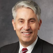 Michael T. Longaker合作论文数Department of Materials Science & Engineering, Stanford University350
 Michael Januszyk合作论文数Department of Surgery, Stanford University196
 Kellen Chen合作论文数Stanford Univ, Surg, Stanford, CA 94305 USA108
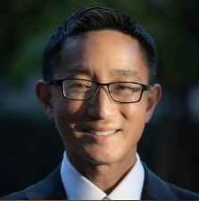 Derrick Wan合作论文数Department of Surgery, School of Medicine, Stanford University103
 Michael Sorkin合作论文数95
 Zeshaan N Maan合作论文数Maan Plastic Surgery94Rennert Robert C合作论文数89Victor W. Wong合作论文数89
 Dominik Duscher合作论文数Tomorrowlabs GmbH;MoleQlar86
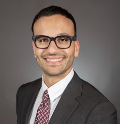 Dominic Henn合作论文数Stanford Univ, Stanford, CA 94305 USA80
 Jason P Glotzbach合作论文数Hagey Lab Pediat Regenerat Med, Stanford Univ79Michael Hu合作论文数78
 Melanie Rodrigues合作论文数Department of Surgery, Stanford University66
 Dharshan Sivaraj合作论文数Stanford Univ, Surg, Stanford, CA 94305 USA63
 Walmsley Graham G合作论文数62
 Padmanabhan Jagannath合作论文数Department of Biomedical Engineering, Yale University;Center for Research on Interface Structures and Phenomena, Yale University;Yale University, Yale University61Clark A. Bonham合作论文数58
 Jayakumar Rajadas合作论文数Biomaterials and Advance Drug Delivery Lab, School of Medicine;Stanford University;Stanford;School of Medicine, Stanford University55
 Artem A Trotsyuk合作论文数Stanford Univ, Surg, Stanford, CA 94305 USA52H Peter Lorenz合作论文数50Kristine C. Rustad合作论文数Stanford University49
 Janos A. Barrera合作论文数Department of Surgery, Stanford University43Sun Hyung Kwon合作论文数Stanford Univ, Stanford, CA 94305 USA37Katharina Fischer合作论文数Department of Surgery, University of Arizona College of Medicine37
 Alexander Whittam合作论文数37
 Michelle Griffin合作论文数Gynaecological Cancer Research Centre, UCL EGA Institute for Women's Health37Hudson C. Kussie合作论文数Dept Surg, Stanford Univ36
 Robert D Galiano合作论文数Department of Dermatology, Feinberg School of Medicine, Northwestern University36
 Tevlin Ruth合作论文数34Elizabeth R. Zielins合作论文数30
 Edward I Chang合作论文数30
 Mimi R. Borrelli合作论文数Stanford, Menlo Pk, CA USA29
 Shamik Mascharak合作论文数Stanford Univ, Sch Med, Stanford, CA 94305 USA28
 Arash Momeni合作论文数Department of Statistical;Physics and Nonlinear Dynamics;Institute of Theoretical Physics;Clausthal University of Technology;Institute of Theoretical Physics, Clausthal University of Technology28Ivan N Vial合作论文数27kevin paik合作论文数27Adrian Mcardle合作论文数27
 David Atashroo合作论文数26
 Michael W Findlay合作论文数25Britta Kuehlmann合作论文数25
 Jamie P. Levine合作论文数Department of Plastic Surgery, Grossman School of Medicine, New York University25Benjamin Levi合作论文数24
 David Perrault合作论文数Division of Plastic and Reconstructive Surgert, Stanford University School of Medicine23Hong Wan Xing合作论文数23V.W. Wong合作论文数22
 Michael Galvez合作论文数Stanford University22
 Michael Hoonbae Chung合作论文数21
 Deshka Foster合作论文数Dept Surg, Stanford Univ21
 Irving Weissman合作论文数Department of Developmental Biology, Stanford University;Department of Biology and Neurosurgery, Stanford University;Department of Pathology, Stanford University21




