Bone marrow endothelial cells (BM-ECs) are the essential components of the BM niche and support the function of hematopoietic stem cells (HSCs). However, conditioning for HSC transplantation causes damage to the recipients' BM-ECs and may lead to transplantation-related morbidity. Here, we investigated the cellular and clonal mechanisms of BM-EC regeneration after irradiative conditioning. Using single-cell RNA sequencing, imaging, and flow cytometry, we revealed how the heterogeneous pool of BM-ECs changes during regeneration from irradiation stress. Next, we developed a single-cell in vitro clonogenic assay and demonstrated that all EC fractions hold a high potential to reenter the cell cycle and form vessel-like structures. Finally, we used Rainbow mice and a machine-learning-based model to show that the regeneration of BM-ECs after irradiation is mostly polyclonal and driven by the broad fraction of BM-ECs; however, the cell output among clones varies at later stages of regeneration.
Abstract Chemotherapy exposure induces cellular stress and can upregulate ‘Eat me’ signals on the surface of cancer cells thereby targeting these cells for clearance by macrophage phagocytosis. However, this phagocytosis can be inhibited by ‘Don’t eat me’ (DEM) signals like CD24 on the surface of tumor cells. The clinical-stage anti-CD24 monoclonal antibody PHST001 blocks the CD24 DEM signal and promotes macrophage phagocytosis of target cells, raising the possibility that cotreatment with PHST001 and chemotherapy could provide combinatorial benefit to patients. To identify potential PHST001 and chemotherapy combinations, we screened 17 standard-of-care chemotherapy agents with PHST001 in coculture of peripheral monocyte-derived human macrophages and each of 13 cell lines representing 6 different cancer indications. We identified several common classes of chemotherapy drugs, including taxanes, platins, topoisomerase inhibitors, and anti-metabolites that enhance PHST001-induced macrophage clearance of cancer cells. We found that while cancer cells are generally sensitive to chemotherapy, macrophages display remarkable resistance to many chemotherapy agents. We further validated PHST001 combination with several chemotherapies in two-way dose-response experiments and identified chemotherapies that synergize with PHST001 to clear cancer cells in macrophage coculture. We subsequently validated select chemotherapy and PHST001 combinations (e.g. cisplatin) in vivo with mouse xenograft studies. Mechanistically, we discovered that certain chemotherapy agents increased the surface expression of the ‘Eat me’ signal phosphatidylserine (PS) on cancer cells, while others directly enhanced the phagocytosis capacity of macrophages. Taken together, these data support therapeutic combination of PHST001 with multiple standard-of-care chemotherapies to deepen tumor-killing response in patients. Citation Format: Giovanni C. Forcina, Kelsey E. Hart, Joseane Sampaio, Joshua D. Rudolph, Suzana A. Kahn, Raphaël F. Rousseau, Amira A. Barkal, Ravindra Majeti, Irving L. Weissman, Roy L. Maute, Jennifer Yinuo Cao, . PHST001 combination with standard-of-care chemotherapy enhances macrophage-mediated elimination of tumor cells [abstract]. In: Proceedings of the American Association for Cancer Research Annual Meeting 2026; Part 1 (Regular Abstracts); 2026 Apr 17-22; San Diego, CA. Philadelphia (PA): AACR; Cancer Res 2026;86(7 Suppl):Abstract nr 1557.
Hematopoietic stem cells (HSCs) are multipotent self-renewing cells that give rise to all types of blood cells. Past research has identified that long-term hematopoietic stem cells in young mice and humans produce a balanced output of lymphoid and myeloid cells, while in old age, they are largely replaced by myeloid-biased HSCs (My-HSC). It has not yet been determined whether this transition results from epigenetic changes in a single population of HSC or if two or more subsets of HSCs exist that gain or lose dominance with age via processes of selection. Whether epigenetic change or competition, several characteristics of each may exist to ensure that the appropriate subset is placed in niches that support them. HSC can be mobilized into the blood and home selectively to target tissues via expression of "homing receptors," but these molecules do not determine their intraorgan migration to appropriate niches. Chemokines are the class of molecules that determine intraorgan migration of cells. Here, we show that the chemokine receptor CCR5 is mainly expressed on My-HSCs, and therefore, the frequency of CCR5+ HSCs increases with age. Aged HSCs negative for CCR5 expression generate lower frequency of myeloid cells than lymphoid cells upon transplantation into recipients. Additionally, disruption of the CCL5-CCR5 signaling axis changes frequency of lymphoid populations in peripheral blood of aged mice, supporting research that shows the depletion of My-HSCs can result in the rejuvenation of adaptive immunity.
Messenger RNA (mRNA) transfection enables rapid, transient protein expression without nuclear entry, providing a powerful alternative to DNA or viral delivery in post-mitotic and otherwise difficult-to-transfect cells. Although in vitro transcribed (IVT) mRNAs have revolutionized therapeutic applications, their adoption in experimental biology remains limited by challenges in synthesis, variability across cell types, and concerns about cytotoxicity. Here, we define design principles that maximize IVT mRNA performance across diverse cellular and organismal systems. Through systematic comparison of capping strategies and base modifications, including N1-methyl-pseudouridine, 5-methylcytidine, and 5-methoxyuridine, we identify modifications that enhance translation while minimizing activation of cellular stress responses. Optimized transcripts drive robust protein expression within four hours, persist for up to one week, and support multiplexed expression of structurally and functionally distinct proteins in mammalian cells, including cancer cell lines, iPSC-derived systems, primary cells, and organoids, as well as in vivo in zebrafish embryos and in less genetically tractable models such as Danionella cerebrum and sea urchin embryos. To further expand accessibility for community use, we developed mRNAbow , a platform for generating low-toxicity mRNAs encoding organelle-targeted fluorescent proteins and biosensors for multiplex imaging, with corresponding plasmids made publicly available. Together, these advances establish a generalizable framework for IVT mRNA design and expand experimental access to synthetic mRNA technologies for dissecting cellular architecture and dynamics. ### Competing Interest Statement The authors have declared no competing interest. Howard Hughes Medical Institute
Microglia are the resident hematopoietic cells of the central nervous system 1 . In mice, microglia seed the brain during embryogenesis and can be maintained throughout life with minimal input from adult hematopoiesis 2-4 . The origins of human microglia are less clear, but recent evidence suggests that marrow-derived cells may be able to supplement the human microglial pool in certain individuals 5,6 . Here, to investigate the ontogeny of human microglia, we develop a method that uses the collection of accumulated somatic mutations which uniquely labels each clone of cells to track the infiltration of marrow-derived cells into the human brain. Applying this method to 20 aged individuals, we find evidence of an influx of marrow-derived cells into the brain in all examined individuals. Single cell analysis, including single cell lineage tracing using mitochondrial DNA variants, demonstrates that these infiltrating cells are nearly identical to microglia and can comprise a large fraction of the microglial pool. Analysis of large-scale sequencing cohorts demonstrates a protective association between most types of clonal hematopoiesis and Alzheimer's disease. In sum, this work uncovers a widespread influx of myeloid cells into the healthy human brain which serves to reinforce the pool of human microglia and becomes common with aging.
PURPOSE:CD24 is a "don't eat me" signal overexpressed across multiple solid tumors and contributes to immune evasion by suppressing macrophage-mediated phagocytosis. Targeting the CD24/SIGLEC-10 axis represents a novel immuno-oncology strategy to restore innate immune surveillance. EXPERIMENTAL DESIGN:We developed PHST001, a humanized IgG4 monoclonal antibody targeting CD24, and evaluated its activity using in vitro phagocytosis assays, xenograft and immunocompetent syngeneic mouse models, and ex vivo systems incorporating human immune cells and tumor samples. Nonclinical safety parameters were assessed to evaluate translational feasibility. RESULTS:PHST001 binds CD24 with high affinity and blocks SIGLEC-10 engagement, resulting in enhanced macrophage-mediated phagocytosis across multiple tumor indications and subtypes, inhibition of primary and metastatic tumor growth, and prolonged survival in preclinical models. PHST001 demonstrated a favorable nonclinical safety profile and exhibited antitumor activity as both monotherapy and in combination with standard-of-care treatments, including chemotherapy, radiotherapy, and antibody-drug conjugates. Antitumor responses were associated with engagement of tissue-resident macrophages and in syngeneic models, induction of tumor-reactive T-cell responses, supporting a role for CD24 in coordinating innate and adaptive immunosuppression. CONCLUSIONS:These findings establish CD24 as a critical regulator of tumor immune evasion and support the clinical development of PHST001 as a CD24-targeted immunotherapy. A phase I clinical study (NCT06840886) evaluating the safety and tolerability of PHST001 in adult patients with relapsed or refractory solid tumors is ongoing.
While it was once thought that neurogenesis is complete by birth, it is now apparent that the human brain continues to generate new neurons postnatally, at least into childhood. While much attention has been focused on postnatally-born neurons, their presumed progenitor - the postnatal neural stem cell (NSC) - remains poorly characterized. Using index sorting, we identify and prospectively isolate two subsets of NSCs from the postnatal human brain, and describe their differentiation dynamics using clonal barcoding and in vivo xenotransplantation. We demonstrate an A2B5 + EGFR + population biased towards interneuron and oligodendrocyte fates (NINO), and an A2B5 - EGFR hi population biased towards an astrocyte fate (NAC). Profiling of human brains across lifespan shows that the frequency of NSCs declined exponentially across the first two decades of life, but stabilized thereafter, still present in the brains of donors as old as 90 years. Our study provides a framework for the functional study of postnatal human NSCs and their potential roles in development, aging, and disease.
Microglia are the resident macrophages of the central nervous system1. In mice, microglia seed the brain during embryogenesis and can be maintained throughout life with minimal input from adult haematopoiesis2-4. The origins of human microglia are less clear, but recent evidence suggests that bone-marrow-derived cells contribute to the human microglial pool in certain individuals5-9. Here, to investigate the ontogeny of human microglia, we develop an approach that uses the collection of accumulated somatic mutations that uniquely labels each clone of cells to track the infiltration of bone-marrow-derived cells into the human brain. Applying this approach to 20 older individuals, we find evidence of an influx of bone-marrow-derived cells into the brain in all examined individuals. Single-cell analysis, including single-cell lineage tracing using mitochondrial DNA variants, demonstrates that these infiltrating cells are similar to microglia and can comprise a large fraction of the microglial pool. Analysis of human cohort data demonstrates a protective association between most types of clonal haematopoiesis and Alzheimer's disease. Together, we identify a widespread influx of myeloid cells into the healthy human brain that contributes to the pool of human microglia and becomes common with ageing.
Abstract Background: CD24 is a highly glycosylated tumor antigen with a restricted expression profile that acts as macrophage “don’t eat me” signal. CD24 is highly expressed by many human cancers and interacts with the macrophage receptor Siglec-10 to protect cancer cells from phagocytosis. PHST001 is a clinical-stage humanized anti-CD24 monoclonal antibody of IgG4 isotype currently in clinical evaluation. In vivo, PHST001 shows significantly greater efficacy against solid tumors than the anti-CD47 antibody Magrolimab. We assessed PHST001's preclinical efficacy against metastatic tumors and the role of Fc receptor engagement in its anti-tumor activity. Results and Methods: BT474, a human Her2+ breast tumor cell line, metastasizes to the axillary lymph nodes when engrafted in the mammary fat pad of NSG mice. In contrast to control-treated mice, PHST001 treatment inhibited the formation of lymph node metastases in orthotopic xenograft models of BT474. To generate a disseminated metastatic model, BT474 cells were engrafted intracardially. A metastatic model was also generated in a syngeneic system by expressing human CD24 in MC38 cells (MC38-huCD24) and implanting tumor cells intracardially. PHST001 treatment significantly reduced the number and size of metastatic lesions in both xenograft BT474 and immune competent MC38-huCD24 models. To define PHST001’s mechanism of action, a variant antibody bearing a LALAGANA mutation (Fc-inert) was generated. This variant induced phagocytosis of tumor cells to a lesser extent than PHST001 (human IgG4), demonstrating the contribution of Fc receptor engagement to PHST001 efficacy. When combined with a companion agent, such as the anti-Her2 ADC trastuzumab deruxtecan, the Fc-inert version of PHST001 dramatically increased phagocytosis, demonstrating the contribution of CD24 blockade to PHST001 efficacy in absence of direct Fc receptor engagement. In vitro, PHST001 induced phagocytosis of human neutrophils, however this effect was completely inhibited by the IgG present in serum at physiological concentrations (50%). PHST001 anti-tumor efficacy against BT474 in NSG mice was maintained even when mice were supplemented with polyclonal IgG to match levels found in immunocompetent mice. Conclusions: PHST001 prevents metastatic spread and demonstrates anti-tumor activity against established metastases. In vitro, peripheral blood cells expressing CD24 are protected from PHST001-mediated phagocytosis due to competition for Fc receptors by high levels of endogenous IgG, predicting protection in treated patients. PHST001 retains anti-tumor effects in vivo despite polyclonal IgG presence, indicating that competition for Fc receptors by endogenous IgG is not a barrier for efficacy in the tumor microenvironment. Citation Format: Suzana A. Kahn, Joseane Sampaio, Douglas V. Faget, Rachel E. Brewer, Giovanni C. Forcina, Blacker Grace, Priyanka R. Malusare, Alexandria Beans, Seth D. Ludwig, John S. Burg, Jennifer Yinuo Cao, Raphaël F. Rousseau, Amira A. Barkal, Ravi Majeti, Irving L. Weissman, Roy L. Maute. PHST001, a humanized anti-CD24 hIgG4 antibody, is effective against metastatic tumors and retains its anti-tumor activity in the presence of competing IgG [abstract]. In: Proceedings of the American Association for Cancer Research Annual Meeting 2026; Part 1 (Regular Abstracts); 2026 Apr 17-22; San Diego, CA. Philadelphia (PA): AACR; Cancer Res 2026;86(7 Suppl):Abstract nr 4353.
ABSTRACT Haematopoietic stem cells (HSC), while usually quiescent, can rapidly divide following specific stimuli (mobilization). These HSC can seed additional niches, allowing for the swift generation of essential blood cells. However, studies in mice and humans have clearly demonstrated that cycling bone marrow (BM) HSC (cells in the G1/S/G2/M phases) engraft and reconstitute the haematopoietic system poorly compared with HSC in the G0 phase 1 . This raises the question why mobilized HSC, immediately following 3 or more cell divisions 2 , efficiently reconstitute the haematopoietic system. We studied this phenomenon in human HSC using scRNAseq analysis. We found that mobilized HSC rapidly start transcribing genes associated with quiescence, specific for the G0 phase of the cell cycle. We hypothesize that this rapid switch from actively dividing to quiescent cells combined with our extensive RNA expression data will allow us to better define pathways involved in this process.
Organ shortage remains a major challenge in transplantation medicine. Interspecies blastocyst complementation offers a promising strategy to generate human organs in livestock. However, efficient xenogeneic donor cell engraftment remains challenging. Here, we identify an innate immune barrier wherein host macrophages selectively eliminate viable xenogeneic donor cells, a process we term xenophagocytosis. Mechanistically, xenogeneic cells display elevated phosphatidylserine, an "eat-me" signal recognized by host macrophages through phagocytic receptor Axl. We demonstrate three orthogonal strategies for xenophagocytosis blockade: genetic ablation of macrophages or the Axl receptor in the host embryo or overexpression of the "don't-eat-me" signal CD47 or the phosphatidylserine-regulating flippase ATP11C in donor cells. Xenophagocytosis blockade enhances rat and human donor chimerism in mouse embryos and improves interspecies pancreas complementation efficiency. These findings reveal a previously unrecognized innate immune barrier that safeguards species integrity during early embryogenesis and provide mechanistic insights to enhance xenogeneic chimerism for generating human organs in livestock.
Applicable methods of rejuvenating organisms and improving resistance to environmental stimuli are needed. During attempts to synchronize heart rates in unhealthy colonial chordates, we observed morphological rejuvenation. While the importance of endogenously generated bioelectric currents in development is well-established1,2, and exogenously applied current has shown promise in regenerative medicine3,4,5,6,7,8,9, a model that robustly increases longevity and fertility while providing detailed mechanistic insights has not been reported. Here, we report the establishment of such a model using pulsatile electrical current (PEC) in Botryllus schlosseri, an established colonial chordate model10,11,12,13,14,15. PEC treatment significantly improved survival, morphological integrity, stem cell mediated regeneration, and gonad production in Botryllus. Transcriptomic analysis revealed pathway changes associated with cellular metabolism, cell cycle, stem cell activity, DNA repair, and immune modulation. Notably, PEC-induced expression patterns resemble the exercise-induced macrophage-associated transcriptional response previously observed across several mammalian species16,17. This transcriptomic signature correlated with an increase in immune-cell-containing populations. These findings demonstrate that PEC can improve longevity, vitality, and reproduction in an established model renowned for defining broadly applicable biological principles. These studies offer insights into novel strategies for promoting healthy aging and organismal survival.
Macrophage-based cancer cellular therapy has gained substantial interest. However, the capability of engineered macrophages to target cancer heterogeneity and modulate adaptive immunity remains unclear. Here, exploiting the myeloid antibody-dependent cellular phagocytosis biology and phagocytosis checkpoint blockade, we report the enhanced synthetic phagocytosis receptor (eSPR) that integrate FcRγ-driven phagocytic chimeric antigen receptors (CAR) with built-in secreted CD47 blockers. The eSPR engineering empowers macrophages to combat tumor antigen heterogeneity. Transduced by adenoviral vectors, eSPR macrophages are intrinsically pro-inflammatory imprinted and resist tumoral polarization. Transcriptomically and phenotypically, eSPR macrophages elicit a more favorable tumor immune landscape. Mechanistically, eSPR macrophages in situ stimulate CD8 T cells via phagocytosis-dependent antigen cross-presentation. We also validate the functionality of the eSPR system in human primary macrophages.
Ebola and Lassa viruses require biosafety-level-4 (BSL4) containment, infect the liver, and cause deadly hemorrhagic fevers. The cellular effects of these viruses, and whether different families of hemorrhagic-fever viruses elicit similar effects, remain fundamental questions in BSL4 virology. Here, we introduce a new metabolic selection approach to create nearly-pure hepatocytes from human pluripotent stem cells, killing non-liver cells by withholding essential nutrients. Unexpectedly, Ebola and Lassa exerted starkly different effects on human hepatocytes. Ebola infection activated the integrated stress response (ISR) and WNT pathways in hepatocytes in vitro and killed them, whereas Lassa did not. Within non-human primates, Ebola likewise infected hepatocytes and activated ISR signaling in vivo . In summary, we present a single-cell transcriptional and chromatin accessibility roadmap of human hepatocyte differentiation, purification, and viral infection.
Calreticulin (CALR) is primarily an endoplasmic reticulum chaperone protein that also plays a key role in facilitating programmed cell removal (PrCR) by acting as an "eat-me" signal for macrophages, directing their recognition and engulfment of dying, diseased, or unwanted cells. Recent findings have demonstrated that macrophages can transfer their own CALR onto exposed asialoglycans on target cells, marking them for PrCR. Despite the critical role CALR plays in this process, the molecular mechanisms behind its secretion by macrophages and the formation of binding sites on target cells remain unclear. Our findings show that CALR undergoes C-terminal cleavage upon secretion, producing a truncated form that functions as the active eat-me signal detectable on target cells. We identify cathepsins as potential proteases involved in this cleavage process. Furthermore, we demonstrate that macrophages release neuraminidases, which modify the surface of target cells and facilitate CALR binding. These insights reveal a coordinated mechanism through which lipopolysaccharide (LPS)-activated macrophages regulate CALR cleavage and neuraminidase activity to mark target cells for PrCR. How they recognize the cells to be targeted remains unknown.
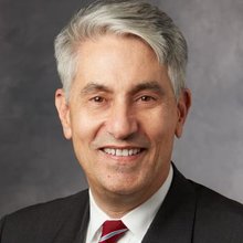
 Ravi Majeti合作论文数Institute for Stem Cell Biology and Regenerative Medicine, Cancer Institute, Division of Hematology, Department of Medicine, Stanford University82
 Volkmer Jens合作论文数69Rahul Sinha合作论文数59
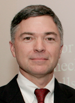
 Michael T. Longaker合作论文数Department of Materials Science & Engineering, Stanford University58
 Eugene Butcher合作论文数Department of Pathology, School of Medicine, Stanford University;Laboratory of Immunology and Vascular Biology, Palo Alto VA Medical Center;Leuvas Therapeutics, Inc55
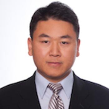 Charles Chan合作论文数Department of Orthopaedic Surgery, Stanford University49
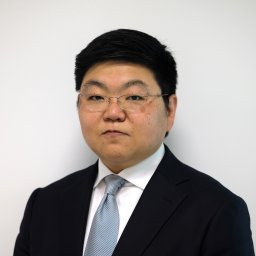 Jun Seita合作论文数Health Data Deep Learning Team, RIKEN48
 Mark Chao合作论文数Scorpion Therapeutics;IconOVir Bio47Kipp Weiskopf合作论文数Inst Stem Cell Biol & Regenerat Med, Stanford Univ46
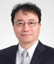 Koichi Akashi (赤司浩一)合作论文数Department of Medicine and Biosystemic Science, Faculty of Medical Sciences, Kyushu University;Department of Medicine, School of Medicine, Kyushu University39
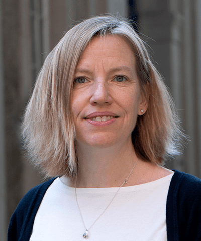 Amy Wagers合作论文数Department of Stem Cell and Regenerative Biology, Faculty of Arts and Sciences, Harvard University39
 Siddhartha Mitra合作论文数USGS38Samuel Cheshier合作论文数Department of Health Services, California;Human Stem Cell Research37
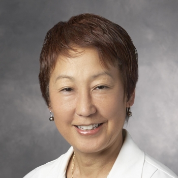 Judith Shizuru合作论文数Pediatrics Department, Stanford University36
 Debashis Sahoo合作论文数Boolean Lab, Department of Pediatrics / Department of Computer Science and Engineering, University of California San Diego36
 Aaron Ring合作论文数Translational Science and Therapeutics Division, Fred Hutch;Immunotherapy Integrated Research Center, Fred Hutch;Ariax Bio;Stipple Bio;Seranova Bio;Simcha Therapeutics32
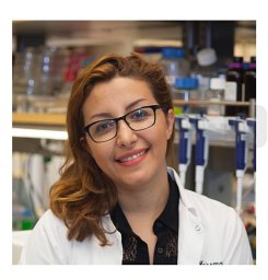 Sharareh Gholamin合作论文数Shahid Modarres Hospital, Shahid Beheshti University (MC),31
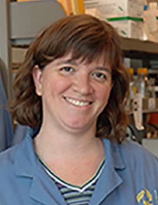 Laurie Ailles合作论文数Department of Medical Biophysics, Temerty Faculty of Medicine, University of Toronto30
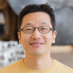 Christopher Y. Park合作论文数Department of Pathology, Stanford University30
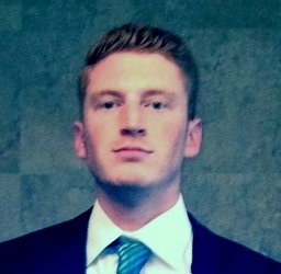 Walmsley Graham G合作论文数29Derrick J. Rossi合作论文数29Matthew A. Inlay合作论文数29
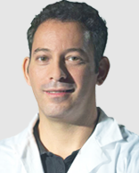 Yuval Rinkevich合作论文数Institute for Regenerative Biology and Medicine, Chinese Institutes for Medical Research, Beijing;Capital Medical University28Susan S Prohaska合作论文数27
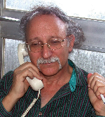 Baruch Rinkevich合作论文数National Institute of Oceanography;Israel Oceanographic and Limnological Research27
 Catriona Jamieson合作论文数Moores Cancer Center, University of California San Diego;Division of Regenerative Medicine, University of California San Diego27Lagasse Eric合作论文数26
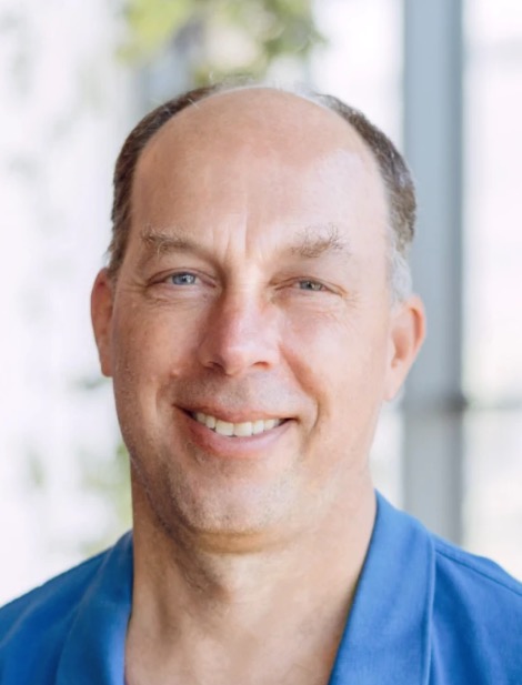 Stephen Quake合作论文数School of Engineering, Stanford University;Department of Bioengineering, Stanford University;Department of Applied Physics, Stanford University;Department of Physics, Stanford University;Cornell University26Wendy W Pang合作论文数25Bhattacharya Deepta合作论文数Dept Pathol & Immunol, Washington Univ25
 Ayelet Voskoboynik合作论文数Institute for Stem Cell Biology and Regenerative Medicine, Stanford University School of Medicine25Holger Karsunky合作论文数25
 Siddhartha Jaiswal合作论文数Department of Pathology, School of Medicine, Stanford University;Institute for Stem Cell Biology and Regenerative Medicine, School of Medicine, Stanford University24L Jerabek合作论文数24Michael W. Gallatin合作论文数23
 Amira Barkal合作论文数Stanford Univ, Sch Med, Inst Stem Cell Biol & Regenerat Med, Stanford, CA 94305 USA23Stephen B. Willingham合作论文数23
 David Traver合作论文数Division of Biological Sciences, Section of Cell and Developmental Biology, School of Biological Sciences, University of California;Department of Cell and Developmental Biology, School of Biological Sciences, University of California23N Uchida合作论文数22
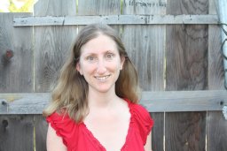 Michal Tal合作论文数Stem cell and Regenerative Medicine, Stanford School of Medicine22
 Matt Van De Rijn合作论文数Maternal & Child Health Research Institute, School of Medicine, Stanford University;Stanford Cancer Institute, Stanford University22Thomas Serwold合作论文数22
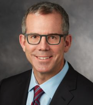 Geoffrey Gurtner合作论文数Department of Surgery, Stanford University;Department of Surgery, College of Medicine, The University of Arizona21Nobuko Uchida合作论文数21
 Gunsagar S Gulati合作论文数Stanford Univ, Inst Stem Cell Biol & Regenerat Med, Sch Med, Lorry Lokey Stem Cell Res Bldg, Stanford, CA 94305 USA20Micha Drukker合作论文数20
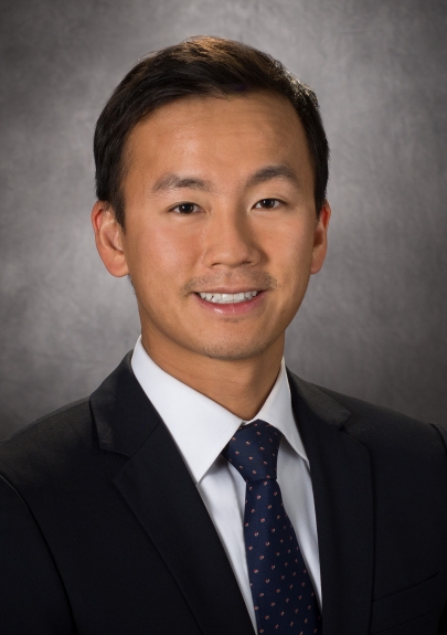 Chad Tang合作论文数Department of Radiation Oncology, Division of Radiation Oncology, The University of Texas MD Anderson Cancer Center20
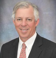 Robert C. Robbins合作论文数Stanford University School of Medicine20
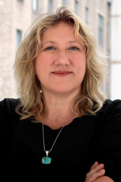 Passegué Emmanuelle合作论文数Columbia Stem Cell Initiative, Department of Genetics and Development, Irving Medical Center, Columbia University19




