Septic shock is associated with over 40% mortality. The immune response in septic shock is tightly regulated by cellular metabolism and transitions from early hyper-inflammation to later hypo-inflammation. Patients are susceptible to secondary infections during hypo-inflammation. The magnitude of the metabolic dysregulation and the effect of plasma metabolites on the circulating immune cells in septic shock are not reported. We hypothesized that the accumulated plasma metabolites affect the immune response in septic shock during hypo-inflammation. Our study took a unique approach. Using peripheral blood from adult septic shock patients and healthy controls, we studied: (i) Whole blood stimulation +/- E. Coli lipopolysaccharide (LPS: endotoxin) to analyze plasma TNF protein, and (ii). Plasma metabolomic profile by Metabolon. Inc. (iii) We exposed peripheral blood mononuclear cells (PBMCs) from healthy controls to commercially available carbohydrate, amino acid, and fatty acid metabolites and studied the response to LPS. We report that: (i) The whole blood stimulation of the healthy control group showed a significantly upregulated TNF protein, while the septic shock group remained endotoxin tolerant, a biomarker for hypo-inflammation. (ii) A significant accumulation of carbohydrate, amino acid, fatty acid, ceramide, sphingomyelin, and TCA cycle pathway metabolites in septic shock plasma. (iii) In vitro exposure to 5 metabolites repressed while 2 metabolites upregulated the inflammatory response of PBMCs to LPS. We conclude that the endotoxin-tolerant phenotype of septic shock is associated with a simultaneous accumulation of plasma metabolites from multiple metabolic pathways, and these metabolites fundamentally influence the immune response profile of circulating cells. Metabolic chaos in septic shock: Simultaneous dysregulation of multiple metabolic pathways such as carbohydrate, branched-chain amino acid, fatty acid, tissue turnover related, and ceramide metabolism in septic shock.
Transceptors, solute transporters that facilitate intracellular entry of molecules and also initiate intracellular signaling events, have been primarily studied in lower-order species. Ammonia, a cytotoxic endogenous metabolite, is converted to urea in hepatocytes for urinary excretion in mammals. During hyperammonemia, when hepatic metabolism is impaired, nonureagenic ammonia disposal occurs primarily in skeletal muscle. Increased ammonia uptake in skeletal muscle is mediated by a membrane-bound, 12 transmembrane domain solute transporter, Rhesus blood group-associated B glycoprotein (RhBG). We show that in addition to its transport function, RhBG interacts with myeloid differentiation primary response-88 (MyD88) to initiate an intracellular signaling cascade that culminates in activation of NFκB. We also show that ammonia-induced MyD88 signaling is independent of the canonical toll-like receptor-initiated mechanism of MyD88-dependent NFκB activation. In silico, in vitro, and in situ experiments show that the conserved cytosolic J-domain of the RhBG protein interacts with the Toll-interleukin-1 receptor (TIR) domain of MyD88. In skeletal muscle from human patients, human-induced pluripotent stem cell-derived myotubes, and myobundles show an interaction of RhBG–MyD88 during hyperammonemia. Using complementary experimental and multiomics analyses in murine myotubes and mice with muscle-specific RhBG or MyD88 deletion, we show that the RhBG–MyD88 interaction is essential for the activation of NFkB but not ammonia transport. Our studies show a paradigm of substrate-dependent regulation of transceptor function with the potential for modulation of cellular responses in mammalian systems by decoupling transport and signaling functions of transceptors.
Supplementary Fig S1. CBL0137 in combination with cisplatin attenuates SCLC tumor growth. H82 SCLC cells, mixed with Matrigel (1:1), were inoculated subcutaneously into the flanks of NSG mice (n=8). Once the tumors reached â^¼50 mm3, the mice were randomized to treatment with vehicle control, CBL0137 (CBL) alone (60 mg/kg i.v., weekly), cisplatin (Cis) alone (5 mg/kg i.p., weekly), or CBL0137 plus cisplatin (CBL+Cis). Tumor diameters were measured 3 times a week for 32 days. The results are represented as means {plus minus} SE.
Supplementary Table 1. Characteristics of the African-American prostate cancer patients in the Cleveland Clinic Cohort according to overall survival; Supplementary Table 2. IRDS Probesets (n=49) for the Affymetrix U133A 2.0 array
Acute exposure of cancer cells to high concentrations of type I interferon (IFN-I) drives growth arrest and apoptosis, whereas chronic exposure to low concentrations provides important prosurvival advantages. Tyrosine-phosphorylated IFN-stimulated gene (ISG) factor 3 (ISGF3) drives acute deleterious responses to IFN-I, whereas unphosphorylated (U-)ISGF3, lacking tyrosine phosphorylation, drives essential constitutive prosurvival mechanisms. Surprisingly, programmed cell death-ligand 1 (PD-L1), often expressed on the surfaces of tumor cells and well recognized for its importance in inactivating cytotoxic T cells, also has important cell-intrinsic protumor activities, including dampening acute responses to cytotoxic high levels of IFN-I and sustaining the expression of the low levels that benefit tumors. More thorough understanding of the newly recognized complex roles of IFN-I in cancer may lead to the identification of novel therapeutic strategies.
<p>Supplementary Fig S3. CBL0137 inhibits FACT function in SCLC TICs. A, SSRP1 levels were determined by Western analysis in CD133high or CD133low cells from H82 or H526. β-actin was used as a loading control. B, H82 CD133high cells were treated with 1 µM CBL0137 for different times. Soluble and chromatin bound SSRP1 levels were detected by immunoblotting.</p>
This file contains detailed materials & methods in addition to the materials & methods section in the main text.
In cancer cells, endogenous or therapy-induced DNA damage leads to the abnormal presence of DNA in the cytoplasm, which triggers the activation of cGAS (cyclic GMP-AMP synthase) and STING (stimulator of interferon genes). STAT2 suppresses the cGAMP-induced expression of IRF3-dependent genes by binding to STING, blocking its intracellular trafficking, which is essential for the full response to STING activation. STAT2 reshapes STING signaling by inhibiting the induction of IRF3-dependent, but not NF-κB-dependent genes. This noncanonical activity of STAT2 is regulated independently of its tyrosine phosphorylation but does depend on the phosphorylation of threonine 404, which promotes the formation of a STAT2:STING complex that keeps STING bound to the endoplasmic reticulum (ER) and increases resistance to DNA damage. We conclude that STAT2 is a key negative intracellular regulator of STING, a function that is quite distinct from its function as a transcription factor.
<p>Supplementary Fig S4. FACT is involved in the expression of regulatory stem cell transcription factors in TICs. A and B, CD44high cells derived from H446 and CD133high cells derived from H526 were treated with 500 nM (H446) or 1µM (H526) CBL0137 for 24 h. The mRNA levels of SOX2 were analyzed by qPCR (A), and SOX2 protein levels were determined by Western analysis (B).</p>
Supplementary Figure 3 from p53-Mediated Growth Suppression in Response to Nutlin-3 in Cyclin D1–Transformed Cells Occurs Independently of p21
Supplementary Figure 1. Expression of IFNL4 after transfection of human prostate cancer cell lines with the IFNL4-Halo construct; Supplementary Figure 2. Expression of two interferon signatures, IRG and IRDS, in cultured prostate cancer epithelial cells from 14 African-American and 13 European-American men; Supplementary Figure 3. Association of IFNL4 SNP rs12979860-T allele with decreased overall survival among African-American prostate cancer patients in the Cleveland Clinic cohort (n = 197).
Due to their broad functional plasticity, myeloid cells contribute to both liver injury and recovery during acetaminophen overdose-induced acute liver injury (APAP-ALI). A comprehensive understanding of cellular diversity and intercellular crosstalk is essential to elucidate the mechanisms and to develop therapeutic strategies for APAP-ALI treatment. Here, we identified the function of IFN-I in the myeloid compartment during APAP-ALI. Utilizing single-cell RNA sequencing, we characterized the cellular atlas and dynamic progression of liver CD11b+ cells post APAP-ALI in WT and STAT2 T403A mice, which was further validated by immunofluorescence staining, bulk RNA-seq, and functional experiments in vitro and in vivo. We identified IFN-I-dependent transcriptional programs in a three-way communication pathway that involved IFN-I synthesis in intermediate restorative macrophages, leading to CSF-1 production in aging neutrophils that ultimately enabled Trem2+ restorative macrophage maturation, contributing to efficient liver repair. Overall, we uncovered the heterogeneity of hepatic myeloid cells in APAP-ALI at single-cell resolution and the therapeutic potential of IFN-I in the treatment of APAP-ALI.
 Ian M. Kerr合作论文数40
 Xiaoxia Li合作论文数Lerner Research Institute, Cleveland Clinic;Department of Molecular Medicine, School of Medicine, Case Western Reserve University;Department of Pathology, School of Medicine, Case Western Reserve University31
 Mark Jackson合作论文数26
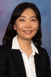 Cheon HyeonJoo合作论文数Division of Rheumatology, Allergy, and Immunology, University of California at San Diego School of Medicine23
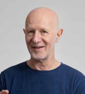 Richard Ransohoff合作论文数Third Rock Ventures;Abata Therapeutics19Tao Lu合作论文数19Munna L. Agarwal合作论文数19William R. Taylor合作论文数18Sarmishtha De合作论文数18
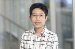 Josephine Kam Tai Dermawan合作论文数Department of Molecular Genetics, Lerner Research Institute16
 Yuxin Wang合作论文数Lanzhou University, Lanzhou University16
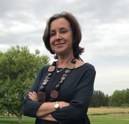 Katerina Gurova合作论文数Dept Cell Stress Biol, Roswell Pk Canc Inst14
 Elise Holvey-Bates合作论文数13
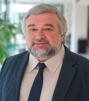 Andrei Gudkov合作论文数Department of Cell Stress Biology, Roswell Park Cancer Institute13
 Ganes C. Sen合作论文数Department of Molecular Medicine, School of Medicine, Case Western Reserve University;Department of Biochemistry, School of Medicine, Case Western Reserve University;Department of Inflammation and Immunity, Lerner Research Institute, Cleveland Clinic13
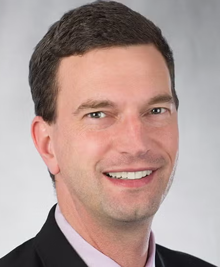 Jeremy Rich合作论文数School of Medicine, University of North Carolina12
 Anette H. H. van Boxel-Dezaire合作论文数Department of Molecular Genetics, Lerner Research Institute11
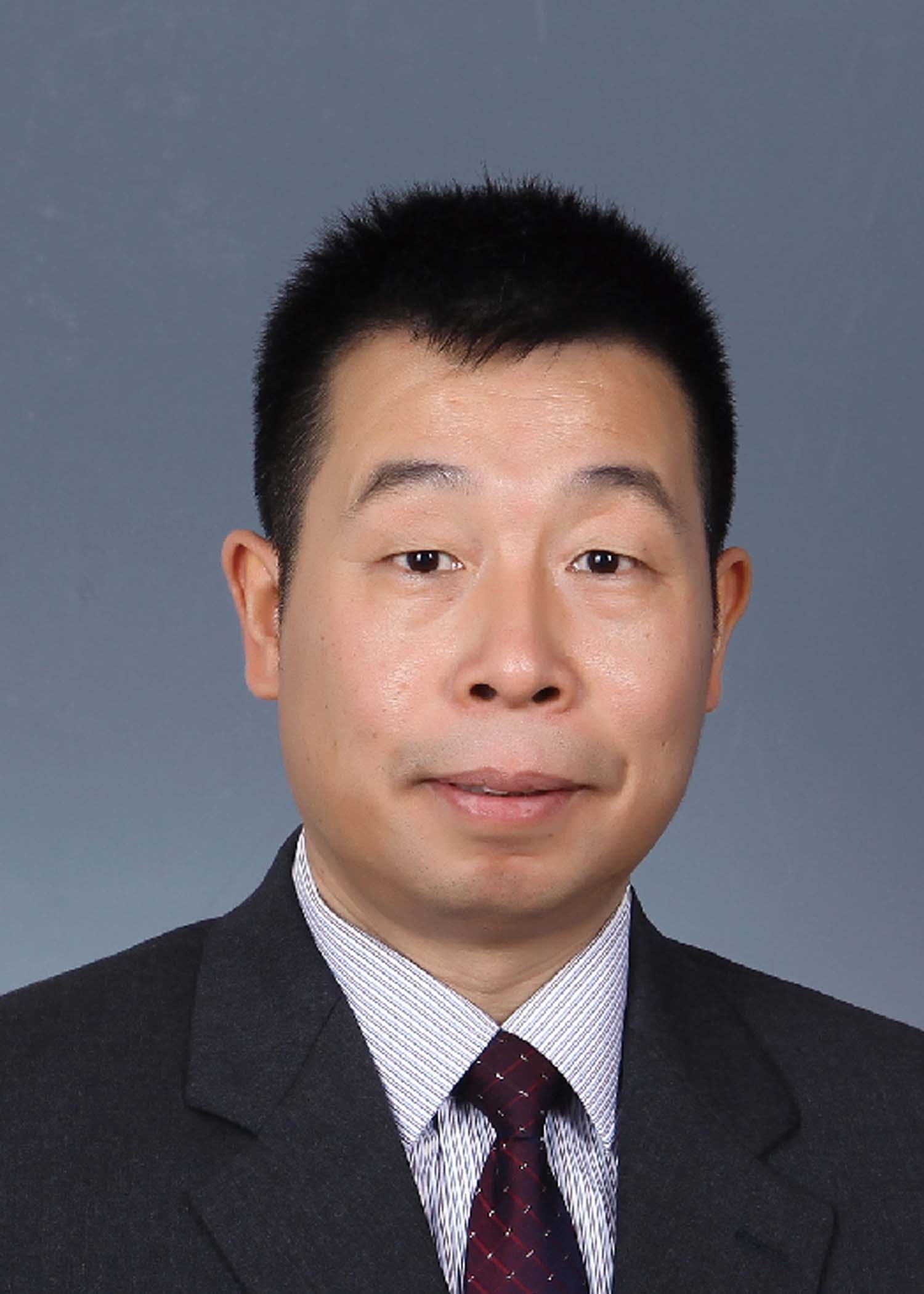 Jinbo Yang (杨金波)合作论文数School of Medicine and Pharmacy, Ocean University of China11
 Justin D. Lathia合作论文数Department of Cardiovascular and Metabolic Sciences, Lerner Research Institute, Cleveland Clinic;Department of Molecular Medicine, School of Medicine, Case Western Reserve University11Stewart Leung合作论文数11Qiulian Wu合作论文数11Moitreyee Chatterjee-Kishore合作论文数11
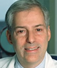 Robert H. Silverman合作论文数Department of Molecular Medicine, School of Medicine, Case Western Reserve University;Department of Biochemistry, School of Medicine, Case Western Reserve University;Case Comprehensive Cancer Center;Department of Cancer Biology Lerner Research Institute, Cleveland Clinic10
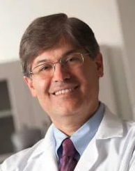 Afshin Dowlati合作论文数Department of Medicine, School of Medicine, Case Western Reserve University;University Hospitals10Mairead Commane合作论文数10
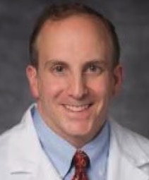 Andrew E. Sloan合作论文数Department of Neurosurgery, School of Medicine, Case Western Reserve University;Piedmont Healthcare;Department of Neurological Surgery University Hospitals Cleveland Medical Center10
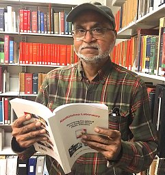 Chilakamarti V. Ramana合作论文数Lerner Research Institute, The Cleveland Clinic Foundation10
 Monica Venere合作论文数James Cancer Hospital and Comprehensive Cancer Center, The Ohio State University College of Medicine9Agarwal Mukesh K合作论文数9Masahiro Hitomi合作论文数9A. Purmal合作论文数9N Sizemore合作论文数8M. R. Sandhya Rani合作论文数8Neil C Rogers合作论文数8
 Poorva Sandlesh合作论文数UPMC Hillman Canc Ctr, UPCI Res Pavil8Maojing Yang合作论文数8Daniel J. Lindner合作论文数7O B Chernova合作论文数7
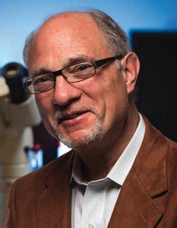 Geoffrey Wahl合作论文数Gene Expression Laboratory, Salk Institute for Biological Studies7
 Gary Wildey合作论文数7
 Xing Chen合作论文数Department of Chemical and Biomolecular Engineering, National University of Singapore7
 James E. Darnell Jr.合作论文数Laboratory of Molecular Cell Biology, The Rockefeller University7
 Belinda Willard合作论文数7
 Elena Giulotto合作论文数Istituto di Genetica e Microbiologia Adriano Buzzati Traverso, University of Pavia7D Watling合作论文数6
 Matthias Muller合作论文数Univ Vet Med Vienna, Inst Anim Breeding & Genet, Vienna, Austria6Kandel Eugene S合作论文数6
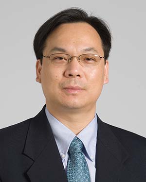 Shideng Bao合作论文数Department of Cancer Biology, Cleveland Clinic Lerner Research Institute;Center for Cancer Stem Cell Research, Cleveland Clinic Lerner Research Institute6
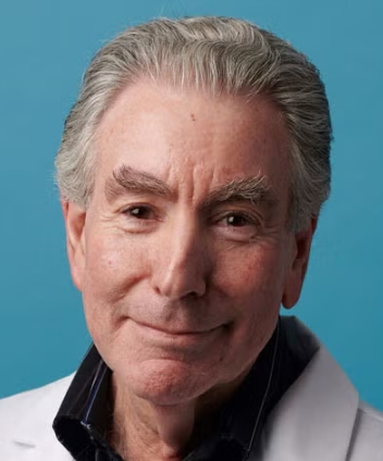 Jerry Silver合作论文数Department of Neurosciences, School of Medicine, Case Western Reserve University6




