Background/Objectives: Contemporary patient-specific subperiosteal implants have re-emerged as graftless solutions for oral and maxillofacial rehabilitation, driven by advances in digital planning, CAD/CAM workflows, additive manufacturing, and biomaterial engineering. Their indications have progressively expanded from severely atrophic edentulous jaws to segmental defects, single-tooth replacement, congenital craniofacial anomalies, salvage situations, and oncologic reconstruction. This scoping review aimed to map the current evidence on modern patient-specific subperiosteal implants, focusing on indications, workflow, design principles, materials, outcomes, complications, and maintenance. Methods: A scoping review was conducted according to PRISMA-ScR principles to identify clinical studies, case series, case reports, systematic and scoping reviews, technical notes, finite element analyses, in vitro studies, and relevant translational investigations dealing with contemporary custom-made or CAD/CAM subperiosteal implants. The evidence was narratively synthesized according to clinical indication and thematic domains, including full-arch rehabilitation, sectional and single-tooth applications, congenital and post-oncologic defects, rescue indications, biomechanics, material selection, surface response, prosthetic protocols, and complication management. No quantitative meta-analysis was performed because of the scoping design and the substantial heterogeneity of study types, indications, implant systems, outcome definitions, and follow-up durations. Results: The final evidence map included 116 records, of which 56 were unique human clinical records with extractable denominators and 60 were biomechanical, in vitro, surface-biology, review, consensus, historical, or conceptual records. Of the 56 unique clinical records, 49 were mapped within the six indication-level clinical sections, while seven were retained as cross-cutting clinical evidence addressing patient-reported outcomes, design-related complications, bone apposition, anchorage strategy, comparative graftless rehabilitation, or reconstructive/prosthetic principles. The six indication-level sections included 52 clinical-record assignments: 15 for full-arch rehabilitation, 13 for segmental or sectional rehabilitation, one for single-tooth rehabilitation, four for congenital or craniofacial indications, 13 for post-oncologic or post-ablative reconstruction, and six for rescue or salvage indications. Because three records addressed more than one indication, these counts represent indication-level assignments rather than mutually exclusive clinical records. Reported survival in most short- to mid-term clinical series was generally high, commonly ranging from 90% to 100%, although lower values of 70-80% were reported in selected longer-term cohorts and survival clearly overestimated clinical success in some studies. Expanding applications include posterior mandibular and maxillary defects, lateral incisor agenesis, cleft-related or syndromic deformities, maxillectomy reconstruction, obturator support, and hybrid rehabilitation with endosseous implants; however, evidence for the indications at the extremes of this spectrum-single-tooth replacement and primary oncologic reconstruction-remains limited to small, largely single-group case series and reports. Soft-tissue events, including dehiscence, mucositis, recession, and framework exposure, were the dominant complications and showed wide variability, with reported recession/exposure rates ranging from approximately 10% in some sectional and full-arch series to as high as 65% in bilateral maxillary cohorts; their clinical significance varied from asymptomatic stable findings to progressive inflammatory complications requiring revision. Conclusions: Patient-specific subperiosteal implants represent a promising and increasingly versatile reconstructive option; however, the present findings should be interpreted as evidence mapping rather than as definitive comparative evidence. Their clinical use should remain highly selective, prosthetically driven, and supported by meticulous planning, rigid fixation, soft-tissue management, and structured maintenance. Standardized success criteria, longer follow-up, and comparative prospective studies are required.
Topic This comprehensive narrative review summarizes the available evidence regarding neonates with congenital epulis, the epidemiologic profile, prenatal imaging findings, histopathologic features, management strategies (surgical excision versus observation), and clinical outcomes. Clinical relevance Congenital epulis is a rare benign gingival tumor of the newborn that can impair feeding and, occasionally, breathing. Because evidence is scattered across isolated case reports, clinicians lack clear guidance for counselling and perinatal management. Methods A comprehensive literature search of MEDLINE, Embase, Scopus, and Web of Science was conducted from database inception to the final search date. Published case reports and case series involving neonates with congenital epulis were reviewed. Data regarding epidemiology, prenatal imaging, histopathology, management strategies, and outcomes were extracted and synthesized descriptively. Results One hundred twenty-four publications reporting 147 neonates were included. Most infants were female and had a solitary mass arising from the anterior maxillary alveolar ridge. Prenatal detection occurred in a minority of pregnancies. Early neonatal surgical excision was the predominant management, under general or local anesthesia, with minimal perioperative morbidity. No recurrences or malignant transformations were documented. Spontaneous regression was described only in a few small, conservatively managed lesions. Conclusion Evidence, restricted to retrospective case reports, consistently indicates an excellent prognosis for congenital epulis. Simple early excision appears safe and curative for most lesions, whereas careful observation may be reasonable for selected small tumors. Further prospective, standardized reporting is needed to refine prenatal counselling and postnatal management.
Background/Objectives: Natural head position (NHP) is an important reference for orthodontic and orthognathic assessment. Although postoperative changes in head posture have been reported previously, simultaneous three-dimensional changes across yaw, pitch, and roll in different skeletal classes remain incompletely characterized. This exploratory study quantified six-month changes in three-dimensional head orientation and compared their direction and magnitude between skeletal Class II and Class III patients. Methods: Fifty-four adults (17 Class II, 37 Class III) undergoing Le Fort I osteotomy and bilateral sagittal split osteotomy were evaluated preoperatively (T0) and 6 months postoperatively (T1). Standardized 3D facial surface acquisitions were obtained using a calibrated structured-light scanner and a fixed external Cartesian laser reference. T0 and T1 datasets were registered using stable upper-facial landmarks (nasion and bilateral exocanthia), and changes in yaw, pitch, and roll were calculated within the common external reference system. Results: Statistically significant T0-T1 changes were observed across all three axes in both skeletal groups (p < 0.01). Class II patients showed mean changes of +2.28° in yaw, -1.30° in pitch, and +2.89° in roll, whereas Class III patients showed +4.61°, +1.96°, and +4.04°, respectively. Between-group differences were significant for yaw, pitch, and roll, with an opposite mean pitch direction and larger mean yaw and roll changes in Class III. Conclusions: At six months, this retrospective cohort demonstrated direction-specific changes in three-dimensional head orientation. Because repeated acquisitions, study-specific observer-reliability and measurement-error analyses, an untreated comparator, an independent validation cohort, and functional or patient-reported outcomes were not available, the findings should be interpreted as exploratory measurements rather than evidence of physiological normalization, neuromuscular adaptation, or clinical benefit. Formal prospective validation is required before the workflow can support predictive or patient management recommendations.
A pyolaryngocele is a rare, potentially life-threatening complication of laryngocele, resulting from secondary infection and obstruction of the saccular neck. Clinical severity ranges from mild dysphonia to acute airway compromise. We report a 51-year-old man with a sore throat, dysphagia, and dyspnea. Flexible laryngoscopy and contrast-enhanced CT revealed an internal pyolaryngocele. After emergency tracheostomy and intravenous antibiotics, the patient underwent CO2 laser microlaryngoscopic excision and marsupialization. Postoperative recovery was uneventful, with complete healing and no recurrence at follow-up. This case underscores the importance of early diagnosis and highlights CO2 laser surgery as an effective, minimally invasive option. Internal pyolaryngocele is a rare airway emergency that requires prompt management. CO2 laser excision appears to be a safe and effective first-line approach in selected internal cases.
Background: Smile aesthetics arise from the interaction between objective dentofacial characteristics and subjective patient perception, and discrepancies between the two may influence treatment planning and postoperative satisfaction. This exploratory study aimed to describe how adults perceive variations in maxillary incisor exposure, sagittal incisor projection, and dental midline deviation, and to identify the self-reported levels of perceptual tolerance at which these variations become consciously noticeable. Methods: Eighty-three adult volunteers (20–65 years; 59.6% female) underwent standardised photographic analysis, three-dimensional facial scanning, and completion of a purpose-built self-perception questionnaire. Objective morphometric measurements were compared with subjective self-estimates, and the difference (delta) was computed for each parameter. Analyses were descriptive; the association between the magnitude of objective–subjective discrepancy and aesthetic dissatisfaction was assessed with Spearman’s rank correlation. No inferential threshold-modelling was performed. Results: Self-reported perceptual tolerance levels were 3.2 mm for incisor exposure at rest, 9.4 mm during smiling, 3.6 mm for midline deviation, and 4.7 mm (protrusion)/3.9 mm (retrusion) for sagittal projection. The proportion of participants whose objective measurement exceeded their own tolerance level was 23.1% (smiling), 17.3% (rest), 15.4% (sagittal), and 3.8% (midline). Objective and subjective assessments were concordant in 60% of cases (27% underestimation, 13% overestimation). A moderate, statistically significant correlation was observed between the magnitude of objective–subjective discrepancy and aesthetic dissatisfaction (Spearman’s ρ = 0.67, p < 0.01). Sagittal discrepancies beyond approximately ±4 mm relative to the Barcelona line were most consistently associated with negative self-perception. Conclusions: Objectively measurable dentofacial discrepancies are not uniformly perceived as unattractive and may remain within individual perceptual tolerance. These descriptive findings are hypothesis-generating and should be interpreted in light of the single-centre design and the non-validated questionnaire; they nonetheless suggest that integrating digital facial analysis with patient-reported perception may support communication and expectation management in aesthetic planning.
Objective: Mucoepidermoid carcinoma (MEC) is the most common malignant tumour of the parotid gland. This systematic review and meta-analysis aims to evaluate treatment strategies, survival, recurrence, and prognostic factors in primary parotid MEC. Materials and Methods: A systematic review was conducted following PRISMA guidelines. PubMed/MEDLINE, the Cochrane Library, Scopus, and Google Scholar were searched to identify eligible observational studies and clinical trials on primary parotid MEC. Pooled estimates of overall survival (OS), disease-specific survival (DSS), and local, regional, and distant recurrence rates were calculated. Prognostic factors associated with survival and recurrence were analysed. Results: Twenty-one studies involving 7192 patients were analysed. Histologic grade was low in 32.2%, intermediate in 41.8%, and high in 26.1%. Surgical treatment included total parotidectomy (2606 patients) and superficial parotidectomy (1642), with facial nerve preservation achieved in 1993 of 4111 reported cases. Positive margins occurred in 18% of patients, and postoperative radiotherapy was administered in 50%. Mean follow-up was 72.6 months. Pooled OS rates were 100% at 1 year, 90% at 5 years, and 70% at 10 years; DSS was 100% at 5 years and 90% at 10 years. Recurrence rates were 10% local, 0% regional, and 10% distant. High-grade histology, advanced T/N stage, positive surgical margins, and intraparotid lymph node metastasis were associated with poorer outcomes. Conclusions: Parotid MEC generally has favourable short- and intermediate-term outcomes. Based on evidence and institutional experience, we propose a grade-based workflow integrating tumour grade, T/N status, and adverse pathological features to guide surgical extent, elective neck dissection, and adjuvant radiotherapy. Prospective studies with standardized reporting are needed.
Severe alveolar bone atrophy represents a major challenge for implant-supported rehabilitation of the maxilla and mandible. While custom-made subperiosteal implants and conventional endosseous implants have each been proposed as graftless solutions in selected clinical scenarios, the outcomes of hybrid rehabilitations combining both implant systems within the same jaw have not yet been investigated. The aim of this retrospective study was to evaluate the clinical and radiological outcomes of hybrid implant-supported rehabilitations using custom-made subperiosteal implants in combination with conventional endosseous implants in patients with severe maxillary and/or mandibular atrophy.Fourteen consecutive patients affected by advanced jaw atrophy and heterogeneous residual bone availability were included. A total of 20 custom-made subperiosteal implants and 48 conventional endosseous implants were placed to support fixed full-arch or segmental prosthetic rehabilitations. Implant survival, success, biological and mechanical complications, and peri-implant soft tissue conditions were assessed over a mean follow-up of 22.1 months.At the last follow-up, survival of both subperiosteal and endosseous implants was 100%, with all rehabilitations remaining functional. All endosseous implants fulfilled established success criteria, while all subperiosteal implants met a composite functional success endpoint. Peri-implant soft tissue conditions remained stable, and no prosthetic complications were observed. However, these findings should be interpreted in light of the retrospective design, limited sample size, and relatively short follow-up.Within the limitations of this retrospective case series, hybrid rehabilitations combining custom-made subperiosteal and conventional endosseous implants may represent a promising and flexible graftless treatment option for severely atrophic jaws with heterogeneous bone availability.
Contemporary patient-specific subperiosteal implants (SPIs) have re-emerged as digitally planned, additively manufactured solutions for oral and maxillofacial rehabilitation when conventional endosseous implants are limited by severe atrophy, anatomical constraints, or reconstructive defects. Unlike conventional implants, SPIs behave as fixation-based skeletal frameworks whose performance depends on passive fit, screw fixation, anchorage, framework architecture, material properties, manufacturing accuracy, and prosthetic load transfer. This scoping review evaluated the maturity of mechanical design and validation evidence for contemporary SPIs. Following a predefined internal protocol and PRISMA-ScR, MEDLINE/PubMed, Scopus, Web of Science, Embase, and the Cochrane Library were searched from inception to 13 June 2026. Reference-list screening and citation tracking supplemented the electronic search. Two reviewers independently screened records against predefined eligibility criteria. Data were charted using a predefined extraction form and synthesized descriptively by evidence type, engineering domain, validation stage, and translational status. No meta-analysis was undertaken because of methodological heterogeneity, and no formal risk-of-bias grading was applied. Across 65 included records, the evidence was dominated by descriptive technical studies and comparative computational analyses, whereas direct mechanical testing, fatigue assessment, manufacturing verification, and clinical correlation were limited. Finite element analysis was useful for comparing design alternatives and identifying stress concentrations, but models were heterogeneous and often insufficiently validated. Design modifications generally redistributed stress across the implant–prosthesis–bone system rather than reducing it globally. Titanium and Ti6Al4V were the most established framework materials, whereas polymeric, ceramic, and scaffold-assisted strategies remained preliminary. The principal contribution of this review is a cross-domain appraisal of progression from anatomical feasibility and comparative modeling to manufacturing verification, experimental testing, and clinical validation. An evidence map, minimum reporting checklist, and integrated validation pathway are provided to support reproducible device development.
BACKGROUND:Orbital floor fractures are among the most frequent facial injuries, and precise reconstruction is essential to prevent both functional and aesthetic complications. Virtual Surgical Planning (VSP) combined with pre-shaped titanium mesh has transformed orbital reconstruction. This study aimed to evaluate whether VSP-assisted surgery with pre-modeled mesh provides greater accuracy and reduced operative times compared with conventional intraoperative mesh shaping. METHODS:A case-control study was performed on 52 patients treated at the "Federico II" University Hospital of Naples for large pure orbital floor fractures. Twenty-four patients (Group A) underwent VSP-assisted reconstruction using a preformed titanium mesh, while twenty-eight patients (Group B) received conventional intraoperative mesh modeling. Operative time, postoperative diplopia, infraorbital sensory disturbance, mesh malposition, screw loosening, and infection were compared in the two groups. Clinical and radiological follow-up was conducted at 12 months. RESULTS:Median operative time was significantly shorter in Group A compared with Group B (55 min [IQR 50-65] vs. 110 min [IQR 98-125]; p < 0.001). Persistent diplopia occurred in 4 % of patients in Group A and 28.6 % in Group B (p = 0.028). Implant malposition was observed exclusively in Group B (25 % vs. 0 %; p = 0.011). Other postoperative complications, including infraorbital nerve alterations, eyelid malposition, wound dehiscence, screw loss, and infection, were more frequent in Group B but did not reach statistical significance. No cases of postoperative ocular motility limitation or iatrogenic fracture were observed in either group. CONCLUSIONS:VSP with pre-modeled titanium mesh significantly reduces operative time and postoperative complications, enhancing reconstructive precision and outcomes in orbital floor fracture management. This virtual protocol is useful for reducing the costs and waiting times of custom-made implants, manufactured by external companies.
OBJECTIVE:To evaluate the occurrence of MRONJ after tooth extraction in patients with cancer and to explore factors associated with its development. MATERIALS AND METHODS:A systematic review was conducted following PRISMA guidelines. PubMed/MEDLINE, the Cochrane Library, Scopus, Embase, and Ovid MEDLINE was conducted for studies published between 1970 and 2026. Studies including oncology patients undergoing tooth extraction while receiving antiresorptive and/or antiangiogenic therapy were considered eligible. A random-effects meta-analysis of proportions was conducted to estimate the pooled occurrence of MRONJ after tooth extraction. Exploratory pooled odds ratios were calculated for selected risk factors. In addition, a random-effects meta-analysis of continuous data was performed to compare bone-modifying agent (BMA) therapy duration between MRONJ-positive and MRONJ-negative patients. RESULTS:Seven studies were included, comprising 497 patients with cancer. The pooled proportion of MRONJ after tooth extraction was 27.6% (95%CI16.0%-43.3%). Exploratory pooled analyses showed no significant association between MRONJ and female sex or chemotherapy. By contrast, patients who developed MRONJ had a significantly longer duration of BMA therapy than those who did not, with a pooled mean difference of 8.38 months (95%CI2.44-14.32). Qualitative synthesis suggested that local infection, inflammatory dental disease, and extraction timing may be more consistently associated with MRONJ risk than tooth extraction alone. CONCLUSIONS:MRONJ after tooth extraction is not uncommon in oncology patients. However, the available evidence does not support considering tooth extraction as an isolated and uniformly avoidable trigger. Rather, MRONJ risk appears to be influenced by local infectious burden, timing of extraction, and cumulative BMA exposure.
BACKGROUND:Cervical lymph node metastasis is a major adverse prognostic factor in primary parotid gland malignancies. However, the incidence, level-specific distribution, and independent predictors of nodal metastasis remain incompletely defined in large contemporary multicenter cohorts. METHODS:A retrospective longitudinal multicenter study was conducted across 17 tertiary referral centers. Adult patients with histopathologically confirmed primary parotid gland carcinoma who underwent parotidectomy with concurrent neck dissection were included. The primary outcome was pathologically confirmed cervical lymph node metastasis. Secondary outcomes included level-specific nodal distribution, identification of independent risk factors, overall survival (OS), and disease-specific survival (DSS). RESULTS:A total of 380 patients were included. Pathological cervical lymph node metastasis was identified in 94 patients (24.7%). Metastases most frequently involved Level II (IIa 17.1%; IIb 11.1%). Salivary duct carcinoma demonstrated the highest rate of nodal metastasis (58.7%). On multivariate analysis, high histological grade and lymphovascular invasion (LVI) were independent predictors of nodal metastasis (p < 0.05). Five-year OS for the entire cohort was 64.4%. Patients with nodal metastasis had significantly worse survival compared with node-negative patients (p < 0.001). CONCLUSIONS:Cervical lymph node metastasis occurs in approximately one-quarter of surgically staged primary parotid gland malignancies and predominantly involves Levels II-III. High-grade histology and LVI independently predict nodal involvement. These findings support a risk-adapted approach to elective neck management based on histological and pathological risk stratification.
Major maxillofacial oncologic surgery requires not only advanced technical expertise but also effective perioperative coordination among multidisciplinary teams. Virtual reality (VR), especially when combined with artificial intelligence (AI), offers new opportunities for team-based surgical training. We developed a multiplayer VR platform with AI-driven procedural supervision (VR/AI Lembo) to simulate perioperative workflow in maxillofacial oncologic surgery. Usability was evaluated in a multicenter study involving 35 healthcare professionals (17 physicians, 18 nurses) from four Italian tertiary referral centers using the System Usability Scale (SUS). Overall usability was high, with a mean SUS score of 81.0 ± 11.1, indicating excellent user acceptance. Most participants achieved scores ≥ 68 (88.6
Orthognathic surgery involves repositioning the skeletal bases to correct dentoskeletal deformities and malocclusions. While its impact on occlusion is well documented, its effects on masticatory muscle activity remain under investigation. This pilot study evaluates changes in static electromyographic (sEMG) patterns of masseter and temporal muscles, and explores correlations with TMJ-related muscular symptoms before and after surgery. A surface electromyography (sEMG), by employing TeethanTM, based on occlusal contacts, was performed recording the activation patterns of masseter and temporal muscles, in ten patients with dentoskeletal malocclusions, undergoing orthognathic surgery. The registration assessed the muscles patterns sEMG preoperatively (T0), postoperatively at one month (T1), and at six months (T2) after surgery. Across the cohort, temporal POC increased by 6.76 ± 8.36 from T0 to T1 and by 5.68 ± 5.33 from T1 to T2. Masseter POC rose by 8.74 ± 7.38 (T0–T1) and 8.12 ± 7.42 (T1–T2). Barycentre values shifted by 7.36 ± 4.13 (T0–T1) and 10.65 ± 9.17 (T1–T2), indicating progressive rebalancing of occlusal force distribution. Patients with Class III malocclusion showed greater masseter activation post-surgery, while Class II patients exhibited increased temporal muscle activity. Three patients with preoperative muscular discomfort reported complete symptom resolution at T2, while one patient experienced mild residual symptoms. Data obtained from this preliminary study seem to confirm that skeletal bases repositioning modifies the occlusal contacts and, consequently, the neuromuscular proprioceptive stimuli and the activation of the masticatory muscles. sEMG may provide useful complementary information on neuromuscular adaptation following orthognathic surgery. However, larger controlled studies with standardized outcomes and statistical analyses are required before drawing definitive conclusions or recommending its routine clinical use.
Background/Objectives: Osteoradionecrosis of the jaws remains a severe late complication of head and neck radiotherapy. Current preventive strategies are still frequently based on population-level dose thresholds and broadly standardized dental-clearance protocols, despite substantial heterogeneity in individual risk. This comprehensive review aimed to integrate established preventive guidance with emerging tooth-level, spatial-dosimetric, prognostic, and causal approaches within an integrative conceptual framework for personalized osteoradionecrosis prevention. Methods: Structured searches of five databases were completed on 30 May 2026. Evidence sources were selected and mapped across six a priori thematic domains, with the selection process summarized in a simplified flow diagram and an evidence map. Priority was given to guidelines, systematic reviews, large cohorts, externally validated prediction models, and clinically actionable studies. Results: Osteoradionecrosis risk is determined by interactions among tumor site, surgical anatomy, mandibular dose distribution, dental and periodontal disease, smoking, diabetes, nutritional status, biological susceptibility, and expected survival. Tooth extraction is not uniformly protective, and its potential benefit depends on tooth prognosis, local radiation dose, oncological urgency, and patient-level vulnerability. Dose-volume parameters and site-specific mapping provide more clinically relevant information than prescribed dose alone. Contemporary normal tissue complication probability and machine-learning models increasingly support individualized risk estimation, although calibration, external validation, data quality, and workflow integration remain limiting. Competing-risk and causal-inference approaches may further distinguish baseline risk from the expected benefit of specific preventive interventions. Conclusions: The evidence reviewed can be organized within an integrative conceptual framework combining tooth-level prognosis, spatial dosimetry, systemic susceptibility, competing mortality, and intervention burden. This framework synthesizes and reorganizes existing evidence; it is not a validated clinical model and requires prospective validation and clinical-impact evaluation before routine implementation. Until then, available prediction models should support, rather than replace, multidisciplinary clinical judgement.
Osteonecrosis of the jaw is most commonly discussed in relation to antiresorptive or antiangiogenic medications and head and neck radiotherapy. However, a heterogeneous group of jaw osteonecrosis cases occurs in the absence of these recognized triggers and remains poorly systematized. This review summarizes the current evidence on jaw osteonecrosis occurring outside the classical settings of antiresorptive/antiangiogenic medication exposure and head and neck radiotherapy, with the aim of clarifying its etiological spectrum, clinical presentation, diagnostic challenges, and management principles. The available literature is largely composed of case reports, small case series, and retrospective studies, reflecting the rarity and heterogeneity of these conditions. Reported causes include local traumatic or idiopathic oral ulceration with bone sequestration, viral infections, immunosuppression, chemotherapy, hematological malignancies, hematopoietic stem cell transplantation, hemoglobinopathies, metabolic or genetic bone disorders, vascular and thrombotic abnormalities, and toxic or recreational drug exposure. Clinical manifestations range from small, self-limited sequestra over bony prominences to extensive necrosis with tooth exfoliation, fistulae, infection, pathological fracture, and midfacial involvement. Management should be tailored to the underlying cause and disease extent, ranging from conservative sequestrectomy to radical debridement or resection combined with systemic treatment. Recognition of these entities is essential to avoid misdiagnosis and to guide appropriate multidisciplinary care.
Objectives: Telemedicine has rapidly expanded in oral and maxillofacial surgery (OMFS), especially during the COVID-19 pandemic, but its specific roles and limitations across the care pathway remain unclear. This narrative review aimed to map telemedicine modalities and indications in OMFS, summarize reported outcomes, and identify priorities for future research. Methods: A narrative synthesis was undertaken after a systematic search of medical and engineering databases to 10 October 2025. Studies applying telemedicine, telehealth, telepresence or teleradiology to OMFS practice were eligible, including trials, observational cohorts, technical reports and surveys. Data were extracted in duplicate and organized thematically; heterogeneity precluded meta-analysis. Results: Fifty studies met the inclusion criteria. Telemedicine was mainly used for preoperative consultation and triage, postoperative follow-up, trauma teleradiology and tele-expertise, oncologic and oral medicine follow-up, temporomandibular disorders, and education or humanitarian work. In low-risk outpatient and postoperative settings, remote consultations showed high concordance with in-person plans, similar complication or reattendance rates, reduced travel, and high satisfaction. In trauma networks, telemedicine supported timely triage and reduced unnecessary inter-hospital transfers. Evidence in oral oncology and complex mucosal disease was more cautious, favouring hybrid models and escalation to face-to-face assessment. Data on cost-effectiveness and impacts on equity were limited. Conclusions: Telemedicine in OMFS has moved from niche innovation to a pragmatic adjunct across the clinical pathway. Current evidence supports its use for selected pre- and postoperative care and trauma triage within risk-stratified hybrid models, while underscoring the need for stronger comparative and implementation studies, clear governance on equity and data protection, and alignment with wider digital and AI-enabled health systems.
Background: Microvascular osseous free flaps play a central role in head and neck reconstruction; surgeons often rely on fragmented and inconsistently reported data when counselling patients and planning reconstructive strategies. This systematic review and meta-analysis aimed to quantify postoperative complication rates and to evaluate complication patterns according to flap type. Methods: The study protocol was registered in PROSPERO (CRD420251237516). Studies published between January 2000 and November 2025 reporting postoperative complications following mandibular or maxillary reconstruction with osseous free flaps were identified. Eligible studies included adult cohorts with a minimum sample size of twenty patients. Random-effects meta-analyses of proportions were conducted. Risk of bias was assessed using the ROBINS-I tool. Results: Fourteen retrospective studies encompassing 1198 flaps were included. The pooled incidence of total flap loss was 6% (95% CI 3-9%), and partial flap loss was 6% (95% CI 3-10%). The pooled rates for postoperative infection, fistula formation, and wound dehiscence were 7% (95% CI 2-22%), 12% (95% CI 7-20%), and 16% (95% CI 8-31%), respectively, with substantial heterogeneity. Fibular free flaps demonstrated pooled rates of 6.1% for total flap loss, 6.6% for partial flap loss, 9.0% for infection, 10.4% for fistula formation, and 17.1% for wound dehiscence. For scapular free flaps, pooled total flap loss was 5% (95% CI 1-29%). DCIA flaps demonstrated hardware-related complications (8.1%), fistulas (16.7%), bone exposure (4.2%), and wound dehiscence (29.7%). Donor site morbidity was inconsistently reported and could not be quantitatively synthesized. Conclusions: Osseous free flap reconstruction shows relevant complication rates, highlighting the need for standardized reporting to support evidence-based decision-making.
Objectives: To synthesize evidence on artificial intelligence (AI)-enabled medical history taking (anamnesis)-beyond large language models (LLMs) alone-and to translate findings into implications and research priorities for head and neck surgery. Methods: We performed a PRISMA-informed narrative review. Searches from database inception to 31 December 2025 (updated 3 January 2026) were conducted in MEDLINE (PubMed), Embase, Scopus, Web of Science Core Collection, IEEE Xplore, and ACM Digital Library, supplemented by medRxiv/arXiv screening and citation chasing. We included studies evaluating or describing AI-supported history capture/summarization, conversational interviewing, symptom checker/digital triage, EHR-integrated intake-to-decision support pipelines, voice interviewing, education/training systems, and governance/ethical considerations related to digital anamnesis. Findings were synthesized by system category and by cross-cutting outcome domains, with a head and neck surgery interpretive lens. Results: Fifty studies (2014-2025) were included. Evidence most consistently suggested feasibility and acceptability of pre-consultation computer-assisted history taking and the potential to reduce documentation burden and improve structured capture. In contrast, symptom checkers and digital triage tools showed highly variable diagnostic/triage performance and prominent safety concerns, highlighting the importance of conservative red-flag escalation strategies, continuous monitoring, and clear accountability. LLM-based diagnostic dialogue demonstrated strong performance in controlled evaluations, but prospective real-world validation, governance, and workflow integration remain limited. Conclusions: AI-enabled anamnesis comprises heterogeneous tools with uneven evidence. For head and neck surgery, potential near-term applications may include structured pre-visit intake, clinician-facing summarization, and training applications, whereas autonomous triage warrants harm-oriented, specialty-calibrated validation and robust governance prior to broader clinical reliance.
OBJECTIVE:TMJ arthrocentesis is a minimally invasive treatment for temporomandibular disorders, but its complication profile is not fully defined. This study evaluated the incidence and spectrum of associated adverse events. METHODS:PubMed/MEDLINE, Embase, and Scopus were systematically searched for human studies reporting complications, including trials, observational studies, case series, and case reports. All were qualitatively reviewed; only studies with extractable numerical data were meta-analyzed. RESULTS:Forty-six studies were included; 23 reported numerical data and 13 entered the meta-analysis. The pooled complication rate was ~11% (95% CI 5-18%) with high heterogeneity; excluding isolated post-procedural pain gave ~9%. Most events were minor and transient, especially periarticular swelling and temporary facial nerve dysfunction. Clinically significant complications (hematoma, infection, vascular injury) were rare (<1%), while severe events were limited to case reports. CONCLUSION:TMJarthrocentesis is a low-risk procedure. Substantialheterogeneity warrant cautious interpretation and highlight the needfor standardized assessment.
Background: The optimal management of the clinically node-negative (cN0) neck in maxillary squamous cell carcinoma (MSCC) remains controversial due to the relatively low yet clinically relevant risk of occult cervical metastasis. While elective neck dissection (END) has been proposed to improve oncologic outcomes, others advocate observation to avoid overtreatment. This study aimed to evaluate the impact of END versus observation on overall survival (OS) in cN0 MSCC patients. Methods: A systematic literature search was conducted in PubMed, Embase, and Scopus according to the PRISMA 2020 statement. The protocol was registered in PROSPERO (CRD420261345038). Studies including patients with maxillary SCC and clinically N0 neck comparing END with observation were eligible. Fourteen studies were included in the qualitative synthesis, and five were suitable for quantitative meta-analysis. The primary endpoint was OS. Pooled hazard ratios (HRs) with 95% confidence intervals (CIs) were calculated using a random-effects model. Risk of bias was assessed using ROBINS-I, and certainty of evidence was evaluated using the GRADE framework. Results: The meta-analysis demonstrated a statistically significant survival benefit associated with END. The pooled HR for OS was 0.76 (95% CI 0.67-0.86; p < 0.001), indicating a 24% relative reduction in the hazard of death compared with observation. Importantly, statistical heterogeneity was negligible (I2 = 0%), although interpretation should be cautious given the limited number of included studies. Despite this, most included studies were retrospective in design. Conclusions: END appears to provide a survival advantage in selected patients with cN0 MSCC. However, the evidence is largely derived from retrospective data. High-quality prospective multicenter studies are needed to better define the role of elective neck management in this population.
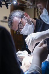 Luigi Angelo Vaira合作论文数Dept Maxillofacial Surg, Univ Hosp Sassari101
 Fabio Maglitto合作论文数73
 Giovanni Dell'Aversana Orabona合作论文数69
 Luigi Califano合作论文数65
 De Riu Giacomo合作论文数65
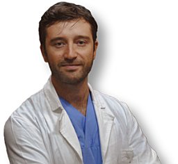 Vincenzo Abbate合作论文数Department of NeurosciencesReproductive and Odontostomatological Sciences, University of Naples “Federico II58
 Jerome R. Lechien合作论文数48
 Francesco Antonio Salzano合作论文数Department of Surgical, Oncologic and Oral Sciences, University of Palermo45Umberto Committeri合作论文数Maxillofacial Surgery Operative Unit, University Hospital of Naples "Federico II",42Petrocelli Marzia合作论文数40
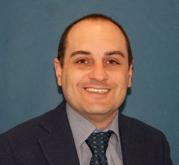 Piombino Pasquale合作论文数Department of Otorhinolaryngology, University of Naples;SUN", Naples;Department of Otorhinolaryngology, University of Naples "SUN"36
 Paola Bonavolonta合作论文数35
 Stefania Troise合作论文数Dept Neurosci Reprod & Odontostomatol Sci, Federico II Univ Naples32Giorgio Iaconetta合作论文数School of Medicine, University ‘Federico II’30Franco Ionna合作论文数Istituto dei Tumori “Pascale25
 Paolo Boscolo-Rizzo合作论文数University of Padua-School of Medicine, Treviso Regional Hospital24
 Antonio Romano合作论文数22
 Alfonso Scarpa合作论文数20Antonio Arena合作论文数20
 Antonino Maniaci合作论文数Department of Biomedical and Biotechnological Sciences (BIOMETEC),, University of Catania20
 Arianna Di Stadio合作论文数20
 Pietro De Luca合作论文数19
 Francesco Perri合作论文数IRCCS Ospedale Casa Sollievo della Sofferenza17
 Sven Saussez合作论文数University of Mons (UMons);Faculty of Medicine UMONS Research Institute for Health Sciences and Technology;Department of Human and Experimental Oncology16
 Simona Barone合作论文数15Biglio Andrea合作论文数14
 Massimo Ralli合作论文数ENT Department;Catholic University of the Sacred Heart Medical School;ENT Department, Catholic University of the Sacred Heart Medical School14
 Pasquale Viola合作论文数Regional Centre for Cochlear Implants and ENT Diseases, Magna Graecia University13
 Miguel Mayo Yanez合作论文数Otorhinolaryngol Head & Neck Surg Dept, Complexo Hosp Univ A Coruna13
 Roberta Fusco合作论文数Department of Biomedical, Electronic and Telecommunication Engineering, University Federico II of Naples13
 Filippo Ricciardiello合作论文数12
 Claire Hopkins合作论文数Guy's and St Thomas' NHS Foundation Trust;King's College London12Maria Grazia Maglione合作论文数12Giulia Togo合作论文数10
 Giovanni Audino合作论文数University Federico II10Giovanna Deiana合作论文数10
 Federico Biglioli合作论文数10
 Giuseppe Chiarella合作论文数Regional Center for Cochlear Implants and ENT Diseases, “Magna Graecia” University10Francesco Giovacchini合作论文数9Francesco Longo合作论文数9
 Giannicola Iannella合作论文数9Cassandro Claudia合作论文数9
 Carlos Miguel Chiesa Estomba合作论文数Servicio ORL, Complejo Hospitalario Universitario de Vigo9Emanuele Carraturo合作论文数8
 Francesco Maffia合作论文数Department of Neurosciences, Reproductive and Odontostomatological Sciences, University Federico II8
 Alessandro Tel合作论文数Academic Hospital of Udine, University of Udine8L Califano合作论文数8
 Agostino Guida合作论文数8
 Giovanni Cammaroto合作论文数8




