KRAS4a and KRAS4b are important regulators of signaling, and their interactions with the plasma membrane are dynamic and influenced by lipid composition. KRAS 4a and 4b have nearly identical globular domains but differ in their membrane-associated hyper variable region (HVR). The functional distinctions between these isoforms remain unclear, particularly with regards to their dependence on specific lipids and the membrane environment. Previous work showed that the membrane orientation of KRAS4b affects its ability to bind to RAF kinase RBDCRD and that the KRAS-RBDCRD complex adopts different poses on the membrane as well as influences the size and composition of the lipid environment. To model differences between KRAS 4a and 4b protein-lipid interactions, we extended the Multiscale Machine-Learned Modeling Infrastructure (MuMMI) to incorporate continuum simulations in the grand canonical ensemble, enabling sampling across macroscopic, coarse-grained, and all-atom resolutions. Using this framework, we systematically altered PIP2 concentrations, KRAS 4a versus 4b, and RAF RBDCRD complexation to assess impacts on membrane-protein interactions and dynamics. Our results reveal that reducing PIP2 shifts and broadens the membrane orientational preference of both KRAS 4b and 4a, with stronger effects on 4b HVR localization versus 4a. We demonstrate that with depletion of the strong negatively charged PIP2 lipid, the less charged phosphatidylserine replaces PIP2. Our findings highlight similarities and distinctions in the dynamics and lipid dependency of KRAS isoforms and suggest that ordering of the local lipid composition by HVRs is a shared property and key modulator of RAS-mediated signaling at the plasma membrane.
To gain molecular and mechanistic insights into initiation of the RAS-RAF signaling cascade, we developed and used a combination of multiscale simulation and experimental approaches. The influence and impact of the membrane on RAS and RAF proteins is a factor we are just beginning to understand and appreciate in more detail. Molecular simulation is an ideal methodology to further study this complicated relationship between the membrane and associated proteins. Our previous work using Multiscale Machine-learned Modeling Infrastructure investigated different lipid compositions solely around the KRAS4b protein and the interplay between protein behavior and these membrane environments. Multiscale Machine-learned Modeling Infrastructure uses machine learning to couple adjacent simulation scales and has been efficiently scaled across some of the world’s largest high-performance computers. Recently, we have expanded this multiresolution framework to include the all-atom simulation scale and to incorporate the RAF RBDCRD domains. Here, we present the overall analysis results from this new simulation campaign comprising a mixture of RAS and RAF RBDCRD proteins. Approximately 35,000 coarse-grained and 10,000 all-atom molecular dynamics simulations were completed, sampled from a variety of protein/lipid composition configurations that were generated from a micron-scale continuum simulation containing hundreds of copies of the proteins.Our studies suggest that orientations of the RAS-RBDCRD complex on the membrane occupy distinct configurational states, and the spatial patterns of lipid arrangements around these different protein states are unique to each state. The extent and size of lipid “fingerprints” imposed on the membrane by the RAS-RBDCRD protein complex are significantly larger than observed for just the RAS protein on its own. These protein complexes strongly associate, but we do not observe statistically significant preferred protein-protein orientations. These observations indicate that spatial colocalization of RAS-RBDCRD proteins in the same vicinity may be assisted by specific membrane environments, acting to increase the probability of signaling complex formation.
<p>Role of metabolic pathways imparting gemcitabine resistance in pancreatic cancer</p>
Interdependence across time and length scales is common in biology, where atomic interactions can impact larger-scale phenomenon. Such dependence is especially true for a well-known cancer signaling pathway, where the membrane-bound RAS protein binds an effector protein called RAF. To capture the driving forces that bring RAS and RAF (represented as two domains, RBD and CRD) together on the plasma membrane, simulations with the ability to calculate atomic detail while having long time and large length- scales are needed. The Multiscale Machine-Learned Modeling Infrastructure (MuMMI) is able to resolve RAS/RAF protein-membrane interactions that identify specific lipid-protein fingerprints that enhance protein orientations viable for effector binding. MuMMI is a fully automated, ensemble-based multiscale approach connecting three resolution scales: (1) the coarsest scale is a continuum model able to simulate milliseconds of time for a 1 μm2 membrane, (2) the middle scale is a coarse-grained (CG) Martini bead model to explore protein-lipid interactions, and (3) the finest scale is an all-atom (AA) model capturing specific interactions between lipids and proteins. MuMMI dynamically couples adjacent scales in a pairwise manner using machine learning (ML). The dynamic coupling allows for better sampling of the refined scale from the adjacent coarse scale (forward) and on-the-fly feedback to improve the fidelity of the coarser scale from the adjacent refined scale (backward). MuMMI operates efficiently at any scale, from a few compute nodes to the largest supercomputers in the world, and is generalizable to simulate different systems. As computing resources continue to increase and multiscale methods continue to advance, fully automated multiscale simulations (like MuMMI) will be commonly used to address complex science questions.
Protein nanoclusters (PNCs) are dynamic collections of a few proteins that spatially organize in nanometer-length clusters. PNCs are one of the principal forms of spatial organization of membrane proteins, and they have been shown or hypothesized to be important in various cellular processes, including cell signaling. PNCs show remarkable diversity in size, shape, and lifetime. In particular, the lifetime of PNCs can vary over a wide range of timescales. The diversity in size and shape can be explained by the interaction of the clustering proteins with the actin cytoskeleton or the lipid membrane, but very little is known about the processes that determine the lifetime of the nanoclusters. In this paper, using mathematical modeling of the cluster dynamics, we model the biophysical processes that determine the lifetime of actin-dependent PNCs. In particular, we investigated the role of actin aster fragmentation, which had been suggested to be a key determinant of the PNC lifetime, and we found that it is important only for a small class of PNCs. A simple extension of our model allowed us to investigate the kinetics of protein-ligand interaction near PNCs. We found an anomalous increase in the lifetime of ligands near PNCs, which agrees remarkably well with experimental data on RAS-RAF kinetics. In particular, analysis of the RAS-RAF data through our model provides falsifiable predictions and novel hypotheses that will not only shed light on the role of RAS-RAF kinetics in various cancers, but also will be useful in studying membrane protein clustering in general.
<p>Metabolic Pathway Enrichment in PDAC Patients Treated with Gemcitabine (Data Pooled from TCGA)</p>
RAS is a signaling protein associated with the cell membrane that is mutated in up to 30% of human cancers. RAS signaling has been proposed to be regulated by dynamic heterogeneity of the cell membrane. Investigating such a mechanism requires nearatomistic detail at macroscopic temporal and spatial scales, which is not possible with conventional computational or experimental techniques. We demonstrate here a multiscale simulation infrastructure that uses machine learning to create a scale-bridging ensemble of over 100,000 simulations of active wild-type KRAS on a complex, asymmetric membrane. Initialized and validated with experimental data (including a new structure of active wild-type KRAS), these simulations represent a substantial advance in the ability to characterize RAS-membrane biology. We report distinctive patterns of local lipid composition that correlate with interfacially promiscuous RAS multimerization. These lipid fingerprints are coupled to RAS dynamics, predicted to influence effector binding, and therefore may be a mechanism for regulating cell signaling cascades.
Single particle tracking (SPT) of fluorescent molecules provides significant insights into the diffusion and relative motion of tagged proteins and other structures of interest in biology. However, despite the latest advances in high‐resolution microscopy, individual particles are typically not distinguished from clusters of particles. This lack of resolution obscures potential evidence for how merging and splitting of particles affect their diffusion and any implications on the biological environment. The particle tracks are typically decomposed into individual segments at observed merge and split events, and analysis is performed without knowing the true count of particles in the resulting segments. Here, we address the challenges in analyzing particle tracks in the context of cancer biology. In particular, we study the tracks of KRAS protein, which is implicated in nearly 20% of all human cancers, and whose clustering and aggregation have been linked to the signaling pathway leading to uncontrolled cell growth. We present a new analysis approach for particle tracks by representing them as tracking graphs and using topological events – merging and splitting, to disambiguate the tracks. Using this analysis, we infer a lower bound on the count of particles as they cluster and create conditional distributions of diffusion speeds before and after merge and split events. Using thousands of time‐steps of simulated and in‐vitro SPT data, we demonstrate the efficacy of our method, as it offers the biologists a new, detailed look into the relationship between KRAS clustering and diffusion speeds.
Single particle tracking (SPT) of fluorescent molecules provides significant insights into the diffusion and relative motion of tagged proteins and other structures of interest in biology. However, despite the latest advances in high-resolution microscopy, individual particles are typically not distinguished from clusters of particles. This lack of resolution obscures potential evidence for how merging and splitting of particles affect their diffusion and any implications on the biological environment. The particle tracks are typically decomposed into individual segments at observed merge and split events, and analysis is performed without knowing the true count of particles in the resulting segments. Here, we address the challenges in analyzing particle tracks in the context of cancer biology. In particular, we study the tracks of KRAS protein, which is implicated in nearly 20% of all human cancers, and whose clustering and aggregation have been linked to the signaling pathway leading to uncontrolled cell growth. We present a new analysis approach for particle tracks by representing them as tracking graphs and using topological events – merging and splitting, to disambiguate the tracks. Using this analysis, we infer a lower bound on the count of particles as they cluster and create conditional distributions of diffusion speeds before and after merge and split events. Using thousands of time-steps of simulated and in-vitro SPT data, we demonstrate the efficacy of our method, as it offers the biologists a new, detailed look into the relationship between KRAS clustering and diffusion speeds. CCS Concepts • Human-centered computing → Scientific visualization; • Applied computing → Computational biology;
Abstract Pancreatic cancer is an extremely aggressive and deadly malignancy. Despite a detailed understanding of pancreatic ductal adenocarcinoma (PDAC)'s biology, the outcome of the current therapeutic regimen remains grim. Chemotherapeutic treatment is one of the first-line systemic treatments for PDAC. However, creating resistance against chemotherapy remains a major challenge. Oncogenic K-Ras mutation is one of the most common events responsible for the initiation and progression of PDAC. K-Ras mutation also promotes the activation of a transcription factor called NRF2 that is crucial for tumor development and pancreatic cancer proliferation. Our study detects differential basal levels of NRF2 in a panel of PDAC cell lines. We hypothesized that this phenomenon might reflect a greater intrinsic capacity of PDAC cells to become resistant to chemotherapy. We then demonstrated that up-regulation of NRF2 creates resistance against chemotherapy in PDAC cells. Consequently, NRF2 activation rewires metabolic pathway networks. Importantly, our metabolomics analysis revealed that, upon NRF2 activation, several metabolic pathways are reprogrammed. Recent advances in tumor metabolism signify the critical role of glutamine as a key regulator of cancer therapy. Moreover, we observed that inhibiting glutamine metabolism enhances the efficacy of chemotherapeutic drugs in PDAC cells. Taken together, our finding holds promise as a novel combination therapy for patients with pancreatic cancer marked by developed resistance against chemotherapy. Finally, this treatment strategy further establishes the potential for exploiting metabolic changes in RAS-driven cancer cells that confer novel therapeutic opportunities. Citation Format: Suman Mukhopadhyay, Debanjan Goswami, Dwight V. Nissley, Frank McCormick. Targeting glutaminolysis potentiates the efficacy of chemotherapy in RAS-driven pancreatic cancers [abstract]. In: Proceedings of the Annual Meeting of the American Association for Cancer Research 2020; 2020 Apr 27-28 and Jun 22-24. Philadelphia (PA): AACR; Cancer Res 2020;80(16 Suppl):Abstract nr 238.
Abstract Rat sarcoma (RAS) family of proteins function as GTP/GDP-dependent control switch to regulate Raf-MAPK effector pathway for cell proliferation and are frequently mutated in human cancer. RAS is anchored to the inner leaflet of the plasma membrane (PM) via C-terminal lipid-tether(s) and hypothesized to concentrate in distinct locations in membrane nano-domains to control the activation step. However, very little is known about the molecular dynamics of RAS on PM and the precise mechanism of activation. Here we characterize the molecular mobility of RAS variants by single-molecule tracking (SMT) method and estimate underlying mobility states. KRAS4b exhibit confined mobility with three diffusive states, a unique characteristic compared to all the other RAS isoforms (KRAS4a, NRAS, and HRAS). Although the major difference across RAS isoforms lies within the C-terminus HVR (hypervariable region) and its post-translational lipid modifications, the additional confinement for KRAS4b is contributed by the G-domain (globular domain) of the protein. Simulation of KRAS4b on membrane revealed a detailed atomistic mechanism that corroborates with the experimental results. Importantly, the oncogenic mutant of KRAS4b shows an altered mode of diffusion, implicating a plausible attribution to/from enhanced effector interaction. This study uncovers a novel underlying principle of KRAS4b functionality on living cell membrane with a potential for innovation in therapeutic strategies against its oncogenic variant. Citation Format: Debanjan Goswami, De Chen, John Columbus, Thomas Turbyville. Cooperative membrane interaction between G-domain and HVR defines unique diffusion behavior of KRAS4b [abstract]. In: Proceedings of the AACR Special Conference on Targeting RAS-Driven Cancers; 2018 Dec 9-12; San Diego, CA. Philadelphia (PA): AACR; Mol Cancer Res 2020;18(5_Suppl):Abstract nr A09.
 Dwight V Nissley合作论文数19
 Frank McCormick合作论文数Helen Diller Family Comprehensive Cancer Center, University of California, San Francisco;Department of Cellular and Molecular Pharmacology, University of California, San Francisco18
 Suman Mukhopadhyay合作论文数Department of Biological Sciences, Hunter College of the City University of New York14William E. Burgan合作论文数13Theresa M Guerin合作论文数12Ming Yi合作论文数12Pavan P. Adiseshaiah合作论文数12Serguei Kozlov合作论文数12
 Satyajit Mayor合作论文数National Centre for Biological Sciences8
 Robert Malenka合作论文数Wu Tsai Neurosciences Institute, Stanford University;Department of Psychiatry and Behavioral Sciences, Stanford University School of Medicine7
 Felice C. Lightstone合作论文数Department of Chemistry, University of California7
 Harsh Bhatia合作论文数University of Utah7
 Thomas Sudhof合作论文数Department of Molecular and Cellular Physiology, School of Medicine, Stanford University6
 Timo Bremer合作论文数Center for Applied Scientific Computing, Lawrence Livermore National Laboratory;Scientific Computing and Imaging Institute, Center of Extreme Data Management Analysis and Visualization, University of Utah6Rebika Shrestha合作论文数5
 Xiaohua Zhang (张晓华)合作论文数5Que N Van合作论文数Leidos Biomed Res Inc, Frederick Natl Lab Canc Res, Canc Res Technol Program, NCI RAS Initiat, Frederick, MD 21702 USA5
 Thomas J Turbyville合作论文数5Gulcin Gulten合作论文数5
 Helgi I. Ingolfsson合作论文数Tri-Institutional Training Program in Computational Biology and Medicine, New York, NY, USA5Tomas Oppelstrup合作论文数5
 Timothy Carpenter合作论文数Department of Biochemistry, University of Oxford5
 Andrew G Stephen合作论文数Protein Chemistry Laboratory, National Cancer Institute at Frederick5S. Gnanakaran合作论文数Department of Chemistry, University of Pennsylvania5
 Francesco Di Natale合作论文数Lawrence Livermore National Laboratory4
 Gautham Dharuman合作论文数Michigan State Univ.;c;Michigan State Univ.4
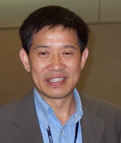 De Chen (陈德)合作论文数Department of Chemical Engineering, Norwegian University of Science and Technology4
 Yue Yang合作论文数BridgeBio Oncology Therapeutics4
 Dhirendra Simanshu合作论文数Struct Biol Program, Mem Sloan Kettering Canc Ctr4
 Christopher Stanley合作论文数4
 Fred Streitz合作论文数Lawrence Livermore National Laboratory4
 Brian Van Essen合作论文数Lawrence Livermore National Laboratory4John Columbus合作论文数3Joseph Y. Moon合作论文数3
 Kien Nguyen合作论文数Dept Phys, Northeastern Univ3
 Cesar Lopez Bautista合作论文数Center for Nonlinear Studies, Los Alamos National Laboratory3
 Arvind Ramanathan合作论文数3Aydin Fikret合作论文数Department of Chemical and Biochemical Engineering, Rutgers The State University of New Jersey3
 Madan Rao合作论文数Raman Research Institute National Centre for Biological Sciences3Sameera Bilgrami合作论文数3Wade Morishita合作论文数3
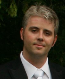 Chris Neale合作论文数Department of Biochemistry, University of Toronto3
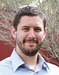 Liam Stanton合作论文数Department of Applied Mathematics;Northwestern University;Department of Applied Mathematics, Northwestern University3Jim Glosli合作论文数Lawrence Livermore National Laboratory3Constance Agamasu合作论文数2
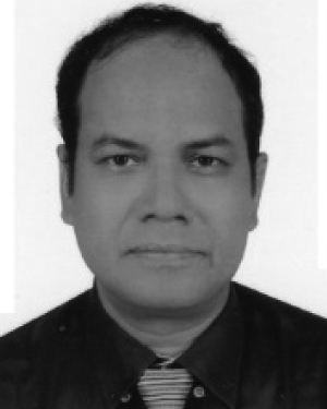 Mohiuddin Ahmad合作论文数2
 Konstantia Georgouli合作论文数Lawrence Livermore National Laboratory2Torin McDonald合作论文数SCI Institute, University of Utah2Xiyu Yi合作论文数2




