The capacity of cells to sense and respond to nutrient availability is essential for metabolic homeostasis. Failure in this process may cause cell death and associated diseases. While nutrient sensing in metabolic pathways is well understood, the mechanisms linking nutrient signals to cell death remain unclear. Here, we show that RIPK1, a key mediator of cell death and inflammation, senses methionine and its metabolite, S-adenosylmethionine (SAM), to dictate cell survival and death. SAM-mediated symmetrical dimethylation at RIPK1 Arg606 by PRMT5 functions as a physiological protective brake against RIPK1 activation. Metabolic perturbations, such as methionine restriction or disrupted one-carbon flux, reduce SAM levels and unmask Arg606, promoting RIPK1 self-association and trans-activation, thereby triggering apoptosis and inflammation. Thus, RIPK1 is a physiological SAM sensor linking methionine and one-carbon metabolism to the control of life-or-death decisions. Our findings suggest that RIPK1 could be a potential target for diseases associated with disrupted SAM availability.
Tumor necrosis factor α (TNFα) maintains homeostasis through promoting cell survival or cell death; however, how this process is regulated by metabolic pathways remains largely unknown. Here, we identify adenosine kinase (ADK), the key enzyme for catalyzing the conversion of adenosine to AMP, as an endogenous suppressor of RIPK1 kinase. ADK-mediated adenosine metabolic clearance is a prerequisite for transmethylation reactions on various cellular targets. We found that ADK licenses constitutive R606 symmetric dimethylation in RIPK1 death domain (DD), which is catalyzed by protein arginine methyltransferase 5. Upon TNFα stimulation, DD-mediated RIPK1 dimerization is inhibited by R606 methylation, preventing RIPK1 kinase activation and keeping cell death in check. Both hepatocyte-specific ADK knockout and systemic ADK inhibition cause spontaneous RIPK1-driven hepatocyte death, which leads to hepatic homeostasis disruption. Furthermore, ADK is reduced in hepatic ischemia-reperfusion, aggravating hepatic injury during liver surgery. Thus, this study reveals a mechanism of adenosine metabolism-dependent homeostasis maintenance that is implicated in both physiological and pathological conditions.
Ischemia-reperfusion injury (IRI) is a major obstacle in liver transplantation, especially with steatotic donor livers. Dysbiosis of the gut microbiota has been implicated in modulating IRI, and Lachnospiraceae plays a pivotal role in regulating host inflammatory and immune responses, but its specific role in liver transplantation IRI remains unclear. This study explores whether Lachnospiraceae can mitigate IRI and its underlying mechanisms. We found Lachnospiraceae-bacterium (Lachn.) abundance was significantly reduced in rats with liver cirrhosis. Lachn.-treated rats exhibited improved intestinal permeability, reduced IRI severity in both normal and steatotic donor livers, and decreased levels of neutrophil and macrophage infiltration, and inflammatory cytokines. Multi-omics analysis revealed elevated pyruvate levels in transplanted livers after Lachn. treatment, alongside reduced Alox15 and Foxo3 expression. Mechanistically, Lachn.-derived pyruvate inhibited Alox15 expression and reduced ferroptosis in normal and steatotic donor livers. Furthermore, reduced nuclear translocation of Foxo3 further suppressed Alox15 expression, alleviating IRI, especially in steatotic donor livers. Clinical samples confirmed reduced donor livers IRI in cirrhotic recipients with high Lachn. abundance after liver transplantation. In conclusion, Lachn. alleviates IRI in steatotic donor liver transplantation by inhibiting ferroptosis via the Foxo3-Alox15 axis, providing a potential therapeutic strategy to modulate gut microbiota to alleviate IRI following liver transplantation.
Background More and more steatotic livers undergo resection or transplantation but they exhibit higher susceptibility to ischemia-reperfusion injury (IRI), which results in increased perioperative complication morbidity and mortality. IRI is driven by various cytokines and receptors, both of which are extensively modified by N-glycosylation. We aim to elucidate susceptibility of steatotic livers to IRI from the perspective of N-glycosylation. Methods Differentially expressed genes and glycoproteins were identified with RNA-seq and N-glycoproteomics. Myeloid LIF or hepatocyte LIFR knockout mice were developed to examine the function of LIF and LIFR. Modalities including phosphoproteomics, ChIP-seq, single cell RNA-seq, metabolomics and immunoblotting were utilized to investigate underlying mechanisms. Results LIF transcription in myeloid cells and LIFR N-glycosylation in hepatocytes were substantially induced by IRI of normal livers. LIF and LIFR protected normal livers from IRI through activating STAT3 and promoting downstream TNFAIP3 expression, which was facilitated by LIFR N-glycosylation. Mechanistically, N-glycosylation at N238 stabilized LIFR protein by disrupting TRIM28-mediated K48 ubiquitination at LIFR K254. Furthermore, N-glycosylation at N358/N658/N675 of LIFR facilitated LIF/LIFR/gp130 complex formation and subsequent signal transduction. However, in steatotic livers, myeloid cell LIF transcription was partially inhibited due to hepatic microenvironment L-arginine insufficiency, while hepatocyte LIFR N-glycosylation was defective due to intracellular UDP-GlcNAc exhaustion. Importantly, combined L-arginine and GlcNAc treatment reversed LIF expression and LIFR N-glycosylation insufficiency, which represents potential therapeutic strategy to protect steatotic livers. Conclusions LIF expression and LIFR N-glycosylation insufficiency aggravates steatotic liver IRI, which can be reversed by combined L-arginine and GlcNAc treatment.
Tumor necrosis factor (TNF)-induced RIPK1-mediated cell death is implicated in various human diseases. However, the mechanisms RIPK1-mediated cell death is regulated by metabolic processes remain unclear. Here, we identify hexokinase 2 (HK2), a critical regulator of glycolysis, as a suppressor of TNF-induced RIPK1 kinase-dependent cell death through its non-metabolic function. HK2 inhibits RIPK1 kinase activity through constitutively phosphorylation at serine 32 of RIPK1. Inhibition of RIPK1 S32-phosphorylation results in RIPK1 kinase activation and subsequent cell death in response to TNFα stimulation. We further show that HK2 is elevated under pathological conditions including liver ischemia-reperfusion (IR) injury and hepatocellular carcinoma (HCC) via the transcriptional factor HMGA1. Moreover, the upregulation of HK2 in the liver confers protection against liver IR injury mediated by RIPK1 kinase, while depleting HK2 in HCC cells enhances TNFα-induced cell death and synergistically improves the efficacy of anti-PD1 therapy in an HCC model. Thus, the findings reveal a potential therapeutic avenue for RIPK1-related diseases through manipulating HK2 non-metabolic function.
Targeted delivery of glutamine metabolism inhibitors holds promise for cholangiocarcinoma therapy, yet effective delivery vehicles remain a challenge. This study reports the development of a biomimetic nanosystem, termed R-CM@MSN@BC, integrating mesoporous organosilicon nanoparticles with reactive oxygen species-responsive diselenide bonds for controlled release of the glutamine metabolism inhibitor bis-2-(5-phenylacetamido-1,3,4-thiadiazol-2-yl) ethyl sulfide (BPTES) and the photosensitizer Ce6. Erythrocyte membrane coating, engineered with Arg-Gly-Asp (RGD) peptides, not only enhanced biocompatibility but also improved tumor targeting and tissue penetration. Upon laser irradiation, R-CM@MSN@BC executed both photodynamic and glutamine-metabolic therapies, inducing necroptosis in tumor cells and triggering significant immunogenic cell death. Time-of-flight mass cytometry analysis revealed that R-CM@MSN@BC can remodel the immunosuppressive tumor microenvironment by polarizing M1-type macrophages, reducing infiltration of M2-type and CX3CR1+ macrophages, and decreasing T cell exhaustion, thereby increasing the effectiveness of anti-programmed cell death ligand 1 immunotherapy. This strategy proposed in this study presents a viable and promising approach for the treatment of cholangiocarcinoma.
Neil, Desley AH; Smith, Maxwell L.; Minervini, Marta; Demetris, A Jake Author Information
Steatotic donor livers are becoming more and more common in liver transplantation. However, the current use of steatotic grafts is less acceptable than normal grafts due to their higher susceptibility to ischemia/reperfusion (I/R) injury. To investigate the mechanism underlying the susceptibility of steatotic liver to I/R injury, we detected cell death markers and inflammation in clinical donor livers and animal models. We found that caspase-8-mediated hepatic apoptosis is activated in steatotic liver I/R injury. However, ablation of caspase-8 only slightly mitigated steatotic liver I/R injury without affecting inflammation. We further demonstrated that RIPK1 kinase induces both caspase-8-mediated apoptosis and cell death-independent inflammation. Inhibition of RIPK1 kinase significantly protects against steatotic liver I/R injury by alleviating both hepatic apoptosis and inflammation. Additionally, we found that RIPK1 activation is induced by Z-DNA binding protein 1 (ZBP1) but not the canonical TNF-α pathway during steatotic liver I/R injury. Deletion of ZBP1 substantially decreases the steatotic liver I/R injury. Mechanistically, ZBP1 is amplified by palmitic acid-activated JNK pathway in steatotic livers. Upon I/R injury, excessive reactive oxygen species trigger ZBP1 activation by inducing its aggregation independent of the Z-nucleic acids sensing action in steatotic livers, leading to the kinase activation of RIPK1 and the subsequent aggravation of liver injury. Thus, ZBP1-mediated RIPK1-driven apoptosis and inflammation exacerbate steatotic liver I/R injury, which could be targeted to protect steatotic donor livers during transplantation.
Tumor necrosis factor (TNF)-induced receptor-interacting serine/threonine protein kinase 1 (RIPK1)-mediated cell death, including apoptosis and necroptosis, is increasingly recognized as a major driver of inflammatory diseases. Cell death checkpoints normally suppress RIPK1 kinase to safeguard the organism from its detrimental consequences. However, the mechanisms licensing RIPK1 kinase activity when a protective checkpoint is disabled remain unclear. Here, we identified S-palmitoylation as a licensing modification for RIPK1 kinase. TNF induces RIPK1 palmitoylation, mediated by DHHC5 and dependent on K63-linked ubiquitination of RIPK1, which enhances RIPK1 kinase activity by promoting the homo-interaction of its kinase domain and promotes cell death upon cell death checkpoint blockade. Furthermore, DHHC5 is amplified by fatty acid in the livers of mice with metabolic dysfunction-associated steatohepatitis, contributing to increased RIPK1 cytotoxicity observed in this condition. Our findings reveal that ubiquitination-dependent palmitoylation licenses RIPK1 kinase activity to induce downstream cell death signaling and suggest RIPK1 palmitoylation as a feasible target for inflammatory diseases.
Background Therapies for cholangiocarcinoma are largely limited and ineffective. Herein, we examined the role of the FGF and VEGF pathways in regulating lymphangiogenesis and PD-L1 expression in intrahepatic cholangiocarcinoma (iCCA). Methods The lymphangiogenic functions of FGF and VEGF were evaluated in lymphatic endothelial cells (LECs) and iCCA xenograft mouse models. The relationship between VEGF and hexokinase 2 (HK2) was validated in LECs by western blot, immunofluorescence, ChIP and luciferase reporter assays. The efficacy of the combination therapy was assessed in LECs and xenograft models. Microarray analysis was used to evaluate the pathological relationships of FGFR1 and VEGFR3 with HK2 in human lymphatic vessels. Results FGF promoted lymphangiogenesis through c-MYC-dependent modulation of HK2 expression. VEGFC also upregulated HK2 expression. Mechanistically, VEGFC phosphorylated components of the PI3K/Akt/mTOR axis to upregulate HIF-1α expression at the translational level, and HIF-1α then bound to the HK2 promoter region to activate its transcription. More importantly, dual FGFR and VEGFR inhibition with infigratinib and SAR131675 almost completely inhibited lymphangiogenesis, and significantly suppressed iCCA tumor growth and progression by reducing PD-L1 expression in LECs. Conclusions Dual FGFR and VEGFR inhibition inhibits lymphangiogenesis through suppression of c-MYC-dependent and HIF-1α-mediated HK2 expression, respectively. HK2 downregulation decreased glycolytic activity and further attenuated PD-L1 expression. Our findings suggest that dual FGFR and VEGFR blockade is an effective novel combination strategy to inhibit lymphangiogenesis and improve immunocompetence in iCCA.
Background Early detection of hepatocellular carcinoma (HCC) is important in order to improve patient prognosis and survival rate. Methylation sequencing combined with neural networks to identify cell-free DNA (cfDNA) carrying aberrant methylation offers an appealing and non-invasive approach for HCC detection. However, some limitations exist in traditional methylation detection technologies and models, which may impede their performance in the read-level detection of HCC. Methods We developed a low DNA damage and high-fidelity methylation detection method called No End-repair Enzymatic Methyl-seq (NEEM-seq). We further developed a read-level neural detection model called DeepTrace that can better identify HCC-derived sequencing reads through a pre-trained and fine-tuned neural network. After pre-training on 11 million reads from NEEM-seq, DeepTrace was fine-tuned using 1.2 million HCC-derived reads from tumor tissue DNA after noise reduction, and 2.7 million non-tumor reads from non-tumor cfDNA. We validated the model using data from 130 individuals with cfDNA whole-genome NEEM-seq at around 1.6X depth. Results NEEM-seq overcomes the drawbacks of traditional enzymatic methylation sequencing methods by avoiding the introduction of unmethylation errors in cfDNA. DeepTrace outperformed other models in identifying HCC-derived reads and detecting HCC individuals. Based on the whole-genome NEEM-seq data of cfDNA, our model showed high accuracy of 96.2%, sensitivity of 93.6%, and specificity of 98.5% in the validation cohort consisting of 62 HCC patients, 48 liver disease patients, and 20 healthy individuals. In the early stage of HCC (BCLC 0/A and TNM I), the sensitivity of DeepTrace was 89.6 and 89.5% respectively, outperforming Alpha Fetoprotein (AFP) which showed much lower sensitivity in both BCLC 0/A (50.5%) and TNM I (44.7%). Conclusions By combining high-fidelity methylation data from NEEM-seq with the DeepTrace model, our method has great potential for HCC early detection with high sensitivity and specificity, making it potentially suitable for clinical applications. DeepTrace: https://github.com/Bamrock/DeepTrace
Hepatocyte apoptosis plays an essential role in the progression of nonalcoholic steatohepatitis (NASH). However, the molecular mechanisms underlying hepatocyte apoptosis remain unclear. Here, we identify UDP-glucose 6-dehydrogenase (UGDH) as a suppressor of NASH-associated liver damage by inhibiting RIPK1 kinase-dependent hepatocyte apoptosis. UGDH is progressively reduced in proportion to NASH severity. UGDH absence from hepatocytes hastens the development of liver damage in male mice with NASH, which is suppressed by RIPK1 kinase-dead knockin mutation. Mechanistically, UGDH suppresses RIPK1 by converting UDP-glucose to UDP-glucuronate, the latter directly binds to the kinase domain of RIPK1 and inhibits its activation. Recovering UDP-glucuronate levels, even after the onset of NASH, improved liver damage. Our findings reveal a role for UGDH and UDP-glucuronate in NASH pathogenesis and uncover a mechanism by which UDP-glucuronate controls hepatocyte apoptosis by targeting RIPK1 kinase, and suggest UDP-glucuronate metabolism as a feasible target for more specific treatment of NASH-associated liver damage.
肝脏具有强大的再生能力,肝再生对于接受肝切除、肝移植以及急性肝损伤患者快速恢复肝功能、维持机体内稳态十分重要.长久以来,普遍认为心理情绪与人体器官尤其是肝脏之间存在着某种联系.传统中医理论有"怒伤肝"的说法,而西医也在半个世纪前认识到不良情绪可能导致肝损伤.循证医学研究已给出了心理应激与肝脏病理相关的流行病学证据,显示恶性心理应激与肝病患者死亡率升高具有密切的关系.然而,至今鲜见研究明确指出心理应激能直接影响肝脏的病理生理过程(如肝再生),并深入阐释其潜在的机制.
OBJECTIVES:The aim of the study was to evaluate the association between the radiomics-based intratumoral heterogeneity (ITH) and the recurrence risk in hepatocellular carcinoma (HCC) patients after liver transplantation (LT), and to assess its incremental to the Milan, University of California San Francisco (UCSF), Metro-Ticket 2.0, and Hangzhou criteria. METHODS:A multicenter cohort of 196 HCC patients were investigated. The endpoint was recurrence-free survival (RFS) after LT. A CT-based radiomics signature (RS) was constructed and assessed in the whole cohort and in the subgroups stratified by the Milan, UCSF, Metro-Ticket 2.0, and Hangzhou criteria. The R-Milan, R-UCSF, R-Metro-Ticket 2.0, and R-Hangzhou nomograms which combined RS and the four existing risk criteria were developed respectively. The incremental value of RS to the four existing risk criteria in RFS prediction was evaluated. RESULTS:RS was significantly associated with RFS in the training and test cohorts as well as in the subgroups stratified by the existing risk criteria. The four combined nomograms showed better predictive capability than the existing risk criteria did with higher C-indices (R-Milan [training/test] vs. Milan, 0.745/0.765 vs. 0.677; R-USCF vs. USCF, 0.748/0.767 vs. 0.675; R-Metro-Ticket 2.0 vs. Metro-Ticket 2.0, 0.756/0.783 vs. 0.670; R-Hangzhou vs. Hangzhou, 0.751/0.760 vs. 0.691) and higher clinical net benefit. CONCLUSIONS:The radiomics-based ITH can predict outcomes and provide incremental value to the existing risk criteria in HCC patients after LT. Incorporating radiomics-based ITH in HCC risk criteria may facilitate candidate selection, surveillance, and adjuvant trial design. KEY POINTS:• Milan, USCF, Metro-Ticket 2.0, and Hangzhou criteria may be insufficient for outcome prediction in HCC after LT. • Radiomics allows for the characterization of tumor heterogeneity. • Radiomics adds incremental value to the existing criteria in outcome prediction.
BACKGROUND AND AIMS:The liver has the unique ability of regeneration, which is extremely important for restoring homeostasis after liver injury. Although clinical observations have revealed an association between psychological stress and the liver, whether stress has a causal influence on the liver regeneration remains markedly less defined.APPROACH AND RESULTS:Rearing rodents in an enriched environment (EE) can induce eustress or positive psychological stress. Herein, EE-induced eustress was found to significantly enhance the ability of liver regeneration after partial hepatectomy or carbon tetrachloride-induced liver injury based on the more rapid restoration of liver/body weight ratio and the significantly increased number of proliferating hepatocytes in EE mice. Mechanistically, the cytokine array revealed that IL-22 was markedly increased in the regenerating liver in response to EE. Blockade of IL-22 signaling abrogated the enhanced liver regeneration induced by EE. Group 1 innate lymphoid cells (ILCs), including type 1 ILCs (ILC1s), have been identified as the major sources of IL-22 in the regenerating liver. EE housing led to a rapid accumulation of hepatic ILC1s after partial hepatectomy and the EE-induced enhancement of liver regeneration and elevation of IL-22 was nearly eliminated in ILC1-deficient Tbx21-/- mice. Chemical sympathectomy or blockade of β-adrenergic signaling also abolished the effect of EE on ILC1s and attenuated the enhanced liver regeneration of EE-housed mice.CONCLUSION:The study findings support the brain-liver axis and suggest that environment-induced eustress promotes liver regeneration through the sympathetic nerve/ILC1/IL-22 axis.
Ischemia-reperfusion injury can be divided into two phases, including insufficient supply of oxygen and nutrients in the first stage and then organ injury caused by immune inflammation after blood flow recovery. Hepatic ischemia-reperfusion is an important cause of liver injury post-surgery, consisting of partial hepatectomy and liver transplantation, and a central driver of graft dysfunction, which greatly leads to complications and mortality after liver transplantation. Natural killer (NK) cells are the lymphocyte population mainly involved in innate immune response in the human liver. In addition to their well-known role in anti-virus and anti-tumor defense, NK cells are also considered to regulate the pathogenesis of liver ischemia-reperfusion injury under the support of more and more evidence recently. The infiltration of NK cells into the liver exacerbates the hepatic ischemia-reperfusion injury, which could be significantly alleviated after depletion of NK cells. Interestingly, NK cells may contribute to both liver graft rejection and tolerance according to their origins. In this article, we discussed the development of liver NK cells, their role in ischemia-reperfusion injury, and strategies of inhibiting NK cell activation in order to provide potential possibilities for translation application in future clinical practice.
Objective:To investigate the diagnosis and treatments of Abernethy malformation complicated with hepatopulmonary syndrome in children.Methods:Clinical data of 1 child with Abernethy malformation complicated with hepatopulmonary syndrome who underwent liver transplantation in Xinhua Hospital Affiliated to Shanghai Jiaotong University School of Medicine on June 5, 2020 were retrospectively analyzed. Relevant literature review was performed. A 12-year-old boy was admitted to our hospital due to "perioral cyanosis for 2 years and liver cirrhosis for 3 months". Three-dimensional reconstruction of portal vein showed severe stenosis of the left and right branches of hepatic portal vein and the left and right branches adjacent to the common trunk. Echocardiography showed a positive result for the bubble test. Preoperative diagnosis: Abernethy malformation, stage Ⅳ hepatic fibrosis, portal hypertension and hepatopulmonary syndrome. The informed consents of the guardians of this child were obtained and the local ethical committee approval was received.Results:After comprehensive preoperative preparations, living related liver transplantation combined with splenic artery ligation were performed. Postoperatively, tacrolimus and glucocorticoid were adopted for immunosuppressive therapy. The child successfully completed the surgery and liver function recovered properly. High-flow oxygen inhalation was performed postoperatively, with gradually declining oxygen flow and concentration, and was changed to nasal cannula oxygen inhalation at 2 weeks after surgery. The child was discharged at postoperative 3 weeks and treated with nasal cannula oxygen inhalation at home, which was removed at 5 months after surgery. Pathological examination of the liver revealed diffuse hepatic fibrosis complicated with abnormal venous changes.Conclusions:Liver transplantation is an effective treatment for pediatric Abernethy malformation complicated with hepatopulmonary syndrome. Comprehensive preoperative evaluation should be delivered, and high-flow oxygen inhalation can be adoptd after surgery.
Activation of RIPK1-driven cell death and inflammation play important roles in the progression of nonalcoholic steatohepatitis (NASH). However, the mechanism underlying RIPK1 activation in NASH remains unclear. Here we identified SENP1, a SUMO-specific protease, as a key endogenous inhibitor of RIPK1. SENP1 is progressively reduced in proportion to NASH severity in patients. Hepatocyte-specific SENP1-knockout mice develop spontaneous NASH-related phenotypes in a RIPK1 kinase-dependent manner. We demonstrate that SENP1 deficiency sensitizes cells to RIPK1 kinase-dependent apoptosis by promoting RIPK1 activation following TNFα stimulation. Mechanistically, SENP1 deSUMOylates RIPK1 in TNF-R1 signaling complex (TNF-RSC), keeping RIPK1 in check. Loss of SENP1 leads to SUMOylation of RIPK1, which re-orchestrates TNF-RSC and modulates the ubiquitination patterns and activity of RIPK1. Notably, genetic inhibition of RIPK1 effectively reverses disease progression in hepatocyte-specific SENP1-knockout male mice with high-fat-diet-induced nonalcoholic fatty liver. We propose that deSUMOylation of RIPK1 by SENP1 provides a pathophysiologically relevant cell death-restricting checkpoint that modulates RIPK1 activation in the pathogenesis of nonalcoholic steatohepatitis.
 Yitao Ding (丁义涛)合作论文数Nanjing Drum Tower Hospital, The Affiliated Hospital of Nanjing University Medical School27
 Xiao-Lei Shi (施晓雷)合作论文数Nanjing Drum Tower Hospital20
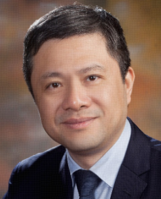 Qiang Xia (夏强)合作论文数Renji Hospital, Shanghai Jiao Tong University School of Medicine;Shanghai Cancer Institute19
 Xiaoni Kong (孔晓妮)合作论文数Renji Hospital, School of Medicine, Shanghai Jiao Tong University;Shanghai University of Traditional Chinese Medicine16
 Yue Zhang (张悦)合作论文数15Bing Han合作论文数12Xuehui Chu (褚薛慧)合作论文数12Jiangqiang Xiao合作论文数11Hailong Wu合作论文数9Xia Lei合作论文数9
 Jian Wang (王坚)合作论文数Renji Hospital, School of Medicine, Shanghai Jiao Tong University9Yichi Zhang合作论文数Shanghai Jiao Tong Univ, Sch Med, Xinhua Hosp, Dept Gen Surg, 1665 Kongjiang Rd, Shanghai 200092, Peoples R China8Ying Tong合作论文数8Huan Cao合作论文数7Shu-Hua Zhang合作论文数6
 Guoliang Wang合作论文数Union Hosp, Huazhong Univ Sci & Technol6Haozhen Ren (任昊桢)合作论文数6Jiajun Tan (檀家俊)合作论文数6Shenghe Deng合作论文数5
 Daichao Xu (许代超)合作论文数Shanghai Institute of Organic Chemistry, Chinese Academy of Science;University of Chinese Academy of Sciences5Tongxi Li合作论文数5Meng Sha合作论文数5
 Hao Cai合作论文数Fujian Med Univ, Dept Bioinformat, Key Lab, Minist Educ Gastrointestinal Canc, Fuzhou 350001, Peoples R China5Seogsong Jeong合作论文数Department of Biomedical Sciences, Seoul National University College of Medicine5
 Zhongze Gu (顾忠泽)合作论文数School of Biological Science and Medical Engineering, Southeast University;State Key Laboratory of Bioelectronics, Southeast University5Shuai Zhao合作论文数4shuai zhao (赵帅)合作论文数4han ding (丁瀚)合作论文数4Miao Huang合作论文数4
 Yankai Wen合作论文数Health Science Center at Houston, The University of Texas4Xue-hua SUN (孙雪花)合作论文数4
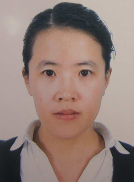 Chang Yu (于畅)合作论文数School of Chemical Engineering, Dalian University of Technology;Carbon Research Laboratory, Dalian University of Technology4
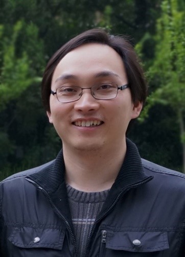 Yu Gan (甘愉)合作论文数Shanghai Cancer Institute4
 Hong Tu (屠红)合作论文数Shanghai Cancer Institute4
 Hualian Hang (杭化莲)合作论文数Renji Hospital, Shanghai Jiao Tong University School of Medicine4Tianhao Zou合作论文数3hao cai (蔡浩)合作论文数3Jiao-jun Tan合作论文数3Yuhan Xia合作论文数3Jichang Li合作论文数3Mengmeng Zhang合作论文数Chinese Acad Med Sci, Inst Mat Med, State Key Lab Bioact Subst & Funct Nat Med, Beijing 100050, Peoples R China3YaFu Wu (吴亚夫)合作论文数3XingYu Wu (吴星宇)合作论文数3Shan Bing合作论文数3
 Jianjun Zhang (张建军)合作论文数College of Stomatology, Shanghai Jiao Tong University3
 Yueqiu Gao (高月求)合作论文数 Shuguang Hospital Affiliated to Shanghai University of Traditional Chinese Medicine3Tian-Fei Lu合作论文数3




