Bone organoids have emerged as transformative models for studying bone development and disease by recapitulating complex cellular ecosystems in vitro. However, unlike soft tissues, the unique mineralized matrix and highly dynamic mechanical environment of bone pose significant challenges to classical self-assembly strategies. Consequently, engineering strategies are not merely auxiliary but essential for constructing functional bone organoids. This review provides a comprehensive overview of advanced engineering strategies designed to overcome these biological hurdles. We critically examine the integration of dynamic mechanical microenvironments and the design of biomimetic topologies for guiding cell fate. Furthermore, we explore the application of bone organoids in disease modeling while addressing inherent limitations. Potential solutions based on the convergence of 3D bioprinting, microfluidic organ-on-a-chip systems, and artificial intelligence are proposed. We anticipate that deep interdisciplinary collaboration will accelerate the transition of bone organoids from theoretical exploration to clinical personalized medicine, bridging the gap between basic research and regenerative therapies.
Organ-on-a-chip systems can replicate human physiological functionsin vitroby simulating the dynamicin vivomicroenvironment, therefore offering great potential for applications in drug screening, disease research, and personalized medicine. Multi-channel microfluidic chips are the core physical components of organ-on-a-chip systems, which often incorporate structures such as stripes, micro-pillars, and porous membranes to confine gels within specific channels, thereby providing a three-dimensional extracellular matrix environment for reconstruction of tissue barrier modelsin vitro. However, current multi-channel microfluidic chips confront challenges such as the unintended absorption of molecules, dependence on complex multi-material and multi-step fabrication processes, and instability in confining liquids. To address these challenges, we propose a multi-channel microfluidic chip with bilateral stripe structures, which can be mass-produced using single cyclic olefin copolymer material through injection molding. The bilateral stripe structures can effectively confine liquids with different wettabilities within the central channel by leveraging the edge effect. To demonstrate the versatility of the microfluidic platform, we successfully constructed tubular endothelial and renal tubule barriers on this chip, showcasing its potential for high-throughput, standardized organoid culture. This innovative microfluidic platform enables the construction of variousin vitroorgan models, offering a powerful tool for preclinical research and drug development.
Respiratory viral infections pose persistent global health threats, yet traditionalin vitroand animal models inadequately recapitulate human tissue microenvironments. Organ-on-a-chip technology integrates microfluidic engineering with cell biology to recreate three-dimensional architectures, mechanical forces, and multicellular interactions of the human respiratory system. This review systematically summarizes recent advances in organ-on-a-chip platforms for modeling respiratory viral infections and host immune responses. We highlight their unique capabilities in simulating alveolar-capillary barriers, lymphoid follicle formation, and multi-organ axes including lung-brain and gut-lung communication. Furthermore, we discuss applications in antiviral drug screening, vaccine evaluation, and personalized medicine, while addressing current challenges and future directions toward standardized, multi-organ integrated, and intelligently monitored systems.
Femtosecond laser three-dimensional printing has emerged as a transformative multiscale fabrication strategy for organ-on-a-chip systems, enabling hierarchical integration of functional nanomaterials with subcellular precision. This review highlights the synergistic convergence of ultrafast laser processing and nanomaterial engineering, overcoming the limitations of conventional fabrication methods by achieving sub-100-nm resolution via nonlinear optical effects. Key advances include nanoparticle-doped photoresists for mechanically robust, electrically conductive, and optically active microarchitectures, as well as laser-induced self-assembly mechanisms for nanomaterials such as silica nanoparticles, liquid crystals, and carbon nanotubes. The integration of in situ photoreduction, photoexcitation-induced bonding, and programmable optical forces enables precise control over nanomaterial dispersion, interfacial interactions, and multiscale structural integrity. These innovations facilitate the creation of biomimetic vascular networks, three-dimensional tissue scaffolds, and embedded biosensors, while enhancing mechanical strength, stimulus responsiveness, and device functionality. This hybrid fabrication paradigm holds significant potential to advance personalized disease modeling, drug screening, and bioelectronic interfaces via multifunctional organotypic platforms.
Background Exosomes (EXOs) are promising biomarker sources for liquid biopsy. Due to their low abundance in biofluids and the presence of non-target vesicle proteins during the isolation process, traditional methods such as ultracentrifugation (UC) are time-consuming, yield low purity, and can cause damage to EXOs. Achieving rapid, high-purity isolation remains a significant challenge. The magnetic beads (MBs) method is the most effective way to enhance EXO purity, but existing MBs face bottlenecks, including poor suspension stability, limited surface functionalization, slow magnetic response, and increased adsorption of interfering contaminants, ultimately leading to the loss of critical proteomic information. Results In this study, a novel strategy for EXO separation was proposed based on the specific interaction between the tetraspanin protein CD63 on EXOs and CD63-targeted aptamers functionalized on immunomagnetic hydrogel nanofibrils (IMHNFs). The IMHNFs showed streamlined magnetic responsivity with a responsivity rate 8-fold faster than that of conventional magnetic beads and exhibited good suspension stability. The IMHNFs enabled rapid EXO isolation from biofluids within 30 min, faster than UC. Furthermore, IMHNFs offer universality for multiple biofluids, higher purity (3.28-8.05-fold compared with UC), lower pollution protein, richer proteomic information, and higher signal intensity (the signal intensity of the specific protein CD63 in urine-derived EXOs was over 10-fold that achieved with UC). Notably, 332 upregulated proteins were identified in urine-derived EXOs from prostate cancer (PCa) patients compared with normal controls, covering 61 PCa biomarkers confirmed and published in the literature, demonstrating the excellent potential of this method for future clinical research and applications. Significance The proposed IMHNFs method exhibited significant advantages in magnetic separation performance (rapid magnetic response and good suspension stability), high-purity EXO enrichment, and proteomic analysis—especially its universality in various biofluids. It not only provides new insights into immunomagnetic separation technology but also contributes to advancing EXO development in liquid biopsy.
Polarized epithelia integrate barrier sealing, vectorial ion-water transport, and cytoskeletal mechanics, yet scalable assays that report this coupled functional state in real time remain limited. Here, we introduce photonic-crystal hydraulic manometry (PCHM), which quantifies out-of-plane mechanical states from single-frame reflection images in standard culture formats. Using PCHM, we find that epithelial monolayers maintain a kilopascal-scale basal compression (∼3 kilopascals) at the cell-substrate interface. A systematic perturbation panel spanning ion transport and actomyosin contractility defines the sensitivity, dynamic range, and reversibility of the readout, establishing basal compression as an actionable state variable of epithelial physiology. Leveraging this physiology-anchored metric, we detect early infection with coxsackievirus as a collapse of basal compression within 2 hours, well before cytopathic effects become apparent (48 hours). In severe acute respiratory syndrome coronavirus 2 pseudovirus neutralization assays, PCHM provided a 2-hour readout that was consistent with matched 48-hour luciferase results. In viral titration assays, the same 2-hour PCHM readout extended the detectable low-input range by approximately one order of magnitude relative to the matched 48-hour luciferase end point. Together, PCHM links epithelial transport and mechanics to a scalable, stain-free assay framework for epithelial pathophysiology and therapeutic screening.
IntroductionAbnormal pigmentation plays an important role in various skin diseases and in studies of whitening efficacy.Three-dimensional pigmented epidermis-on-a-chip models provide a crucial in vitro platform for exploring melanin production and regulation in skin. However, dynamic and non-invasive quantitative assessment of melanin distribution remains difficult with traditional histological methods.MethodsIn this study, an AI-assisted objective evaluation framework was established for three-dimensional pigmented epidermis-on-a-chip models based on brightfield images. Melanin regions were segmented using the MEM-ViT algorithm, and their morphological features were extracted to build a multi-indicator comprehensive analysis system for determining the “good/poor” status of the model.ResultsThe results showed 98% consistency between algorithmic predictions and manual annotations, demonstrating the reliability and generalization capability of the proposed method. The framework enabled accurate segmentation of melanin regions and standardized evaluation of model quality without staining.DiscussionThis method provides a rapid, non-invasive, and standardized approach for evaluating 3D pigmented epidermis-on-a-chip models. It offers a useful technical pathway for drug efficacy research, whitening mechanism analysis, and objective assessment of skin pigmentation-related disorders.
Hair follicle (HF) organoid transplantation has emerged as a significant advancement for hair regeneration and wound repair. This study developed an efficient and minimally invasive cryo-microneedles array patch (cryo-MAP) technique to transplant self-assembled 3D HF organoids from matrix-free, serum-free culture and low-DMSO cryopreservation. This method successfully avoids the growth suppression caused by hydrogels, allowing HFs to germinate within 3 days. Furthermore, proteomics confirmed that cryopreservation not only maintained high cell viability but also enhanced the organoids' capacity for directional differentiation. The transplanted organoids regenerated complex skin structures, achieving successful hair growth penetrating the skin in approximately 15 days with an over 86% success rate. These results open up new possibilities for organoid transplantation platforms in the fields of tissue engineering and regenerative medicine.
Chronic kidney disease (CKD), driven largely by renal fibrosis, lacks effective therapies due to the limited predictive capacity of existing preclinical models. To address this, we developed a human tubuloid-on-a-chip model integrating tubuloids, endothelial cells, and immune cells within a microfluidic system to recapitulate the key pathophysiology of renal fibrosis. Induction of fibrosis with TGF-β1 in this system recapitulated key pathological features, including extracellular matrix deposition, epithelial-mesenchymal transition, and loss of epithelial polarity. Functional assessments revealed impaired tubular reabsorption, including reduced albumin uptake and glucose transport, alongside elevated oxidative stress, mirroring clinical observations in CKD patients. The model’s pharmacological relevance was validated by the therapeutic effects of nintedanib, which attenuated fibrotic phenotypes. Taken together, this tubuloid-on-a-chip platform demonstrates the potential to model complex fibrotic pathologies in vitro and may serve as a useful tool for CKD research and anti-fibrotic drug development, potentially accelerating therapeutic discovery for renal fibrosis.
Bioelectronics are pivotal to biomedical engineering as they enable seamless communication between electronic devices and living tissues. However, conventional interfaces with static, rigid configurations often suffer from mechanical mismatch, chronic inflammation, and progressive signal degradation. By integrating stimuli-responsive materials with programmable architectures, 4D-printed devices enable in situ shape transformation and modulus adaptation, establishing a new paradigm for adaptive, high-fidelity biointerfaces. This review systematically examines recent advancements in 4D-printed bioelectronics. We first evaluate how smart materials-including hydrogels, liquid crystal elastomers (LCEs), and shape memory polymers (SMPs)-synergize with structural concepts such as auxetic lattices and kirigami/origami geometries to achieve dynamic compliance. Subsequently, we list advanced additive manufacturing strategies, particularly vat photopolymerization and direct ink writing (DIW) as well as hybrid printing strategies, which ensure high-precision fabrication. Furthermore, we highlight transformative biomedical applications, including neural interfaces, wearable devices, soft robotics, and implantable therapeutic devices. Finally, we discuss future directions for 4D-printed bioelectronic devices, focusing on the long-term stability of bioelectronic signal transduction, the integration of multifunctionality, bidirectional bioelectronic modulation, manufacturing scalability and standardization, and artificial intelligence (AI)-driven design and predictive modeling.
Background: Metabolic dysfunction-associated steatohepatitis (MASH) is a progressive form of metabolic liver disease with high clinical relevance, but the lack of physiologically relevant human in vitro models has limited mechanistic studies and therapeutic development. Angelica sinensis polysaccharide (ASP) has shown promising efficacy in preclinical studies. This study aimed to establish a human liver organoid-based MASH model and apply it to investigate ASP as a representative intervention, given its hepatoprotective and metabolic regulatory properties. Methods: Human induced pluripotent stem cells (hiPSCs) were differentiated into liver organoids, and oleic acid was used to induce MASH-like phenotypes. ASP was selected to assess the model's responsiveness to metabolic interventions and was also tested in high-fat diet (HFD)-fed mice. Proteomic analyses were conducted to explore potential molecular targets and pathways. Results: hiPSCs-derived liver organoids formed 3D hollow multicell spheres containing parenchymal and nonparenchymal cells, exhibiting stable hepatic synthetic and metabolic functions. The model successfully recapitulates hallmark MASH features, including disrupted glucose and lipid metabolism as well as inflammatory and immune dysregulation. Leveraging the MASH-like organoid model, we found that ASP intervention alleviated hepatocellular injury and improved glucose and lipid homeostasis, consistent with findings in HFD-fed mouse models. Mechanistically, ASP might exert hepatoprotective effects by indirectly suppressing perilipin 2 (PLIN2) through insulin signaling, thereby linking glucose and lipid metabolism. Conclusion: A human-derived MASH-like organoid model was established as a physiologically relevant platform for mechanistic research and drug screening. The findings highlight the therapeutic potential of ASP and underscore the translational value of organoid-based disease models.
Traditional toxicology, with its reliance on animal models and oversimplified cell cultures, often fails to predict human responses due to interspecies differences and limited physiological relevance. Organ-on-a-chip (OoC) technology, as a microengineering breakthrough, enables reconstruction of human-relevant organ functions, providing a powerful tool for toxicity testing. However, OoC remains largely regarded as a technological platform rather than a distinct research discipline. In this review, we propose organ-on-a-chip toxicology (OCT) as a groundbreaking interdisciplinary paradigm that integrates advanced engineering, toxicological science, and biomedical research to redefine toxicological assessment. OCT transcends conventional OoC technology by providing a unified framework for elucidating toxicity effects and mechanisms at molecular, cellular, and organ levels. It uniquely enables comprehensive systemic toxicity modeling, incorporating full absorption-distribution-metabolism-excretion pathways and inter-organ signaling. Leveraging cutting-edge bioengineering, organoid-driven cellular fidelity, and AI-enhanced data analytics, OCT delivers unparalleled precision in drug safety evaluation, personalized toxicology, environmental hazard assessment, and food health. Despite current challenges in standardization, scalability, and regulatory acceptance, OCT holds the potential to revolutionize toxicological science by offering predictive, ethical, and human-centric insights, minimizing animal testing while advancing global health risk assessments.
Diabetic kidney disease has increasingly emerged as a global public health concern, yet substantial challenges persist in its mechanistic research and drug development. Traditional 2-dimensional cell cultures and animal models frequently lack the capacity to faithfully recapitulate human pathophysiological conditions, and the use of animal models is increasingly limited by ethical and policy-related hurdles. In vitro models such as organoids and organs-on-a-chip have demonstrated remarkable advantages, enabling more faithful simulation of the in vivo microenvironment. The advancement of 3-dimensional bioprinting, microfluidic, and vascularization technologies has further propelled the maturation and progress of in vitro kidney models. This review summarizes and discusses in vitro kidney models and their advances, including kidney cell lines, spheroids, kidney organoids, and kidneys-on-a-chip. It also elaborates on the establishment of diabetic kidney disease models based on these in vitro platforms, which provides robust support for both basic and clinical research.
Superwettability describes an extreme wetting regime in which a solid surface exhibits exceptional affinity for or strong repellency against fluids, including superhydro/superoleo/superaero-phobicity, superhydro/superoleo/superaero-philicity, and directional liquid transport. Recent advances in biomimetics and theoretical modeling reveal that precise and intelligent wettability regulation is governed not only by surface chemistry but, more fundamentally, by microstructural geometry. Over the past two decades, bioinspired reentrant microstructures have exhibited exceptional capability in enhancing liquid repellency and enabling high-performance directional transport through modulation of interfacial wetting physics. In this review, we provide a comprehensive summary on structure-driven superwettability, focusing on symmetric and asymmetric reentrant microstructures. We first elucidate the fundamental physical mechanisms underlying wettability regulation, followed by a critical assessment of state-of-the-art fabrication strategies, including silicon micromachining, replica molding, and advanced 3D printing. We then highlight representative applications in microreactions, oil-water separation, liquid harvesting, evaporation and desalination, and fluidic-electronic systems, and discuss emerging strategies for smart liquid manipulation enabled by stimuli-responsive reentrant microstructures. Finally, we outline key challenges and forward-looking perspectives, emphasizing artificial intelligence-assisted design, novel functional materials, scalable manufacturing, and next-generation applications of superwettable surfaces.
Developing smart conductive hydrogels with excellent physicochemical properties to meet intricate requirements of epidermal and implantable bioelectronics remains a great challenge. Herein, an interface-enhanced polyzwitterionic composite hydrogel (PNTS) was developed via an interfacial fusion cross-linking strategy to achieve high mechanical strength and toughness, low swelling ratio, excellent tissue-adhesion, anti-nonspecific adsorption, and antibacterial properties for soft bioelectronics. By the interfacial fusion cross-linking strategy, PNTS hydrogel forms the interpenetrating network structure with the surface-enriched polyzwitterion and tannic acid, which endow the PNTS hydrogels not only excellent mechanical properties (>700% elongation and > 98% compression) and low swelling ratio (<80%), but also remarkable anti-protein adsorption, robust adhesion to various wet biological tissues and high antibacterial efficiency (>90%). The proposed PNTS displayed superior capability for monitoring of various electrophysiological signals (such as ECG, EEG, and EMG) and dynamic biological tissue motions (such as the contractile movements of pig lungs), as well as the implantable electrodes for neural signal transmission and myocardial infarction diagnosis, indicating their promising applications in epidermal and implantable bioelectronics.
Photonic crystal gels, as structural color materials with broad applications, derive their unique optical properties from the orderly arrangement of nanoparticles within the material. However, the self-assembled nanoparticle arrangement is highly susceptible to disturbances from external environmental stimuli, which significantly constrains their integration with other materials or manufacturing processes. In this study, we developed a strategy for preparing nonclose-packed photonic crystal gels mediated by a degradable gel network. This strategy locks the ordered nonclose-packed arrangement of nanoparticles at the nanoscale via the degradable gel network and enables easy incorporation of diverse functional materials into the photonic crystals through gel network exchange while maintaining structural color integrity throughout the process. This approach allows for facile preparation of photonic crystal gels with various compositions. Furthermore, integration with digital light processing (DLP) systems holds significant application value in the customized fabrication of patterned, multimaterial photonic crystals.
Calciphylaxis (calcific uremic arteriolopathy, CUA) is a rare, fatal disorder primarily affecting chronic kidney disease patients, characterized by microvascular calcification, thrombosis, and skin necrosis. In a discovery cohort (3 CUA, 10 uremic), plasma proteomics identified Thrombospondin-1 (THBS1) as the top upregulated hub in CUA, significantly reduced after human amnion-derived mesenchymal stem cell (hAMSC) therapy, alongside latent TGF-β binding protein 1, both linked to coagulation and wound healing. In vitro proteomics indicated that THBS1/TGF-β1 blockade impaired CUA serum-induced endothelial adhesion and coagulation. ELISA in combined discovery and validation cohorts (8 CUA, 20 uremic) confirmed this reduction post-treatment (6 patients), independent of systemic inflammation. Multiplex immunofluorescence revealed THBS1 and CD47 co-localized with CD31 and integrin β3 in injured microvessels. A human microvascular chip showed that THBS1 inhibition or hAMSC-conditioned medium alleviates injury. These findings implicate THBS1 as a key factor and potential biomarker in calciphylaxis, suggesting hAMSC therapy as a promising mechanism-based approach. Video Abstract:
The pursuit of 3D structures with precise microarchitectures and broad material adaptability has long been a central scientific objective, given their transformative potential to tackle critical challenges across applications such as vascular networks, optical components, soft robotics, energy storage devices, and microfluidics. While conventional additive manufacturing enables unprecedented design complexity and rapid prototyping, it remains limited by architectural instability, resolution constraints, low programmable porosity, and incompatibility with high-performance materials like carbon, glass, ceramics, and metals. By integrating 3D printing technologies with sacrificial strategies (also known as sacrificial 3D printing), these limitations have been largely overcome, where 3D-printed sacrificial templates are thermally decomposed, chemically etched, or photolytically degraded, enabling the deterministic fabrication of microarchitectures with previously unattainable adaptability and precision. Sacrificial 3D printing has made significant progress over the past decade, witnessing a growth in sacrificial materials, removal methods, and applications. This review highlights recent advancements of sacrificial strategies in additive manufacturing, with a further focus on key printing technologies (e.g., vat photopolymerization and material extrusion), sacrificial materials, permanent materials, and extensive applications in tissue engineering, optics, mechanics, electronics, and thermodynamics. Finally, this review critically evaluates the current challenges and future perspectives in materials development, printing technologies, post-processing, structural design and simulation, functions and applications, aiming to inspire further research and innovation to unlock the full potential of sacrificial 3D printing.
Skin aging results from a combination of intrinsic factors and exogenous stimuli, leading to changes in the structure and components of the extracellular matrix (including the skin basement membrane), which directly influence the aging process. In vitro models are powerful tools for exploring skin aging and overcoming inter-species differences and ethical issues associated with animal models, thus demonstrating powerful potential in skin aging research and anti-aging drug development. In this review, the advantages and disadvantages of in vitro models are discussed, including 2D monolayer models, 3D static reconstructed human skin models, 3D bioprinting models, organoid models, and Skin-on-Chip models for studying skin aging and anti-aging drug development. Finally, concepts and perspectives for the next-generation skin aging models are proposed. These models are expected to provide innovative tools for investigating the mechanisms of skin aging in depth, as well as skin aging repair and prevention.
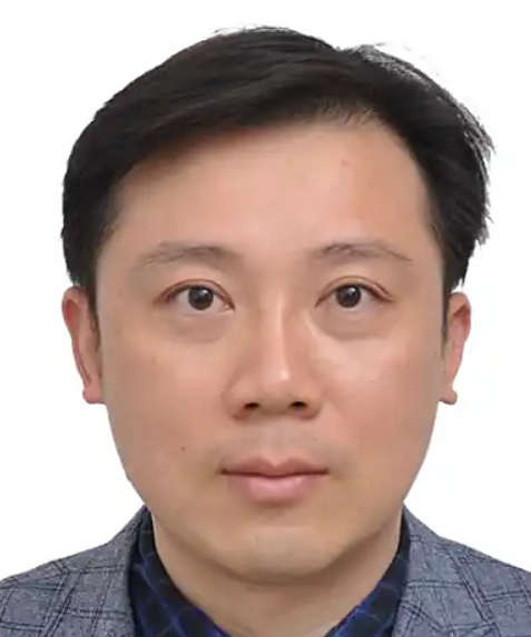 Yuanjin Zhao (赵远锦)合作论文数Medical School of Nanjing University91
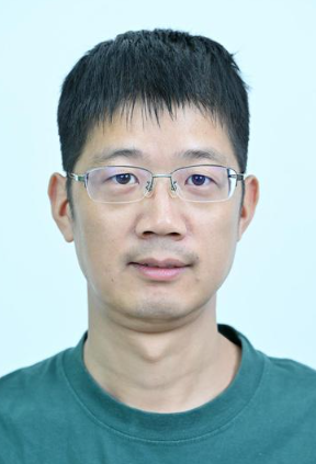 Zhuoying Xie (谢卓颖)合作论文数School of Biological Science & Medical Engineering, Southeast University72
 Xiangwei Zhao (赵祥伟)合作论文数School of Biological Science & Medical Engineering, Southeast University;State Key Laboratory of Digital Medical Engineering, Southeast University57
 Sato Osamu (佐藤治)合作论文数Department of Fundamental Organic Chemistry, Institute for Materials Chemistry and Engineering, Kyushu University53
 Zaozao Chen (陈早早)合作论文数School of Biological Science & Medical Engineering, Southeast University50
 Hua Xu (徐华)合作论文数School of Biological Science & Medical Engineering, Southeast University42
 Ding Haibo合作论文数School of Biological Science and Medical Engineering, Southeast University39
 Xiaojiang Liu (刘小将)合作论文数School of Biological Science & Medical Engineering, Southeast University32
 Bingbing Gao (高兵兵)合作论文数Sch Pharmaceut Sci, Nanjing Tech Univ32
 Yasuaki Einaga (栄長泰明)合作论文数Department of Chemistry, Faculty of Science of Technology, Keio University32
 Hongcheng Gu (顾洪成)合作论文数School of Biological Science & Medical Engineering, Southeast University31
 A. Fujishima合作论文数30
 Xin Du (杜鑫)合作论文数School of Biological Science & Medical Engineering, Southeast University30
 Xuejun Kang (康学军)合作论文数Research Center for Learning Science, Southeast University;School of Biological Science & Medical Engineering, Southeast University29
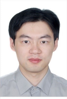 Cun Zhu (朱存)合作论文数School of Biological Science & Medical Engineering, Southeast University28
 Akira Fujishima (藤岛昭)合作论文数Tokyo University of Science;Kanagawa Academy of Science and Technology;University of Shanghai for Science and Technology27
 Luoran Shang (商珞然)合作论文数Institutes of Biomedical Sciences, Fudan University26Yi Zeng合作论文数Southeast Univ, Sch Biol Sci & Med Engn, State Key Lab Bioelect, Nanjing 210096, Jiangsu, Peoples R China25
 Qiwei Li合作论文数25Yao Cheng合作论文数24Chao Pan合作论文数23
 Tomokazu Iyoda合作论文数Tokyo Institute of Technology20
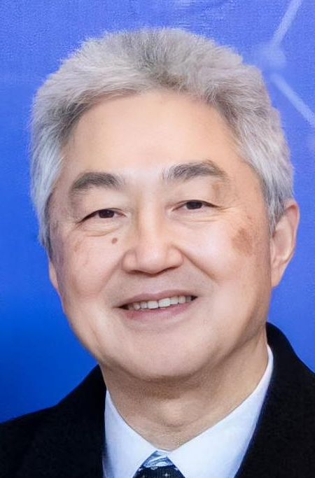 Zuhong Lu (陆祖宏)合作论文数State Key Laboratory of Digital Medical Engineering, School of Biological Science & Medical Engineering, Southeast University;Nanjing Percare Biotechnology Co., Ltd19
 Zhangqi Feng (冯章启)合作论文数5chool of Chemistry and Chemical Engineering, Nanjing University of Science and Technology17
 Zhenzhu He合作论文数15Jie Sun合作论文数Shenyang Ligong University(Shenyang Ligong University,Shenyang Institute of Technology),Shenyang,China15Junlong Liao合作论文数14Junning Zhang合作论文数14
 Fu Fanfan合作论文数14
 Xuemei Wang (王雪梅)合作论文数School of Biological Science & Medical Engineering, Southeast University14
 Liguo Sun合作论文数13
 Wang Wan合作论文数13Mu Zhongde合作论文数13
 Xingran Cui (崔兴然)合作论文数School of Biological Science & Medical Engineering, Southeast University13Ye Baofen合作论文数13
 Jie Wang (王洁)合作论文数College of Artificial Intelligence, Nanjing Agricultural University13
 Degang Fu (付德刚)合作论文数School of Biological Science & Medical Engineering, Southeast University13
 Yitao Ding (丁义涛)合作论文数Nanjing Drum Tower Hospital, The Affiliated Hospital of Nanjing University Medical School13
 Liqin Ge (葛丽芹)合作论文数School of Biological Science and Medical Engineering, Southeast University13
 Abdelrahman Elbaz合作论文数12
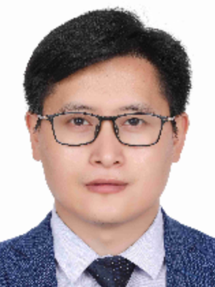 Fuyin Zheng (郑付印)合作论文数School of Biological Science and Medical Engineering, Beihang University12
 Hong Liu (刘宏)合作论文数School of Biological Science and Medical, Southeast University;Chien-Shiung Wu College, Southeast University12
 Liqin Chen (陈利琴)合作论文数School of Public Health, Tianjin Medical University12Min Wang合作论文数12
 Xiao-Lei Shi (施晓雷)合作论文数Nanjing Drum Tower Hospital12Huan Wang合作论文数11
 Guozhi Han (韩国志)合作论文数南京工业大学化学与分子工程学院11
 Fei Rong (戎非)合作论文数School of Biological Science & Medical Engineering, Southeast University11Xuehui Chu (褚薛慧)合作论文数11




