Lesions of the superior parietal lobule are associated with impaired manipulative hand movements. The tight connections of the intraparietal sulcus with the premotor areas could explain the marked deficits in joint kinematic and dynamics occurring in apractic patients with lesions of this part of the parietal lobe. Damage to the posterior parietal cortex may cause the deficit of ideomotor limb apraxia. The observations indicated defects of visuo-kinaesthetic motor representation of learned movements that are stored in the dominant parietal lobe, or alternatively a separation of these representations from premotor or motor areas. Florida Apraxia Screening Test and modified Aachener Apraxia Test revealed signs of an ideomotor apraxia in two patients with left parietal damage and one with a right parietal lesion. The patient with a right parietal lesion without apraxia showed some preshaping and his maximum aperture was moderately increased and delayed.
Objective The progression of Alzheimer’s disease (AD) is associated with impaired nutritional status. New methods, such as deep brain stimulation (DBS), are currently being tested to decrease the progression of AD. DBS is an approved method in the treatment of Parkinson’s disease, and its suitability for the treatment of AD patients is currently under experimental investigation. To evaluate the advantages and disadvantages of this new treatment, it is important to assess potential side effects of DBS regarding the nucleus basalis of Meynert; this new treatment is thought to positively affect cognition and might counteract the deterioration of nutritional status and progressive weight loss observed in AD. This study aims to assess the nutritional status of patients with AD before receiving DBS of the nucleus basalis of Meynert and after 1 year, and to analyze potential associations between changes in cognition and nutritional status. Design A 1-year phase I proof-of-concept study. Setting The Department of Psychiatry and Psychotherapy at the University of Cologne. Participants We assessed a consecutive sample of patients with mild to moderate AD (n=6) who fulfilled the inclusion criteria and provided written informed consent. Intervention: Bilateral low-frequency DBS of the nucleus basalis of Meynert. Measurements Nutritional status was assessed using a modified Mini Nutritional Assessment, bioelectrical impedance analysis, a completed 3-day food diary, and analysis of serum levels of vitamin B12 and folate. Results With a normal body mass index (BMI) at baseline (mean 23.75 kg/m 2 ) and after 1 year (mean 24.59 kg/m 2 ), all but one patient gained body weight during the period of the pilot study (mean 2.38 kg, 3.81% of body weight). This was reflected in a mainly stable or improved body composition, assessed by bioelectrical impedance analysis, in five of the six patients. Mean energy intake increased from 1534 kcal/day (min 1037, max 2370) at baseline to 1736 kcal/day (min 1010, max 2663) after 1 year, leading to the improved fulfillment of energy needs in four patients. The only nutritional factors that were associated with changes in cognition were vitamin B12 level at baseline (Spearman’s rho = 0.943, p = 0.005) and changes in vitamin B12 level (Spearman’s rho = −0.829, p = 0.042). Conclusion Patients with AD that received DBS of the nucleus basalis of Meynert demonstrated a mainly stable nutritional status within a 1-year period. Whether DBS is causative regarding these observations must be investigated in additional studies.
In the past decade deep brain stimulation (DBS)—the application of electrical stimulation to specific target structures via implanted depth electrodes—has become the standard treatment for medically refractory Parkinson's disease and essential tremor. These diseases are characterized by pathological synchronized neuronal activity in particular brain areas. We present an external trial DBS device capable of administering effectively desynchronizing stimulation techniques developed with methods from nonlinear dynamics and statistical physics according to a model-based approach. These techniques exploit either stochastic phase resetting principles or complex delayed-feedback mechanisms. We explain how these methods are implemented into a safe and user-friendly device.
To reveal the dynamic mechanism underlying Parkinsonian resting tremor, we applied a phase dynamics modelling technique to local field potentials and accelerometer signals recorded in three Parkinsonian patients with implanted depth electrodes. We detect a bidirectional coupling between the subcortical oscillation and the tremor. The tremor → brain driving is a linear effect with a small delay corresponding to the neural transmission time. In contrast, the brain → tremor driving is a nonlinear effect with a long delay in the order of 1–2 mean tremor periods. Our results are well reproduced for an ensemble of 41 tremor epochs in three Parkinsonian patients and confirmed by surrogate data tests and model simulations. The uncovered mechanism of tremor generation suggests to specifically counteract tremor by desynchronizing the subcortical oscillatory neural activity.
BACKGROUND AND PURPOSE:Motor hemineglect is characterized by an underutilization of one side of the body. It is a higher-order motor disorder that resembles hemiplegia although being substantially different from it due to a preserved motor output system. Its role for poststroke recovery is still unclear.METHODS:We studied 52 patients presenting with acute hemiparetic stroke over the first 7 days after symptom onset. Nineteen patients had unilateral motor hemineglect. Impairment was clinically assessed with the European Stroke Scale and a multifactorial motor score. It was further assessed quantitatively, as overall arm activity was measured continuously by Actiwatches. Lesion volumes were measured morphometrically within 24 h on perfusion- and diffusion-weighted magnetic resonance images and on average on day 9 by T2-weighted magnetic resonance imaging.RESULTS:Patients with motor hemineglect were characterized by significantly reduced initial arm activity in comparison to patients without motor hemineglect. This was paralleled by larger brain lesions in the patients with motor hemineglect. Patients with motor neglect either recovered virtually completely (5 cases; 2/5 left hemisphere; 3/5 treated with recombinant tissue plasminogen activator, rt-PA) within 7 days or did not improve at all (14 cases; 3/14 left hemisphere; 3/14 rt-PA treated).CONCLUSION:Our data reveal a high incidence of motor hemineglect in patients with acute stroke. They further show that these patients are more severely compromised than those without motor hemineglect. A rapid and near complete recovery was observed in about one fourth of the motor hemineglect patients and may be related to involvement of the left hemisphere or to therapy with thrombolysis.
The history of stereotactic neurosurgery for treatment of Parkinson's disease has a remarkable track record. The traditional lesion method, employed and refined over half a century, seemed to have reached its end with the advent of l-dopa medication. But, paradoxically, levodopa-induced motor fluctuations and dyskinesias led to a renaissance of surgical therapy and the introduction of deep brain stimulation by Alim-Louis Benabid et al . (1987) opened a new therapeutic window for treatment of the late levodopa syndrome. From the beginning it appeared that the effects of stimulation mimic those of stereotactic lesions. Whereas the advantage of the fine tuning of treatment and its reversibility spoke for the new method, its long term efficacy still awaited the test of time. Recently, the Grenoble group reported the results of a 5 year follow-up showing that subthalamic nucleus deep brain stimulation had persistent beneficial effects in 42 patients (Krack et al ., 2003). In this issue of Brain , Rodriguez-Oroz et al . (2005) report results of the first worldwide multicentre study on long term effects of deep brain stimulation. Eight groups enrolled 69 bilaterally implanted patients who were assessed preoperatively and 3–4 years postoperatively. What is particularly valuable is the comparative evaluation of the effects of stimulating …
We used functional magnetic resonance imaging (fMRI) to explore how guidance of motor acts is influenced by the visually perceived body scheme. We found that when subjects view their hand as their opposite hand, i.e., the right hand is seen as the left hand and vice versa, activation in the visual cortex was lateralized opposite to the seen hand. This demonstrates for the first time that our body scheme to which vision relates our environment is already represented at the level of visual cortex.
Dieses Kapitel gibt eine Übersicht über die klinische Wirksamkeit der tiefen Hirnstimulation des Nucleus subthalamicus (STN) in der Behandlung der Parkinsonkrankheit. Dabei ist zu berücksichtigen, dass es sich um ein noch relativ junges Therapieverfahren handelt, das erstmals 1993 von Alim-Louis Benabid eingesetzt wurde [22]. Die beeindruckenden Therapieerfolge führten dazu, dass es rasch von einer steigenden Zahl von Behandlungszentren aufgenommen wurde. Zum Teil wird es bereits jetzt als Therapie der Wahl zur Behandlung des Levodopa-Langzeitsyndroms sowie des medikamentös therapie- refraktären Parkinsontremors angesehen. Um den genauen Stellenwert der STN-Stimulation in der Parkinsonbehandlung zu ermitteln, ist es jedoch wichtig, das gesamte Spektrum der Auswirkungen auf den Parkinsonkranken zu kennen. Hierzu gehören nicht nur die unmittelbaren Effekte auf motorische und nichtmotorische Parkinsonsymptome, sondern auch der Einfluss auf den Krankheitsverlauf und besonders auf die komplexen Aspekte, welche die Lebensqualität der Parkinsonkranken betreffen. In dieser Hinsicht sind bezüglich der kritischen Wertung der STN-Stimulation mehrere Phasen zu erkennen. Während zunächst der Einfluss auf die motorischen Kardinalsymptome bzw. spezifischere motorische Leistungen im Vordergrund stand, folgten zunehmend Untersuchungen zu den neuropsychologischen Auswirkungen. Diesen schließt sich derzeit ein wachsendes Interesse an möglichen Auswirkungen auf neuropsychiatrische Nebenwirkungen an, besonders in Bezug auf das affektive und emotionale Erleben der Betroffenen. Die im Folgenden vorgestellten Ergebnisse geben somit nur eine Momentaufnahme eines wachsenden Kenntnisprozesses über dieses Therapieverfahren wieder. Die Ergebnisse zur bilateralen Nucleus-subthalamicus-Stimulation der eigenen Arbeitsgruppe stammen aus einem Kollektiv von 69 Patienten, das im Zeitraum von 1998 bis 2002 in einer offenen prospektiven Studie über mindestens sechs Monate untersucht wurde. Neun dieser Patienten wurden zudem in eine internationale prospektive Studie der Firma Medtronic (Advanced Parkinson’s disease postmarketing study — APDPMS) eingeschlossen, in die insgesamt 96 Patienten aus 18 Zentren eingingen [9]. Die demographischen Daten dieser Köln-Düsseldorfer Patienten sind in Tabelle 1 wiedergegeben.
In the surrounding of focal ischemic brain lesions dysfunctional neuronal zones emerge often resulting in pathological oscillatory activity. Using whole-head magnetoencephalography we recorded brain activity during rest in 23 patients with ischemic cortical lesions to find out whether we can localise and characterise low-frequency oscillatory activity. We measured patients at different times after stroke and partly in a follow-up approach to determine the time course of slow-wave activity. Using the analysis tool Dynamic Imaging of Coherent Sources we computed tomographic maps of oscillatory power in the delta-band (0.5-3 Hz). Fifteen of 23 patients with cortical strokes showed delta-activity, which was localised in an area not more than 2 cm away from the lesion. We found this perilesional low-frequency activity in the acute as well as in the chronic stage of stroke. Follow-up measurements of individual patients revealed persistence of perilesional low-frequency activity for months and even years. No consistent relation between perilesional activity and clinical symptoms was observed. Our results indicate that perilesional delta activity is common after ischemic cortical stroke. However, the functional significance remains to be elucidated.
Synchronization of neuronal activity appears to be the hallmark of several neurological diseases like Parkinson's disease and essential tremor. In patients who do not respond to medication, permanent electrical high-frequency (>100Hz) deep brain stimulation (DBS) turned out to be the therapeutic gold standard. This standard DBS basically mimics the effect of tissue lesioning. However, its mechanism is still not sufficiently understood. For this reason, we have investigated stimulation-induced dynamics in mathematical models of relevant target areas. We have developed novel stimulation techniques using methods from statistical physics, especially from the field of stochastic phase resetting. The standard DBS seems to block the neuronal firing, in particular neuronal populations. In contrast, our novel DBS techniques work on demand and desynchronize the pathologically synchronized activity. In this way the neuronal firing is maintained and modulated in a way that comes closer to the physiological mode. In this poster we explain how our novel DBS techniques have been optimized by means of real-time model simulations. Furthermore, we show our first successful results obtained in intraoperative test stimulations during deep electrode implantation. We expect our novel, model-based and physiologically oriented DBS techniques to be milder and more effective compared to the standard high-frequency DBS.
Reaching for a target involves integrative coordinate transformation processes between the representation of the target location, the sensorimotor information of limb of reach, and body space. Although right hemisphere dominance for visuospatial information processing is well established, corresponding right hemisphere dominance for kinesthetic spatial information processing remains to be demonstrated. We explored neural mechanisms of encoding target locations using 15 O‐butanol positron emission tomography (PET) in normal volunteers in a factorial experiment, where modality (visual/kinesthetic) and hemispace of target presentation (left/right of midsagittal plane) were varied systematically. After target presentation, subjects reached to the encoded target location. PET data analysis using SPM99 showed increased neural activity ( P < 0.05, corrected) associated with left hemispace target presentation in right hemisphere areas (sensorimotor, anterior cingulate, insular, and temporo‐occipital cortex) only. By contrast, right hemispace target presentation activated bilateral temporo‐occipital cortex, which extended into the right temporo‐parietal cortex and left sensorimotor cortex. A significant interaction of hemispace and modality of target presentation observed in right temporo‐parietal cortex resulted from an increase in neural activity with kinesthetic target presentation in right hemispace. The data support an important role for the right temporo‐parietal area in visuospatial processing and suggest a specific role of the right hemisphere in kinesthetic spatial processing. Hum. Brain Mapp. 21:165–177, 2004. © 2004 Wiley‐Liss, Inc.
To improve deep brain stimulation (DBS), we have studied stimulation-induced dynamics in target areas for DBS in mathematical models. We have implemented these neuronal networks on special fast digital hardware. Our real-time models enable us to simulate stimulation-induced processes in patients under real-time conditions. Our approach makes it possible to evaluate and optimize software and hardware realizing our novel demand-controlled DBS techniques (so-called model in the loop concept). The models and the control algorithms have been coded with the graphical programming language SIMULINK by Mathworks which enables a modification of the model dynamics and the control concepts in a fast and transparent way and thus facilitates and improves the interdisciplinary research in the field of neuromodulation. The goal of our approach is the design of novel stimulation protocols which specifically modulate the affected neuronal dynamics.
Data from experiments in MPTP monkeys as well as from invasive and non-invasive recordings in patients with Parkinson's disease suggest an abnormal synchronization of neuronal activity in the generation of resting tremor in Parkinson's disease. In six patients with tremor-dominant idiopathic Parkinson's disease, we recorded simultaneously surface electromyograms (EMGs) of hand muscles, and brain activity with a whole-head magnetoencephalography (MEG) system. Using a recently developed analysis tool (Dynamic Imaging of Coherent Sources; DICS), we determined cerebro-muscular and cerebro-cerebral coherence as well as the partial coherence between cerebral areas and muscle, and localized coherent sources within the individual MRI scans. The phase lag between the EMG and cerebral activity was determined by means of a Hilbert transform of both signals. After overnight withdrawal from medication, patients showed typical Parkinson's disease resting tremor (4-6 Hz). This tremor was associated with strong coherence between the EMG of forearm muscles and activity in the contralateral primary motor cortex (M1) at tremor frequency but also at double tremor frequency. Phase lags between M1 activity and EMG were between 15 and 25 ms (M1 activity leading) at single, but also at double tremor frequency, corresponding well to the corticomuscular conduction time. Furthermore, significant coherence was observed between M1 and medial wall areas (cingulate/supplementary motor area; CMA/SMA), lateral premotor cortex (PM), diencephalon, secondary somatosensory cortex (SII), posterior parietal cortex (PPC) and the contralateral cerebellum at single tremor and, even stronger at double tremor frequency. Spectra of coherence between thalamic activity and cerebellum as well as several brain areas revealed additional broad peaks around 20 Hz. Power spectral analysis of activity in all central areas indicated the strongest frequency components at double tremor frequency. Partial coherence analysis and the calculation of phase shifts revealed a strong bidirectional coupling between the EMG and diencephalic activity and a direct afferent coupling between the EMG and SII and the PPC. In contrast, the cerebellum, SMA/CMA and PM show little evidence for direct coupling with the peripheral EMG but seem to be connected with the periphery via other cerebral areas (e.g. M1). In summary, our results demonstrate tremor-related oscillatory activity within a cerebral network, with abnormal coupling in a cerebello-diencephalic-cortical loop and cortical motor (M1, SMA/CMA, PM) and sensory (SII, PPC) areas contralateral to the tremor hand. The main frequency of cerebro-cerebral coupling corresponds to double the tremor frequency.
Objectives - To test the hypothesis that cortical plasticity related to destructive tumour growth is functionally relevant. This hypothesis predicts that function is dependent on the intactness of tissue surrounding the tumour. Material and methods - Eight patients underwent laser-induced interstitial thermotherapy (LITT) for minimally invasive palliative treatment of brain tumours located in eloquent frontal motor regions including the primary motor cortex. A multimodal approach was used to assess the functional outcome of patients after LITT in detail. Results - Following LITT, motor function deteriorated in four patients. In three of these four patients the LITT-induced lesion involved minimal parts of adjacent non-tumorous tissue. By contrast, the other four patients whose LITT-induced signal changes were confined to the tumour, showed no functional deficits. Conclusion - These findings support the idea that peri-tumorous neuronal circuitry in motor competent areas may permanently take over those functions that were formerly represented in the neuronal tissue destroyed by the tumour.
BackgroundHigh-frequency electrical stimulation of the subthalamic nucleus is a new and highly effective therapy for complications of long-term levodopa therapy and motor symptoms in advanced Parkinson disease (PD). Clinical observations indicate additional influence on emotional behavior.MethodsElectrical stimulation of deep brain nuclei with pulse rates above 100 Hz provokes a reversible, lesioning-like effect. Here, the effect of deep brain stimulation of the subthalamic nucleus on emotional, cognitive, and motor performance in patients with PD (n = 12) was examined. The results were compared with the effects of a suprathreshold dose of levodopa intended to transiently restore striatal dopamine deficiency. Patients were tested during medication off/stimulation off (STIM OFF), medication off/stimulation on (STIM ON), and during the best motor state after taking levodopa without deep brain stimulation (MED).ResultsMore positive self-reported mood and an enhanced mood induction effect as well as improvement in emotional memory during STIM ON were observed, while during STIM OFF, patients revealed reduced emotional performance. Comparable effects were revealed by STIM ON and MED. Cognitive performance was not affected by the different conditions and treatments.ConclusionsDeep brain stimulation of the subthalamic nucleus selectively enhanced affective processing and subjective well-being and seemed to be antidepressive. Levodopa and deep brain stimulation had similar effects on emotion. This finding may provide new clues about the neurobiologic bases of emotion and mood disorders, and it illustrates the important role of the basal ganglia and the dopaminergic system in emotional processing in addition to the well-known motor and cognitive functions.
To assess the effects on motor functioning, health status and direct medical costs of high-frequency stimulation of the subthalamic nucleus (DBS-STN) in patients with idiopathic Parkinson's disease (PD). In addition, the cost-effectiveness of DBS-STN vs. drug treatment was investigated.
 Rudiger Seitz合作论文数51
 Ferdinand Binkofski合作论文数Department of Neurology, University Hospital44
 Harald Hefter合作论文数40Alfons Schnitzler合作论文数29
 Karlheinz Reiners合作论文数Department of Neurology, Julius-Maximilians-Universität24
 Otto W. Witte合作论文数Department of Neurology Jena University Hospital20
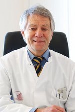 Volker Sturm合作论文数University of Cologne19
 Jens Volkmann合作论文数Neurologische Klinik und Poliklinik des Universitatsklinikums18
 Karl Zilles合作论文数Institut für Neurowissenschaften und Medizin, Forschungszentrum Jülich;Klinik für Psychiatrie, Psychotherapie und Psychosomatik, Uniklinik RWTH Aachen16Stefan Posse合作论文数15Hj Budingen合作论文数14Erwin Kunesch合作论文数14
 Christian Dohle合作论文数Department of Neurology, Heinrich-Heine-University13Volker Homberg合作论文数13K. M. Stephan合作论文数12F. Schmitz合作论文数12
 Volker Dietz合作论文数Spinal Cord Injury Center, Research, University of Zurich;Balgrist University Hospital12
 Peter Tass合作论文数Department of Neurosurgery, School of Medicine, Stanford University;Wu Tsai Neurosciences Institute, Stanford University12
 Giovanni Buccino合作论文数Faculty of Medicine, University San Raffaele;Division of Neuroscience, San Raffaele Scientific Institute12
 Gereon Rudolf Fink合作论文数Institute Cognitive Neuroscience, Forschungszentrum Jülich GmbH;Department of Neurology, Cologne University Hospital & Institute of Neuroscience and Medicine, Cognitive Neuroscience, Research Centre Jülich10
 Hans Herzog合作论文数Institut für Neurowissenschaften und Medizin, Forschungszentrum Jülich9J Herdmann合作论文数8Lutz Tellmann合作论文数Institute of Medicine, Research Centre Jülich8
 Ulla Knorr合作论文数Department of Psychiatry, University of Copenhagen7Gilbert Wunderlich合作论文数7Gabriele Arendt合作论文数6Helmuth Steinmetz合作论文数6
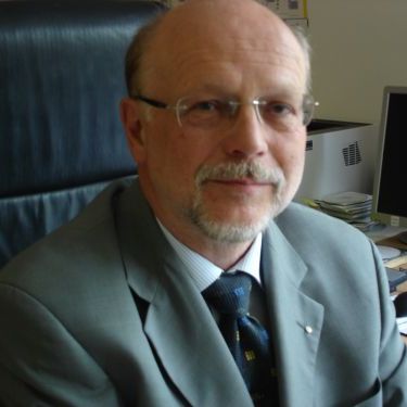 Reiner Benecke合作论文数Zentrum für Nervenheilkunde, Universität Rostock6
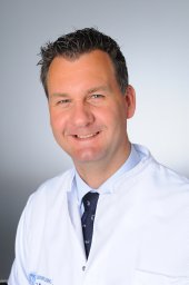 Peter Weiß合作论文数Department of Neurology, University Hospital Cologne6W. Heide合作论文数5K Voigt合作论文数5Joachim Gross合作论文数5Mario Siebler合作论文数5
 Andreas Kleinschmidt合作论文数Neurology Division, Département des Neurosciences Cliniques, Faculty of Medicine, Université de Genève;Faculty of Medicine, Geneva University Hospitals5Georg Hagemann合作论文数5Niels Allert合作论文数5
 N. J. Shah合作论文数Institute of Neurosciences and Medicine, Forschungszentrum Jülich;Faculty for Psychology and Neuroscience, Maastricht University;Department of Neurology, Faculty of Medicine, RWTH Aachen University;Faculty of Natural Sciences, RWTH Aachen University;University of New Brunswick5
 Joseph Classen合作论文数Clinic and Policlinic of Neurology, University Hospital of Leipzig;Department of Neurology, University Hospital Leipzig5
 Christian Hauptmann合作论文数Research Center Juelich, Institute of Medicine and Virtual Institute of Neuromodulation, 52425, Juelich, Germany5Thorsten Schormann合作论文数5
 Utako Barnikol合作论文数5
 Matthias Nitschke合作论文数5E Logigian合作论文数4
 Lars Timmermann合作论文数4
 Lutz Jäncke合作论文数Psychological Institute, University of Zurich;Division Neuropsychology, University of Zurich4Hilperath F合作论文数4Wittsack Hans-Joerg合作论文数4
 Gottfried Schlaug合作论文数Neuroimaging Laboratory, Beth Israel Deaconess Medical Center;Music and Neuroimaging Laboratory, Harvard Medical School;Department of Neurology, University of Massachusetts Medical School4Tobias Neumann-Haefelin合作论文数Institut für Pharmakologie, Universität Bern4




