Cranioplasty restores cranial integrity following decompressive craniectomy or skull trauma. Despite its reconstructive benefits, post-cranioplasty complication rates are high. Post-operative drainage has been proposed to mitigate these risks, yet its effectiveness remains uncertain. This study evaluates the impact of post-cranioplasty drain insertion on surgical outcomes. A systematic literature search of MEDLINE, Embase, and Cochrane CENTRAL Library was conducted in accordance with PRISMA guidelines (PROSPEROID:CRD420251030365). Studies reporting cranioplasty outcomes with post-operative drainage were selected. Primary outcomes were complication rates, including infection, haemorrhage, and cerebrospinal fluid (CSF) leak. Four studies met the inclusion criteria, comprising 522 patients (mean age 43.7 years) who underwent cranioplasty—282 with post-operative drainage and 240 without. Following decompressive craniectomy, the most common indications for cranioplasty were traumatic brain injury (196/514, 38.1
Post-traumatic seizure (PTS) is a significant complication of traumatic brain injury (TBI), associated with unfavourable outcomes. However, reported risk factors for PTS are inconsistent, and considerable variation exists in clinical practice regarding seizure prophylaxis. This systematic review and meta-analysis aimed to identify and evaluate the risk factors for early and late PTS in adults. A systematic search was conducted on PubMed, MEDLINE, EMBASE, and the Cochrane Library for studies published from 1975 to November 2024, investigating risk factors for PTS in adults with TBI. The protocol was registered with PROSPERO (CRD42021287959). Two independent reviewers screened studies, extracted data, and assessed the risk of bias using the Critical Appraisal Skills Programme (CASP) tool. A narrative synthesis and random-effects meta-analysis were performed to pool data on PTS incidence and key risk factors. From 13,999 records identified, 11 studies involving 242,803 patients were included. The pooled incidence of PTS was 7.2
Background:Traumatic acute subdural haematomas often require surgical evacuation via craniotomy or decompressive craniectomy. Decompressive craniectomy may prevent intracranial hypertension; however, it is unclear whether it is associated with better outcomes. Objective:Multicentre, pragmatic, parallel-group randomised trial to compare the clinical and cost-effectiveness of decompressive craniectomy versus craniotomy for evacuation of acute subdural haematomas. Design:International, multicentre, pragmatic, parallel-group randomised trial with additional observational arm. Setting:Hospitals with neurosurgical services in the UK and internationally. Participants:Patients aged ≥ 16 years, with a diagnosis of acute subdural haematomas on a computed tomography scan that required evacuation with a large bone flap either by craniotomy or decompressive craniectomy according to the opinion of the admitting neurosurgeon. Interventions:The enrolled patients underwent acute subdural haematoma evacuation in the operating room under general anaesthesia. A large bone flap ipsilateral to the haematoma was raised, the dura opened and the haematoma evacuated. Other haematomas, such as contusions, were evacuated at the discretion of the surgeon. If clinically appropriate randomisation occurred, the bone flap was either replaced (craniotomy) or not replaced (decompressive craniectomy). Patients who could not be randomised were followed up in the observational arm. Main outcome measures:Primary outcome measure was the extended Glasgow Outcome Scale assessed 12 months post injury. An economic evaluation (based on UK participants) was undertaken to estimate the cost-effectiveness of craniotomy compared to decompressive craniectomy. Results:Four hundred and fifty patients were randomised: 228 to craniotomy and 222 to craniectomy - with the common odds ratio for the differences across the Glasgow Outcome Scale scores of 0.85 [95% confidence interval (0.6 to 1.18; p = 0.324)]. The results were similar at 6 months. At 12 months, death occurred in 30.2% of the craniotomy group versus 32.2% of the decompressive craniectomy group, vegetative state occurred in 2.3% versus 2.8%, and good recovery occurred in 25.6% and 19.9%, respectively. In the observed cohort, those who had a decompressive craniectomy had significantly worse outcomes at 6 and 12 months, but their baseline characteristics were different. Limitations:Clinicians were not blinded to the trial groups. Decompressive craniectomy was performed in 8.8% of patients allocated to the craniotomy group, and 5.4% of patients allocated to the decompressive craniectomy group underwent craniotomy. Intraoperative non-adherence with allocation did not influence the primary analysis, which was based on the intention-to-treat principle. Conclusions:Among patients undergoing evacuation of acute subdural haematomas, the outcomes were similar in both groups. Additional surgery was required in a higher proportion of patients in the craniotomy group, but more wound complications occurred in the decompressive craniectomy group. Future work:Long-term outcomes of patients following decompressive craniectomy, timing and impact of cranial reconstruction on a patient's rehabilitation. Trial registration:This trial is registered as ISRCTN87370545. Funding:This award was funded by the National Institute for Health and Care Research (NIHR) Health Technology Assessment programme (NIHR award ref: 12/35/57) and is published in full in Health Technology Assessment; Vol. 30, No. 66. See the NIHR Funding and Awards website for further award information.
Following calls for more qualitative research in neurosurgery, this scoping review aimed to describe the range and reach of qualitative studies relevant to the field of neurosurgery and the patients and families affected by neurosurgical conditions. A systematic search was conducted in September 2024 across six databases: Medline via Ebsco; Embase via OVID; PsycINFO via Ebsco; Scopus; Web of Science Core Collection; and Global Health via Ebsco. Eligibility criteria were based on Population, Concept, and Context. The search identified 18,809 hits for screening with 812 included in the final analysis. Seven themes were identified from a content analysis of study aims: 1 Perspectives of living with a neurosurgical condition; 2 Family perspectives; 3 Perceptions of neurosurgery; 4 Perceptions of general healthcare care; 5 Decision making; 6 Advancing neurosurgery; and, 7 Understanding neurosurgical conditions. Traumatology was identified as the most researched sub-specialty (43.2%) yet few studies were led explicitly by a neurosurgeon (1.6%) or those with a neurosurgical affiliation (10.5%). Lead authors were predominantly from high income countries (93.7%), as were most multi-author teams (86.6%). There was a trend towards increasing publication over time; however, only 8.4% of papers were published in neurosurgical specific journals. The data set had an average Field Weighted Citation Impact of 0.96 and Field Weighted Views Impact of 1.11, 18.9% were cited in policy documents in 15 countries. This scoping review provides a comprehensive picture of the current qualitative research base in neurosurgery and suggests ways to improve the conduct and reporting of such studies in the future. Addressing these challenges is crucial if qualitative research is to advance the neurosurgical evidence base in a rigorous way.
Objective: For patients undergoing decompressive craniectomy for TBI, cranioplasty facilitates rehabilitation and reintegration into society. Cranioplasty practice is poorly documented globally. Challenges including lack of materials and technical skills disproportionately affect LMICs. This study evaluates global cranioplasty practice and the extent to which barriers preclude patients from accessing cranial reconstruction. Methods: An international survey was disseminated to centres performing cranioplasty for TBI. Survey questions addressed baseline hospital information, cranioplasty indications, barriers and techniques, follow-up. Centres in HICs and LMICs were compared. Results: 101 responses were received (86 individual institutions, 39 countries). Variation in practice was seen globally, and between HICs and LMICs. Autologous bone was the most common material used. Titanium and polymethylmethacrylate were the most used artificial implants. LMIC sites were more likely to store bone flaps in the patient's abdomen, while HICs had more access to 3D printing. Lack of infection and good neurological recovery were the commonest eligibility criteria. Cost of materials/operation and unavailable materials were the commonest barriers. Responders suggested easier access to cheaper materials would significantly improve access. Cranioplasty associated costs were higher than the country's GNI per capita in 7 cases. Less than 50 % of patients without cranioplasty had access to protective equipment. Less than a quarter of respondents stated patients had access to brain injury charities, and over 50 % believed stigma affects their patients. Conclusions: Variation in cranioplasty practice was confirmed. Barriers limiting access were identified, specifically, availability of materials and operation-related costs. These findings can inform context-specific interventions to overcome current challenges.
BACKGROUND:Cranioplasty is performed to repair the cranium after injury or surgery. Cerebrospinal fluid shunts are commonly required to treat associated hydrocephalus. Single-stage shunt and cranioplasty surgery have been associated with increased risks compared with a staged approach. We aimed to assess whether the timing of cerebrospinal fluid (CSF) shunting (pre- or post-cranioplasty) affects complication rates. METHODS:We retrospectively identified all cranioplasty procedures conducted between 11/2017-12/2021 and 1/2004-3/2022 from the Cambridge and Oulu University Hospitals, respectively. The primary and secondary outcomes were implant removal and complications, respectively. RESULTS:Four-hundred-and-thirty-three cranioplasties were performed in 379 patients. Sixty-eight (16 %) cranioplasties were performed in patients requiring a shunt. Forty-three (63 %) shunts were inserted before, three (4 %) during, and 22 (32 %) after cranioplasty. Overall complication rates excluding hydrocephalus were 47 % and 41 % among those shunted before and after cranioplasty, respectively (OR 0,74, 95 % CI 0,24-2,28). SSIs (26 % vs. 18 %) and CSF leaks (7 % vs. 0 %) were slightly more common among those shunted before cranioplasty compared to those shunted after cranioplasty, respectively, but rates of post-operative haematomas were similar (5 % vs. 5 %, respectively). Overall implant removal rates were statistically similar between patients with shunts cited pre-cranioplasty and those with shunts cited after cranioplasty (26 % vs. 32 %, respectively, OR 1,26, 95 % CI 0,44-3,55). CONCLUSION:Although patients who underwent CSF shunting before cranioplasty had 6 % more complications than those who had been shunted after cranioplasty, those shunted after cranioplasty had 6 % more implant failures. Delaying CSF shunt insertion after cranioplasty should be preferable, not least because CSF absorption can improve on cranioplasty insertion. Single-stage surgery should be avoided.
IntroductionMedical helmets (MHs) are used by individuals with an increased vulnerability to falls and are essentially unregulated in the UK; therefore, their impact performance is unproven. This study investigated the performance of a selection of medical helmets available to clinicians using general techniques to determine their protective performance against impacts. Additionally, clinicians have stated that medical helmets need to consider focal vulnerabilities to impact (often a postsurgical site of a decompressive craniectomy); therefore, novel techniques were specifically employed for measuring the protection of a focal site.Materials and MethodsA freefall drop test methodology was used to assess six medical helmets (MH1–6) and two sports helmets (SH1 and SH2). The headform was instrumented with six degrees of freedom instrumentation to quantify global kinematics metrics related to injury risk (peak linear acceleration (PLA), peak angular velocity (PAV), peak angular acceleration (PAA), head injury criterion (HIC), and brain injury criterion (BrIC)), and a thin-film contact pressure measurement system was used to quantify the contact area (above a threshold of 560 kPa) focal to the impact. Due to the advanced nature of these measurements, a novel biofidelic headform was used to more accurately represent local deformation. Additionally, impact performance was plotted against two proxy measures of comfort.ResultsThe difference in performance between the worst and best helmets ranged from 90% to 2844%, showing a substantial variation. HIC, PLA, and PAA showed the largest range, whereas PAV showed the smallest range. Nonetheless, there was good agreement between each kinematic metric regarding the rank order of the medical helmets. The contact pressure was a consistent outlier. Each metric included at least one injury threshold, which MH4 and MH6 consistently exceeded (15/18 occasions).DiscussionMH2 and MH3 were the only medical helmets comparable to sports helmets in terms of both comfort and performance. MH1 showed excellent performance metrics but exhibited possible discomfort, while MH4 was above average across both measurement categories. MH4 and MH6 were significantly deficient compared to the sample of helmets. These results highlight the need for standardisation.
BACKGROUND:Traumatic brain injury (TBI) is associated with an increased risk of venous thromboembolism (VTE), which can complicate recovery from TBI, lead to long-term reductions in quality of life, and occasionally be fatal. There is no high-quality evidence to support recommendations for optimal timing, dosing or type of pharmacological thromboprophylaxis (PTP). This study aimed to characterise current clinical practice among healthcare professionals in the UK and Ireland regarding VTE prophylaxis in patients with TBI, to inform research. METHODS:An online survey was distributed to healthcare professionals across the UK and Ireland. The survey addressed types and timing of PTP in TBI patients, as well as factors influencing decision-making, use of imaging, ward-based scenarios, and local protocols. RESULTS:Responses were obtained from 61 individuals from 26 tertiary centres with neuroscience units. There was a heterogenous response with regards to the timing of PTP following TBI across all clinical scenarios, however, the most common factors contributing to decision making before starting VTE prophylaxis included progression of intracranial haemorrhage, new intracranial haemorrhage and prevention of VTE events. 85% of respondents agreed there is no high-quality evidence on the timing of starting pharmacological thromboprophylaxis after an acute TBI. DISCUSSION:This study underscores the lack of consensus for VTE prophylaxis in TBI patients. There is a pressing need for a randomised control trial to guide the optimal timing of PTP following TBI to improve patient care.
Cranial reconstruction, cranioplasty, is conducted to repair skull defects caused by craniectomy following traumatic brain injury, stroke, or postoperative infection. Complications requiring implant removal occur in 10-20% of cases as the optimal cranioplasty material is not known. We describe the Cambridge University Hospital's (CUH) multidisciplinary cranial reparation service and aim to assess the safety of the workflow compared with existing technologies. We retrospectively analyzed the medical records of all patients who underwent cranioplasty via the CUH cranioplasty pathway with cranioplasty implants manufactured utilizing grade 23 Ti-6Al-4V powder bed fusion (PBF) between December 2017 and December 2021. The primary and secondary outcomes were implant removal and the occurrence of cranioplasty infections, respectively. We identified 107 cranioplasty procedures performed in 105 patients, who were followed for a median time of 34.9 months (interquartile range 46.7-17.7, range 2 days to 60.2 months). Twenty-four (22%) patients had at least one complication, and 11 (10%) cranioplasties had been removed because of complications. Thirteen (12%) patients had surgical site infections, but only eight (7%) cranioplasties had to be removed because of infections. Placement of a cerebrospinal fluid shunt (hazard ratio [HR] 8.57, 95% confidence interval [CI] 2.36-31.12) and high American Society of Anesthesiologists grade (HR 6.87, 95% CI 1.66-28.39) predicted shorter cranioplasty survival. We demonstrated the largest currently published series of titanium cranioplasties produced using PBF-the overall complication and removal rates (22% and 10%, respectively) were comparable with those reported in the literature. We have embedded the key steps and skills in the cranioplasty process in an academic setting allowing for tailored surgery and flexibility to develop further service innovations in the future. Patients with cerebrospinal fluid shunts and those in poor general condition were at increased risk of infections and subsequent cranioplasty failure.
Introduction:Cranioplasty, a surgical procedure to restore skull integrity and aesthetic contour following decompressive craniectomy, poses challenges in material selection and timing, driven by the lack of guidelines and ongoing regulatory changes. Research question:This study aimed to provide an overview of current cranioplasty practices in Europe, explicitly addressing a potential shift towards alloplastic materials and the management of patients with concomitant hydrocephalus. Material and methods:An online survey was conducted among European neurosurgical centers from January to March 2024, collecting data on material preferences, timing of procedures, and management strategies for cranioplasty. Descriptive and statistical analyses were performed on 110 complete responses. Results:Respondents favored alloplastic materials over autologous bone for cranioplasty, citing regulatory constraints and reduced infection risk as primary reasons. Variability was observed in the timing of procedures and the management of patients with hydrocephalus, with most centers adopting staged approaches. Discussion and conclusion:The shift towards alloplastic materials in cranioplasty reflects regulatory pressures rather than material-specific considerations. Despite variability in practice, our findings underscore the need for standardized guidelines and further research to optimize patient outcomes. This study provides valuable insights into current practices and highlights areas for future investigation in cranioplasty.
Introduction Stroke is the second leading cause of death worldwide, with the greatest burden in low- and middle-income countries (LMICs). Haemorrhagic stroke or spontaneous intracranial haemorrhage (sICH), including intraparenchymal haemorrhage (IPH) and subarachnoid haemorrhage (SAH), has the highest mortality and morbidity. Local management practices for haemorrhagic stroke vary greatly between geographical regions. The Planetary Outcomes after Intracranial Haemorrhage study aims to provide a global snapshot of the patient characteristics, processes of care and short-term outcomes of patients being treated for sICH across high- and low-income settings. It will also describe variation seen in care processes and available resources and time delays to receiving care. A greater understanding of the current state of sICH care is essential to identify possible interventions and targets for improved standards of care in all settings.Methods and analysis We describe a planned prospective, multicentre, international observational cohort study of patients admitted to hospital for management of sICH. We will include patients of all ages presenting to hospital with imaging evidence of sICH (IPH, intraventricular haemorrhage and/or SAH). The study will collect patient, care process and short-term outcome data, following patients for up to 30 days (or until discharge or death, whichever occurs first). Any centre globally where patients with sICH are admitted and managed can participate, targeting a sample size of 712 patients. The study will recruit centres worldwide through pre-existing research networks and by dissemination through neurosurgical and stroke conferences and courses. Each participating centre will complete a site questionnaire alongside patient data collection.Ethics and dissemination The study has received ethical approval by the University of Cambridge (PRE.2024.070). Participating centres will also confirm that they have undergone all necessary local governance procedures prior to starting local data collection. The findings will be disseminated via open access peer-reviewed journals, relevant conferences and other professional networks and lay channels, including the study website (https://plotich.org/) and social media channels (@plotichstudy).Trials registration number NCT06731751.
Background: The rapid expansion of the brain–computer interface for patients with neurological deficits has garnered significant interest, and for patients, it provides an additional route where conventional rehabilitation has its limits. This has particularly been the case for patients who lose the ability to communicate. Circumventing neural injuries by recording from the intact cortex and subcortex has the potential to allow patients to communicate and restore self-expression. Discoveries over the last 10–15 years have been possible through advancements in technology, neuroscience, and computing. By examining studies involving intracranial brain–computer interfaces that aim to restore communication, we aimed to explore the advances made and explore where the technology is heading. Methods: For this scoping review, we systematically searched PubMed and OVID Embase. After processing the articles, the search yielded 41 articles that we included in this review. Results: The articles predominantly assessed patients who had either suffered from amyotrophic lateral sclerosis, cervical cord injury, or brainstem stroke, resulting in tetraplegia and, in some cases, difficulty speaking. Of the intracranial implants, ten had ALS, six had brainstem stroke, and thirteen had a spinal cord injury. Stereoelectroencephalography was also used, but the results, whilst promising, are still in their infancy. Studies involving patients who were moving cursors on a screen could improve the speed of movement by optimising the interface and utilising better decoding methods. In recent years, intracortical devices have been successfully used for accurate speech-to-text and speech-to-audio decoding in patients who are unable to speak. Conclusions: Here, we summarise the progress made by BCIs used for communication. Speech decoding directly from the cortex can provide a novel therapeutic method to restore full, embodied communication to patients suffering from tetraplegia who otherwise cannot communicate.
BACKGROUND:Surgical-site infections (SSIs) are the most common cause of failure in cranial repair surgeries. Most cranioplasty-related infections result in implant removal, recreating a problematic cranial bone defect. Implant-retaining cranioplasty infection management has gained clinical interest following initial success in small patient cohorts. We audited the results of implant-retaining cranioplasty infection management at Cambridge University Hospital. METHODS:We retrospectively identified 206 cranioplasty procedures conducted in 195 patients between November 2017 and December 2021 and systematically assessed cranioplasty-related SSI management and outcomes. RESULTS:In total, 201 (98%) cranioplasties were performed with titanium implants. Sixty-seven patients (33%) had one or more complications. SSIs occurred after 34 (17%) cranioplasties. Twenty (10%) cranioplasties were removed because of infections, and in total, 25 (12%) cranioplasties were removed due to complications. Implant-retaining SSI management was attempted in 23 cases, and it was successful in 14 of 23 (61%). Wound discharge was associated with failure of implant-retaining infection management (P<0.003), but wound dehiscence, microbiological culture results, timing of the infection, previous infection of the cranioplasty site, inflammatory parameters, collections in imaging, age, clinical condition and shunt treatment were not. CONCLUSIONS:With appropriate patient selection, implant-retaining management of cranioplasty infections appears to produce viable long-term results. This approach may be considered in reasonably healthy patients whose neurological status can be monitored. These results have influenced our clinical decision-making, and we now aim to retain cranioplasty implants in all patients presenting with suspected SSI with intact or non-discharging wounds who are not systemically unwell. Prospective studies are needed to confirm our results.
Introduction:Planning cranioplasty (CPL) in patients with suspected or proven post-traumatic hydrocephalus (PTH) poses a significant management challenge due to a lack of clear guidance.Research question:This project aims to create a European document to improve adherence and adapt to local protocols based on available resources and national health systems.Methods:After a thorough non-systematic review, a steering committee (SC) formed a European expert panel (EP) for a two-round questionnaire using the Delphi method. The questionnaire employed a 9-point Likert scale to assess the appropriateness of statements inherent to two sections: "Diagnostic criteria for PTH" and "Surgical strategies for PTH and cranial reconstruction."Results:The panel reached a consensus on 29 statements. In the "Diagnostic criteria for PTH" section, five statements were deemed "appropriate" (consensus 74.2-90.3 %), two were labeled "inappropriate," and seven were marked as "uncertain."In the "Surgical strategies for PTH and cranial reconstruction" section, four statements were considered "appropriate" (consensus 74.2-90.4 %), six were "inappropriate," and five were "uncertain."Discussion and conclusion:Planning a cranioplasty alongside hydrocephalus remains a significant challenge in neurosurgery. Our consensus conference suggests that, in patients with cranial decompression and suspected hydrocephalus, the most suitable diagnostic approach involves a combination of evolving clinical conditions and neuroradiological imaging. The recommended management sequence prioritizes cranial reconstruction, with the option of a ventriculoperitoneal shunt when needed, preferably with a programmable valve. We strongly recommend to adopt local protocols based on expert consensus, such as this, to guide patient care.
ObjectiveTo estimate the cost-effectiveness of craniotomy, compared with decompressive craniectomy (DC) in UK patients undergoing evacuation of acute subdural haematoma (ASDH).DesignEconomic evaluation undertaken using health resource use and outcome data from the 12-month multicentre, pragmatic, parallel-group, randomised, Randomised Evaluation of Surgery with Craniectomy for Patients Undergoing Evacuation-ASDH trial.SettingUK secondary care.Participants248 UK patients undergoing surgery for traumatic ASDH were randomised to craniotomy (N=126) or DC (N=122).InterventionsSurgical evacuation via craniotomy (bone flap replaced) or DC (bone flap left out with a view to replace later: cranioplasty surgery).Main outcome measuresIn the base-case analysis, costs were estimated from a National Health Service and Personal Social Services perspective. Outcomes were assessed via the quality-adjusted life-years (QALY) derived from the EuroQoL 5-Dimension 5-Level questionnaire (cost-utility analysis) and the Extended Glasgow Outcome Scale (GOSE) (cost-effectiveness analysis). Multiple imputation and regression analyses were conducted to estimate the mean incremental cost and effect of craniotomy compared with DC. The most cost-effective option was selected, irrespective of the level of statistical significance as is argued by economists.ResultsIn the cost-utility analysis, the mean incremental cost of craniotomy compared with DC was estimated to be −£5520 (95% CI −£18 060 to £7020) with a mean QALY gain of 0.093 (95% CI 0.029 to 0.156). In the cost-effectiveness analysis, the mean incremental cost was estimated to be −£4536 (95% CI −£17 374 to £8301) with an OR of 1.682 (95% CI 0.995 to 2.842) for a favourable outcome on the GOSE.ConclusionsIn a UK population with traumatic ASDH, craniotomy was estimated to be cost-effective compared with DC: craniotomy was estimated to have a lower mean cost, higher mean QALY gain and higher probability of a more favourable outcome on the GOSE (though not all estimated differences between the two approaches were statistically significant).EthicsEthical approval for the trial was obtained from the North West—Haydock Research Ethics Committee in the UK on 17 July 2014 (14/NW/1076).Trial registration numberISRCTN87370545.
AbstractBackgroundChronic subdural haematoma (CSDH) drainage is a common neurosurgical procedure. CSDHs cause excess mortality, which is exacerbated by frailty. Sarcopenia contributes to frailty – its key component, low muscle mass, can be assessed using cross‐sectional imaging. We aimed to examine the prognostic role of temporal muscle thickness (TMT) measured from preoperative computed tomography head scans among patients undergoing surgical CSDH drainage.MethodsWe retrospectively identified all patients who underwent CSDH drainage within 1 year of February 2019. We measured their mean TMT from preoperative computed tomography scans, tested the reliability of these measurements, and evaluated their prognostic value for postoperative survival.ResultsOne hundred and eighty‐eight (122, 65% males) patients (median age 78 years, IQR 70–85 years) were included. Thirty‐four (18%) patients died within 2 years, and 51 (27%) died at a median follow‐up of 39 months (IQR 34–42 months). Intra‐ and inter‐observer reliability of TMT measurements was good‐to‐excellent (ICC 0.85–0.97, P < 0.05). TMT decreased with age (Pearson's r = −0.38, P < 0.001). Females had lower TMT than males (P < 0.001). The optimal TMT cut‐off values for predicting two‐year survival were 4.475 mm for males and 3.125 mm for females. TMT below these cut‐offs was associated with shorter survival in both univariate (HR 3.24, 95% CI 1.85–5.67) and multivariate (HR 1.86, 95% CI 1.02–3.36) analyses adjusted for age, ASA grade and bleed size. The effect of TMT on mortality was not mediated by age.ConclusionsIn patients with CSDH, TMT measurements from preoperative imaging were reliable and contained prognostic information supplemental to previously known predictors of poor outcomes.
Objectives To investigate the longitudinal trends of decompressive craniectomy (DC) following traumatic brain injury (TBI) or stroke and explore whether the timing of cranial reconstruction affected revision or removal rates using Hospital Episode Statistics (HES) between 2014 and 2019.Design Retrospective observational cohort study using HES. The time frame definitions mirror those often used in clinical practice.Setting HES data from neurosurgical centres in England.Participants HES data related to decompressive craniectomy procedures and cranioplasty following TBI or stroke between 2014 and 2019.Main outcome measures The primary outcome was the timing and rate of revision/removal compared with cranioplasty within <12 weeks to ≥12 weeks.Results There were 4627 DC procedures, of which 1847 (40%) were due to head injury, 1116 (24%) were due to stroke, 728 (16%) were due to other cerebrovascular diagnoses, 317 (7%) had mixed diagnosis and 619 (13%) had no pre-specified diagnoses. The number of DC procedures performed per year ranged from 876 in 2014–2015 to 967 in 2018–2019. There were 4466 cranioplasty procedures, with 309 (7%) revisions and/or removals during the first postoperative year. There was a 33% increase in the overall number of cranioplasty procedures performed within 12 weeks, and there were 1823 patients who underwent both craniectomy and cranioplasty during the study period, with 1436 (79%) having a cranioplasty within 1 year. However, relating to the timing of cranial reconstruction, there was no evidence of any difference in the rate of revision or removal surgery in the early timing group (6.5%) compared with standard care (7.9%) (adjusted HR 0.93, 95% CIs 0.61 to 1.43; p=0.75).Conclusions Overall number of craniectomies and the subsequent requirements for cranioplasty increased steadily during the study period. However, relating to the timing of cranial reconstruction, there was no evidence of an overall difference in the rate of revision or removal surgery in the early timing group.
OBJECTIVE:The aim of this study was to compare the outcomes of early (≤ 90 days) and delayed (> 90 days) cranioplasty following decompressive craniectomy (DC) in patients with traumatic brain injury (TBI). METHODS:The authors analyzed participants enrolled in the Collaborative European NeuroTrauma Effectiveness Research in Traumatic Brain Injury (CENTER-TBI) and the Neurotraumatology Quality Registry (Net-QuRe) studies who were diagnosed with TBI and underwent DC and subsequent cranioplasty. These prospective, multicenter, observational cohort studies included 5091 patients enrolled from 2014 to 2020. The effect of cranioplasty timing on functional outcome was evaluated with multivariable ordinal regression and with propensity score matching (PSM) in a sensitivity analysis of functional outcome (Glasgow Outcome Scale-Extended [GOSE] score) and quality of life (Quality of Life After Brain Injury [QOLIBRI] instrument) at 12 months following DC. RESULTS:Among 173 eligible patients, 73 (42%) underwent early cranioplasty and 100 (58%) underwent delayed cranioplasty. In the ordinal logistic regression and PSM, similar 12-month GOSE scores were found between the two groups (adjusted odds ratio [aOR] 0.87, 95% CI 0.61-1.21 and 0.88, 95% CI 0.48-1.65, respectively). In the ordinal logistic regression, early cranioplasty was associated with a higher risk for hydrocephalus than that with delayed cranioplasty (aOR 4.0, 95% CI 1.2-16). Postdischarge seizure rates (early cranioplasty: aOR 1.73, 95% CI 0.7-4.7) and QOLIBRI scores (β -1.9, 95% CI -9.1 to 9.6) were similar between the two groups. CONCLUSIONS:Functional outcome and quality of life were similar between early and delayed cranioplasty in patients who had undergone DC for TBI. Neurosurgeons may consider performing cranioplasty during the index admission (early) to simplify the patient's chain of care and prevent readmission for cranioplasty but should be vigilant for an increased possibility of hydrocephalus. Clinical trial registration nos.: CENTER-TBI, NCT02210221 (clinicaltrials.gov); Net-QuRe, NTR6003 (trialsearch.who.int) and NL5761 (onderzoekmetmensen.nl).
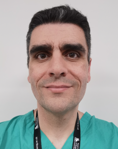 Angelos Kolias合作论文数Department of Clinical Neuroscience, Addenbrooke's Hospital;CRUK Cambridge Centre, Cancer Research UK Cambridge Institute, University of Cambridge33
 Peter J A Hutchinson合作论文数Department of Clinical Neurosciences, Robinson College, University of Cambridge31
 Anwar Fahim合作论文数Institute of Neurological Sciences, Southern General Hospital20
 Ivan Sergeevich Timofeev合作论文数University of Cambridge;Department of Neurosurgery;Addenbrooke ' s Hospital;Addenbrooke ' s Hospital, University of Cambridge20
 Adel Helmy合作论文数Division of Neurosurgery, University of Cambridge19
 Carole L Turner合作论文数Department of Neurosurgery, Addenbrooke’s Hospital13
 Edoardo Viaroli合作论文数8
 Gemma Whiting合作论文数6
 Ellie Edlmann合作论文数5Judith Allanson合作论文数Department of Public Health and Primary Care, University of Cambridge5
 Antonio Belli合作论文数Institute of Inflammation and Ageing, College of Medical and Dental Sciences, University of Birmingham5
 David Menon合作论文数Division of Anaesthesia, Department of Medicine, Queens' College, University of Cambridge5
 Diederik O. Bulters合作论文数Department of Neurosurgery, Addenbrooke’s Hospital5G Whiting合作论文数4Tommi K Korhonen合作论文数4
 Ardalan Zolnourian合作论文数4
 Midhun Mohan合作论文数4
 Bart Depreitere合作论文数Dept Neurosurg, UZ Leuven4
 Jussi Posti合作论文数Department of Neurosurgery, University of Turku;Turku Brain Injury Center, Turku University Hospital;Academy of Finland4
 Charlotte Whiffin合作论文数University of Derby Derbyshire Chambers and Business Link, Canal Wharf, Chesterfield, UK.4
 Tom Bashford合作论文数Addenbrooke's Hospital, University of Cambridge4
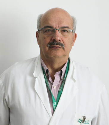 Franco Servadei合作论文数Humanitas University4
 Catherine Mcmahon合作论文数Centre for Emotional Health, Macquarie University4
 Virginia Newcombe合作论文数Division of Anaesthesia, University of Cambridge;National Institute for Health and Care Research;Royal College of Emergency Medicine4E. Viaroli合作论文数3Nicola Owen合作论文数3
 Thomas van Essen合作论文数3
 Sara Venturini合作论文数Department of Clinical Neurosciences, University of Cambridge3
 Jonathan Coles合作论文数Department of Medicine, University of Cambridge3
 Ignatius Esene合作论文数Department of Neurosurgery, Ain Shams University3
 Wilco C Peul合作论文数Department of Neurosurgery, Leiden University Medical Center;Department of Neurosurgery, Medical Center Haaglanden;Medical Center Haaglanden, Leiden University Medical Center3
 Yahia Z. Al-Tamimi合作论文数Department of Neurosurgery, Leeds General Infirmary3
 Niklas Marklund合作论文数Department of Neuroscience, Uppsala University3Mathew R Guilfoyle合作论文数Cambridge University Hospitals NHS Foundation Trust;Addenbrooke's Hospital, Cambridge University3Alexis Joannides合作论文数Division of Neurosurgery, University of Cambridge3Garry R. Barton合作论文数3
 Simon Thomson合作论文数3
 Andrew T King合作论文数2Sarah Pyne合作论文数2M T Barki合作论文数2Carol Davis-Wilkie合作论文数2Godard C W de Ruiter合作论文数2Katie Bond合作论文数2Ranjit Singh合作论文数2Jeroen T. J. M. van Dijck合作论文数2Michael Martin合作论文数2
 Orla Mantle合作论文数2Kirsty Grieve合作论文数2Aref-Ali Gharooni合作论文数2




