The evolving landscape of heart transplantation requires cardiac anesthesiologists to adapt to new paradigms aimed at expanding donor and recipient pools such as utilizing more donor hearts from older donors, those with higher BMI, left ventricular hypertrophy (LVH), or coronary artery disease, as well as grafts obtained through long-distance procurement and from donors with infectious conditions such as hepatitis C, COVID-19, and HIV. The promising use of devices such as Organ Care System (OCS), Paragonix SherpaPak (SCTS), and Hypothermic Oxygenated Machine Perfusion (HOPE) for preservation of donor hearts, as well as recent increases in donation after circulatory death (DCD) transplants, exemplifies these advancements. In particular, the introduction of beating heart DCD transplants offers the opportunity to minimize ischemic times. However, perioperative complications such as catecholamine-sensitive and resistant vasoplegia, acute right-sided heart dysfunction, primary graft dysfunction (PGD), surgical bleeding, and coagulopathy complicate the picture. The expansion of donor and recipient pools increases the risk of ischemia and reperfusion injury. Despite these challenges, cardiac anesthesiologists must be vigilant in recognizing and managing these complications to ensure the best short and long-term outcomes. For example, novel prediction tools for PGD such as RADIAL and PREDICTA may assist in facilitating earlier intervention in high-risk patients. Finally, the interplay between surgical bleeding, vasoplegia, acute RV dysfunction/failure, and PGD immediately post-transplant provides a significant challenge to cardiac anesthesiologists. By addressing these challenges, they can improve outcomes and play a pivotal role in advancing heart transplantation. Their efforts will make the expansion worthwhile, increasing the availability of donor hearts and enhancing patient care.
BACKGROUND:Perioperative cerebral oximetry is widely used during cardiopulmonary bypass (CPB) to monitor cerebral perfusion and guide intraoperative management. However, standard thresholds may not account for disease-specific physiology. In cystic fibrosis (CF), chronic hypoxia and hypercapnia may influence baseline cerebral oxygenation and intraoperative responses. METHODS:This retrospective cohort study included adults who underwent bilateral lung transplantation with intraoperative CPB between January 2014 and February 2022 at a single academic center. Patients with CF (n = 18) were compared with non-CF controls (n = 128). Bilateral near-infrared spectroscopy (NIRS) was used to monitor cerebral oxygenation, with desaturation defined as a ≥20% decline from baseline. Arterial blood gases (ABGs) were collected during CPB, and postoperative neurologic outcomes were assessed through clinical documentation during the index hospitalization. RESULTS:CF patients demonstrated significantly lower intraoperative cerebral oximetry values and higher PaCO2 compared with controls. Prior to CPB and during bypass, model-based mean rSO2 values were significantly lower in CF patients (62.0% vs. 69.1%, p = 0.04; 42.6% vs. 57.0%, p < 0.001; and 40.3% vs 53.9%, p = 0.0001), with a significant group × time interaction (p = 0.002). Imaging-confirmed stroke was infrequent and did not differ significantly between groups (5.6% vs 3.1%, p = 0.487). CONCLUSION:CF patients exhibit lower cerebral oxygen saturation and distinct carbon dioxide physiology during CPB, yet similar short-term neurologic outcomes compared with non-CF patients. Cerebral oximetry values in CF should be interpreted in the context of chronic pulmonary physiology rather than uniform desaturation thresholds.
The Impella 5.5 (Abiomed, Danvers, MA, USA) is the newest iteration of temporary left-ventricular assist devices in the Impella family of devices used for cardiogenic shock. While general guidelines for implantation of mechanical circulatory support devices are available, the authors aim to provide specific and detailed guidance for the Impella 5.5 for the practical application of intraoperative transesophageal echocardiography during placement and transthoracic and transesophageal echocardiography for device monitoring and management. Transesophageal echocardiography is paramount for preimplantation assessment of the right and left ventricles, possible shunt physiology, and valvular pathology. Its use is also helpful in ensuring appropriate device placement, initiating and modifying device settings, troubleshooting device placement, and diagnosing and managing complications.
BACKGROUND:Ischemic spinal cord injury (ISCI) is a serious complication following thoracoabdominal aortic aneurysm (TAAA) repair, whether by open (OR) or endovascular (TEVAR) approaches. Although MRI can detect spinal cord ischemia, it's not routinely used. Emerging evidence suggests MRI may offer insights into ISCI mechanisms, lesion pattern, and prognosis. This narrative review integrates published literature with our illustrative institutional case series to evaluate MRI's diagnostic and clinical role in postoperative ISCI. OBSERVATIONS:Records from October 2011 to August 2023 were reviewed and 10 patients (7 OR, 3 TEVAR) were identified who developed postoperative ISCI and underwent spinal MRI. Diffuse infarction patterns predominated (9/10 cases), and partial neurologic recovery occurred only in TEVAR patients. Prophylactic CSF drains were used in most OR cases (6/7), while rescue drains were more common in TEVAR cases (2/3). Across the literature, MRI classifications by Mawad, Tanaka, Yasuda, and others link lesion pattern and tissue involvement to prognosis, whereas experimental and anatomic studies highlight the influence of vascular variability and watershed physiology. CONCLUSIONS:MRI clarifies the mechanism, extent, and timing of spinal cord ischemia after aortic repair and helps differentiate infarction from other causes of myelopathy. Differences in recovery between OR and TEVAR likely reflect underlying vascular anatomy and ischemic dynamics. Future multicenter studies using standardized MRI and vascular mapping could refine risk stratification, optimize CSF drainage strategies, and improve outcomes in this high-risk population.
Paralysis post TEVAR is a challenging complication, this study discusses its incidence, Risk factors, clinical outcome, reversibility, protective strategy and associated mortality. Retrospective study Conducted on patients underwent thoracic and thoracoabdominal aortic Aneurysm Endovascular Repair in Last 11 years (January 2012 till June 2022) in single center as regard incidence, personal history, medical and surgical risk factors, preventive strategies, reversibility of paralysis and associated mortality. Data was obtained from records; paralysis was diagnosed through clinical examination immediately after emergence from anesthesia and frequently post operatively in SICU or even word and after discharge in follow-up outpatient clinic visits. A study carried on 137 patients, 15 patients of them developed lower limb paralysis with incidence of 10.9%, 3 patients of them (23.1%) developed paralysis immediately post operative, 4 patients developed paralysis in day 1 post operative (30.8%). And the rest of patients developed delayed onset paralysis (more than 24 Hour post procedure), The latest one developed paralysis 26 days post operative, 8 of paralyzed patients had spinal cord imaging (MRI) 4 of them (50%) didn’t show any abnormality and 3 of them (37.5%) showed spinal cord ischemia and 1 patient didn’t tolerate the MRI machine so the study was discontinued. According to reversibility, 9 patients had complete reversal of their motor and sensory symptoms to the baseline preoperative status. 2 patients had partial reversal of their symptoms, and 4 patients had persistent symptoms without any improvement despite the routine management that included elevating MAP and draining CSF to less than 10 mmHg in order to maintain spinal cord perfusion pressure at level of 80 mmHg and giving naloxone. Data showed 12 out of 81 patients (14.8%) who had functioning spinal drain developed paralysis. Duration of procedure is the most affecting risk factor on paralysis, patients with mean duration of 270 minutes had higher risk of paralysis. Prediction of paralysis post TEVAR is still difficult and data analysis showed that the duration of procedure is the only statistically significant variable that we can rely on.
Lumbar cerebrospinal fluid (CSF) drainage is one of the few preventative and therapeutic practices that may reduce spinal cord ischemia in high-risk thoracic endovascular aortic aneurysm repair (TEVAR). Although this is part of clinical guidelines in open thoracoabdominal aortic repair, there are no randomized controlled trials that provide convincing evidence on the protection conferred by CSF drainage in high-risk TEVAR patients. This gap in knowledge obfuscates clinical decision making given the risk of significant complications of CSF drain insertion and management. The current literature is inconclusive and provides conflicting results regarding the efficacy of, and complications from, CSF drainage in TEVAR. Filling the knowledge gap resulting from the limited current state of the literature warrants additional high-quality randomized controlled clinical trials that balance CSF drainage efficacy with potential complications in high-risk TEVAR patients.
Objective Spinal cord ischemia (SCI) is a potentially devastating complication of thoracic and thoraco-abdominal endovascular aortic repair (TEVAR and TAEVAR) that can result in varying degrees of short-term and permanent disability. This study was undertaken to evaluate the incidence, investigate the risk factors of SCI, and describe the clinical outcomes, reversibility, long-term functional impact, and influence on survival of SCI
STROKE IS a primary determinant of morbidity and mortality in patients with nonvalvular atrial fibrillation (AF). Atrial fibrillation causes thrombus formation in the left atrial appendage (LAA) and increases the risk of a cerebrovascular accident 5-fold.1 Systemic oral anticoagulation is the mainstay of therapy for thrombus prevention and stroke reduction in AF patients.2 Barriers to oral anticoagulation therapy include cost, drug-drug interactions, frailty with increased risk of falls, increased risk of bleeding, and noncompliance.
ObjectiveSpinal cord ischemia (SCI) is a potentially devastating complication of thoracic and thoraco-abdominal endovascular aortic repair (TEVAR and TAEVAR) that can result in varying degrees of short-term and permanent disability. This study was undertaken to evaluate the incidence, investigate the risk factors of SCI, and describe the clinical outcomes, reversibility, long-term functional impact, and influence on survival of SCIMethodsThis study employed a retrospective design to investigate patients who underwent TEVAR and TAEVAR successfully within the past 11 years between January 2012 and June 2022 in a single center. The analysis focused on factors such as incidence, personal history, and detailed assessment of medical and surgical risk factors. Data was retrieved from medical records. SCI was defined by any new lower neurologic deficit not attributable to another cause and diagnosed through clinical examination immediately after emergence from anesthesia and frequently postoperatively, as well as during follow-up outpatient clinic visits.ResultsA total of 137 patients were enrolled in the study. Among them, 15 patients developed lower limb paralysis with an incidence of 10.95%, three patients (20.1%) developed paralysis immediately postoperatively, and five patients (33.3%) developed paralysis on day 1 postoperatively (33.3%). Furthermore, the remaining patients experienced delayed onset paralysis, with the most recent case occurring 26 days after the operation. In terms of reversibility, nine patients had complete reversal of their motor and sensory symptoms to the baseline preoperative status. Two patients experienced a partial reversal of their symptoms, and four patients had persistent symptoms without any improvement. Data showed that 12 out of 81 patients (14.8%) who had functioning spinal drains developed paralysis. The duration of the procedure was the most significant risk factor for paralysis. Patients with a mean duration of 270 min had a higher risk of paralysis.ConclusionPrediction of paralysis post-TEVAR and TAEVAR remains challenging, with data analysis indicating that the duration of the procedure is the sole statistically significant variable to consider.
The use of endovascular, percutaneous interventions to treat cardiac, arterial, and venous pathologies is becoming increasingly common in medical practice. While endovascular device placement typically carries a low risk, device migration remains a persistent problem with these procedures for which anesthesia providers must have a high index of suspicion. Anesthesia providers should be aware of the wide range of indications for such devices, potential migration locations, and hemodynamic consequences of both the inciting pathology and device migration so they can safely care for patients in these settings. This article aims to discuss a case series of five different device migration events, review the literature surrounding the migration of each device, and propose unique anesthetic considerations as well as a systematic approach by which to approach these complications.
First international meeting towards eliminating paralysis after aortic aneurysm surgery Professor Hamdy Awad, MD, FASA, from the Ohio State University Wexner Medical Center, examines the dangers of aortic aneurysm surgery and the importance of continued research towards eliminating paralysis. Aortic aneurysmal disease is a rapidly growing problem in the United States. Ruptured aortic aneurysm is the 12th leading cause of death in the U.S. with 43,000–47,000 deaths per year (1). Risk factors associated with aneurysmal disease include old age, smoking, male sex, and a family history of aortic aneurysm. Aortic aneurysms inevitably rupture due to their thin and weakened walls, resulting in massive internal bleeding, which is usually fatal. This necessitates timely surgical intervention.
The presence of a tracheal bronchus, which is often incidentally discovered, complicates endotracheal intubation and lung isolation during thoracic surgery. Prior reports of successful right-sided lung isolation in the presence of tracheal bronchus required utilization of a double lumen tube. Although right-sided lung isolation was required in our case, due to other patient factors, it was determined that a double lumen tube of a suitable size would be unlikely to be placed safely and successfully. We describe the successful use of a Rüsch EZ-Blocker bronchial blocker in obtaining right-sided isolation in a patient with a difficult airway and tracheal bronchus.
 Michael Essandoh合作论文数Ohio State University38
 Tili Esmerina合作论文数Wexner Med Ctr, Ohio State Univ24
 Galina Dimitrova合作论文数13
 Gerard J. Nuovo合作论文数The James Comprehensive Cancer Center, The Ohio State University12
 Michaille Jean-Jacques合作论文数11Bhandary Sujatha P合作论文数Department of Anesthesiology, The Ohio State University Wexner Medical Center11Mohamed Ehab Ramadan合作论文数Department of Anesthesiology, New York Presbyterian Brooklyn Methodist Hospital10Samiya Saklayen合作论文数9Manoj H Iyer合作论文数9Hesham Kelani合作论文数Anesthesiol Dept, Ohio State Univ9Antolin Flores合作论文数7
 Chittoor B Sai-Sudhakar合作论文数7
 Elgharably Haytham合作论文数Department of Surgery, Davis Heart &Lung Research Institute, The Ohio State University;Davis Heart & Lung Research Institute, The Ohio State University7
 Bryan Whitson合作论文数Division of Cardiac Surgery, The Ohio State University Wexner Medical Center;Department of Surgery, The Ohio State University7Karina Anam合作论文数6
 Katja Regina Turner合作论文数6Bhatt Amar合作论文数Wexner Medical Center, The Ohio State University6
 Eric C. Bourekas合作论文数Department of Radiology, The Ohio State University6
 Nicolas Kumar合作论文数5Alexander A Efanov合作论文数5
 David Suster合作论文数5
 Ahmed Ahmed合作论文数Wexner Med Ctr, Ohio State Univ5
 Adam A Dalia合作论文数Harvard Medical School, Massachusetts General Hospital5Perez William J合作论文数5Hussain Nasir合作论文数5Racha Boulos合作论文数4Jayanth Rajan合作论文数4Ryan Dalton合作论文数4
 Thomas Graul合作论文数4Jonathan E Tang合作论文数4Erica Stein合作论文数4Sashwati Roy合作论文数Division of General and Gastrointestinal Surgery, College of Medicine, The Ohio State University4Malik Obaid合作论文数4
 Chandan K. Sen合作论文数McGowan Institute for Regenerative Medicine, University of Pittsburgh School of Medicine;Mary Ann Liebert, Inc.;Bioengineering MDPI;JoVE;Genes and Nutrition;DNA & Cell Biology4Juan A. Crestanello合作论文数Division of Cardiothoracic Surgery, Allegheny University Hospitals4
 Richard Urman合作论文数Wexner Medical Center, The Ohio State University4Leonid Gorelik合作论文数Department of Biochemistry, University of Illinois at Chicago4
 Hosam El Sayed合作论文数Michael E. DeBakey VA Medical Center, Baylor College of Medicine4
 Mahmoud Abdel-Rasoul合作论文数4
 John Hummel合作论文数From the Department of Internal Medicine, The University of Michigan Medical Center4
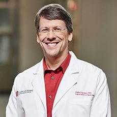 Phillip Popovich合作论文数Department of Neuroscience, College of Medicine, The Ohio State University;Belford Center for Spinal Cord Injury, The Ohio State University;Center for Brain and Spinal Cord Repair, The Ohio State University;Institute for Behavioral Medicine Research, The Ohio State University4Ashley Taylor合作论文数3Kasey Fiorini合作论文数3Amr Elrifay合作论文数3Jasmine Ryu合作论文数3
 Kasturi Ganesh合作论文数Hist, Univ Calicut3
 Sergio D Bergese合作论文数Dept Anesthesiol, Ohio State Univ3
 D. Michele Basso合作论文数School of Health and Rehabilitation Sciences, College of Medicine, The Ohio State University Wexner Medical Center3Magro C M合作论文数3




