This paper will not be able to cover the subject comprehensively, rather it will highlight and explain major lines of thought in Artificial Intelligence (AI).Mathematics will be largely avoided.The paper describes the elementary working principles of today's AI, points to a number of successful applications, and presents perspectives for the future.
To achieve a detailed understanding of processes in biological systems, cellular features must be quantified in the three-dimensional (3D) context of cells and organs. We described use of the intrinsic root coordinate system (iRoCS) as a reference model for the root apical meristem of plants. iRoCS enables direct and quantitative comparison between the root tips of plant populations at single-cell resolution. The iRoCS Toolbox automatically fits standardized coordinates to raw 3D image data. It detects nuclei or segments cells, automatically fits the coordinate system, and groups the nuclei/cells into the root's tissue layers. The division status of each nucleus may also be determined. The only manual step required is to mark the quiescent centre. All intermediate outputs may be refined if necessary. The ability to learn the visual appearance of nuclei by example allows the iRoCS Toolbox to be easily adapted to various phenotypes. The iRoCS Toolbox is provided as an open-source software package, licensed under the GNU General Public License, to make it accessible to a broad community. To demonstrate the power of the technique, we measured subtle changes in cell division patterns caused by modified auxin flux within the Arabidopsis thaliana root apical meristem.
This paper presents one novel spatial geometric constraints histogram descriptors(SGCHD) based on curvature mesh graph for automatic three-dimensional(3D) pollen particles recognition. In order to reduce high dimensionality and noise disturbance arising from the abnormal record approach under microscopy, the separated surface curvature voxels are extracted as primitive features to represent the original 3D pollen particles, which can also greatly reduce the computation time for later feature extraction process. Due to the good invariance to pollen rotation and scaling transformation, the spatial geometric constraints vectors are calculated to describe the spatial position correlations of the curvature voxels on the 3D curvature mesh graph. For exact similarity evaluation purpose, the bidirectional histogram algorithm is applied to the spatial geometric constraints vectors to obtain the statistical histogram descriptors with fixed dimensionality, which is invariant to the number and the starting position of the curvature voxels. Our experimental results compared with the traditional methods validate the argument that the presented descriptors are invariant to different pollen particles geometric transformations(such as posing change and spatial rotation), and high recognition precision and speed can be obtained simultaneously.
This erratum corrects an error in the results section (in-vitro data, measurement 1). Using velocities derived from the phantom geometry and flow rate, the modified and simplified Bernoulli approach revealed values of 6.0 ± 1.0 mmHg and 5.9 ± 1.0 mmHg, respectively. In the original article values of 8.2 ± 1.3 mmHg and 8.1 ± 1.3 mmHg were incorrectly reported. However, none of the conclusions are altered by these new values.
This index covers all technical items - papers, correspondence, reviews, etc. - that appeared in this periodical during the year, and items from previous years that were commented upon or corrected in this year. Departments and other items may also be covered if they have been judged to have archival value. The Author Index contains the primary entry for each item, listed under the first author's name. The primary entry includes the co-authors' names, the title of the paper or other item, and its location, specified by the publication abbreviation, year, month, and inclusive pagination. The Subject Index contains entries describing the item under all appropriate subject headings, plus the first author's name, the publication abbreviation, month, and year, and inclusive pages. Note that the item title is found only under the primary entry in the Author Index.
We propose to use a kernel intensity penalizer (KIP) in the blind maximum likelihood expectation maximization (MLEM) deconvolution scheme. With this very general kernel regularization term, we can stabilize the blind MLEM scheme even for the deconvolution of wide-field microscopic recordings. No complex prior point spread function models are needed. We combine state of the art optimization schemes using Tikhonov-Miller and TV regularization with our new kernel regularization. The proposed method improves the conventional deconvolution methods in terms of SNR on real and simulated datasets.
Nat. Methods 9, 735–742 (2012); published online 17 June 2012; corrected after print 9 July 2012. In the version of this article initially published, two items in the Online Methods section were incorrect. The MATLAB code in the 'ViBE-Z database file' section contained an extraneous semicolon, whichappeared in the HTML only and has been corrected.
Accurate identification of protein secondary structures is beneficial to understand three-dimensional structures of biological macromolecules. In this paper, a novel refined classification framework is proposed, which treats alpha-helix identification as a machine learning problem by representing each voxel in the density map with its Spherical Harmonic Descriptors (SHD). An energy function is defined to provide statistical analysis of its identification performance, which can be applied to all the α-helix identification approaches. Comparing with other existing α-helix identification methods for intermediate resolution electron density maps, the experimental results demonstrate that our approach gives the best identification accuracy and is more robust to the noise.
We present a method for densely computing local rotation invariant image descriptors in volumetric images. The descriptors are based on a transformation to the harmonic domain, which we compute very efficiently via differential operators. We show that this fast voxelwise computation is restricted to a family of basis functions that have certain differential relationships. Building upon this finding, we propose local descriptors based on the Gaussian Laguerre and spherical Gabor basis functions and show how the coefficients can be computed efficiently by recursive differentiation. We exemplarily demonstrate the effectiveness of such dense descriptors in a detection and classification task on biological 3D images. In a direct comparison to existing volumetric features, among them 3D SIFT, our descriptors reveal superior performance.
We propose an algorithm for 3-D multiview deblurring using spatially variant point spread functions (PSFs). The algorithm is applied to multiview reconstruction of volumetric microscopy images. It includes registration and estimation of the PSFs using irregularly placed point markers (beads). We formulate multiview deblurring as an energy minimization problem subject to L1-regularization. Optimization is based on the regularized Lucy-Richardson algorithm, which we extend to deal with our more general model. The model parameters are chosen in a profound way by optimizing them on a realistic training set. We quantitatively and qualitatively compare with existing methods and show that our method provides better signal-to-noise ratio and increases the resolution of the reconstructed images.
The detection of 3D objects and landmarks in arbitrary orientations is one of the most challenging tasks in biomedical 3D image analysis. In this paper we introduce the spherical Bessel Filter (BF) for rotation invariant 3D object detection tasks. The BF is based on the Harmonic Filter (HF) and thus inherits all the gentle properties of the HF, in particular the data driven adaptability and the processing speed. In contrast to the HF the BF benefits from a better object representation based on local spherical Fourier basis functions leading to noticeably better object detections and localizations.
In this paper, we present an algorithm for automatic 3D liver segmentation from CT volumetric dataset. The algorithm begins with the filtering of original volume using anisotropic diffusion. Then, based on a priori knowledge we perform a thresholding process and multi-morphological operations to extract a coarse liver region. In the thresholding process, we preserve all tissues that have intensities similar to the liver and exclude the rest. We perform morphological opening to disconnect small regions that do not belong to liver and connected-component labeling to clear those small regions. Finally, a fine liver region is obtained through graph-cut solver. Our algorithm does not require any user interaction or training datasets. The algorithm has been evaluated on 10 CT scans, and the results are encouraging to poor quality of images.
Accurate identification of protein secondary structures is beneficial to the structural interpretation of low-resolution X-ray and EM electron density maps. Existing alpha helix identification methods mainly focus on locally voxelwise classification and then link the helix-voxels based on post-processing processes. In this paper, a novel alpha helix identification approach, named as SSEPredictor, based on Metropolis-Hastings sampling is proposed, which can provide both locally and globally optimized prediction for alpha helical structures in low-resolution electron density maps. The approach has been tested on X-ray crystallographic electron density maps at 8Å resolution. The experimental results show that the identification accuracy is promising.
A framework for fast multiview fusion of Single Plane Illumination Microscopy (SPIM) images based on a spatially-variant point spread function (PSF) model is presented. For the multiview fusion a new algorithm based on the regularized Lucy-Richardson deconvolution and the Overlap-Save method is developed and tested on SPIM images. In the algorithm the image is decomposed into small blocks which are processed separately thus saving memory space and allowing for parallel processing.
Introduction: MR angiography (MRA) is widely applied in the clinical routine to analyze vascular malformations. While most applications are based on contrast enhanced MRA, Phase Contrast (PC)-MRA has proven to be a useful alternative [1,2]. However, most approaches provide only the time-averaged 3D lumen and thus static 3D vessel segmentation, which does not account for temporal changes such as the substantial motion of the thoracic aorta during the cardiac cycle. It was the purpose of the study to extract time-resolved 3D PC-MR angiography from ECG gated PC-MRI data with 3-directional velocity encoding using fully automated feature based fuzzy clustering for dynamic aortic lumen segmentation. This approach was applied and evaluated in 11 healthy subjects and 12 patients with different cardiovascular pathologies. Methods Data Analysis: Fuzzy clustering algorithms (here fuzzy c-means, FCM) classify the image by grouping similar data points in the feature space into clusters [3]. For the calculation of aortic PC-MRA, the aim was to group voxels of the timeresolved 3D PC volume into 3 clusters labeled as noise, static tissue and flow/vessel lumen for each time frame. To yield more homogeneous regions and to remove outliers, we modified the FCM algorithm by incorporating spatio-temporal information into the membership assignment function and thus allowing the spatio-temporal neighborhood (1 voxel in each +/direction) of a voxel to influence its labeling. To correct for signal variations related to the multi-element coil used for data acquisition, a nonuniformity correction was performed on the magnitude data using a technique described in [4]. To account for possible velocity aliasing all calculations were performed using velocity vector magnitude (speed). For each voxel, four features were chosen for FCM: 1) sum of squares PC-MRA [2], 2) pseudo complex difference PC-MRA [5], 3) non-uniformity corrected magnitude and 4) mean value of the haar wavelet coefficients (for 2 scaling values) of the velocity-time course in a temporal neighborhood of 5 voxels. A spatio-temporal FCM algorithm was applied until the maximal change in values representing cluster membership was less than 0.2%. Subsequently, each voxel was assigned to a specific cluster for which the membership was maximal. Furthermore to improve the segmentation of diastolic time frames, voxels in the peak systolic time frames were labeled according to their distance from the vessel boundaries; core voxels (with distance > 3 voxels) were copied to all diastolic time frames. Additionally, a 3D holes closing algorithm was applied for each time frame. To enable the calculation of hemodynamic parameters (flow, mean velocities, etc.) in the aorta only, automatic removal of the pulmonary system was performed based on vector field homogeneity [6] and a subsequently applied flood fill algorithm. Voxels classified by FCM algorithm as static tissue were used for eddy current correction of the velocity data [7]. The data processing workflow is illustrated in fig. 1. Methods MR Imaging: 11 young healthy subjects (mean age 24.6 years, 4 females) and 12 patients with different cardiovascular pathologies (mean age 29.7 years, 5 females; 8 patients with coarctation, 2 with aortic aneurysm, 1 with bicuspid aortic valve and 1 with aortic insufficiency) were included in our study after approval by the local ethic committee and written informed consent. All patients received contrast agent. Data were acquired on 1.5T and 3T systems (Avanto and TRIO, Siemens, Germany) using an ECG gated and respiration controlled rf-spoiled gradient echo phase contrast sequence with three-directional velocity encoding (spatial / temporal resolutions 1.9-2.9x1.6-1.7x2-3.5 mm / 39.2 – 48.8 ms, venc = 150-230 cm/s). Methods Validation: For validation of the geometric accuracy of the derived time-resolved aortic geometry, 3 planes were placed at anatomical landmarks: 1) in the ascending aorta at the level of the lower edge of the pulmonary artery (AAo), 2) proximal to the first branch of the supra-aortic vessels (FB), 3) and at in the descending aorta at the same height as plane 1 (DAo) as shown in figure 2. As reference standard, the aortic lumen contours in all 3 planes were segmented manually for each time frame in the cardiac cycle. Area, mean velocity and flow were calculated for manual (reference standard), time-resolved and time-averaged (static) PC-MRA. BlandAltman analysis was performed (Tab.1) and flow and area time-curves were plotted for each position (Fig.3, illustration for AAo position only). Results: The time-resolved 3D PC-MR angiography in Figure 2 shows good vessel depiction in peak systolic time frames (180-340 ms). During early systole (100ms) and in diastole (402ms), the vessel boundaries especially in the distal descending aorta are not completely depicted. Similar results were obtained for all other volunteers and patients. Different pathologies had no influence on the segmentation quality. However, in 10 of 12 patients the automatic removal of the pulmonary system was incomplete; in healthy subjects, the removal was incomplete for 3 of 11 data sets. Bland-Altman analysis (Tab.1) revealed that time-resolved PC-MRA in most cases underestimated hemodynamic parameters compared to manual segmentation approach; but in some cases (AAo position in patients), time-resolved PC-MRA overestimated values probably due to incomplete removal of pulmonary system and thus incorporating values from other vessels into the calculations. Time-resolved flow curves (Fig. 3) derived with automatic segmentation were underestimated in most cases compared to reference standard (healthy/patients: 7.5 ± 5.1% / -0.5 ± 3.8% difference in AAo, 8.3 ± 5.6% / 3.5 ± 2.4% difference in FB, 9.5 ± 4.3% / 4.3 ± 4.0% difference in DAo). Time-resolved area were underestimated in healthy subjects (7.8 ± 5.2% difference in AAo, 10.0 ± 4.0% in FB and 14.3 ± 2.5% in DAo). In patients, areas in AAo (-5.5 ± 4.5 % difference) and in FB (-0.7 ± 2.2%) were overestimated and underestimated in DAo (6.9 ± 3.3%) compared to manual segmentation. Discussion and Outlook: Preliminary results indicate the potential of FCM for time-resolved 3D vessel segmentation. Limitations of the current implementation are related to low velocity in early systole and in diastole, which resulted in incomplete depiction of vessel boundaries during these time frames. Thus, time-resolved PC-MRA tends to underestimate the derived hemodynamic parameters. But in comparison to the rigid segmentation, flow could be determined more accurately (s.Fig 3) Advantages of the presented approach are that timeresolved segmentation follows the general motion of the aorta during the cardiac cycle. As a result, the incorporation of the noisy values from outside the vessel is reduced compared to a rigid segmentation. Fig. 1: Flow chart for extraction process of time-resolved PC-MRA
In this paper we introduce a novel way for describing and classifying high angular resolution diffusionweighted magnetic-resonance images (HARDI) of the human brain. Our approach is capable to segment the brain images into gray matter (GM) and white matter (WM) tissue. For the segmentation a two step approach is suggested: The appearance of a training image is described locally at each voxel position in a rotation invariant manner. Then a classifier is trained and used for distinguishing between background (BG), GM and WM in unclassified images. In contrast to existing model-free methods we are not only using the raw measurements at each position, we also comprise neighboring measurements in a rotation invariant way. Experiments show that our method outperforms existing methods significantly. Furthermore, we show that our method gives also reasonable results for brains with pathologies like tumors.
In this work, we present a systematic phantom comparison and clinical application of noninvasive pressure difference mapping in the human aorta based on time-resolved 3D phase contrast data. Relative pressure differences were calculated based on integration and iterative refinement of pressure gradients derived from MR-based three-directional velocity vector fields (flow-sensitive 4D MRI with spatial/temporal resolution ∼ 2.1 mm(3)/40 ms) using the Navier-Stokes equation. After in vitro study using a stenosis phantom, time-resolved 3D pressure gradients were systematically evaluated in the thoracic aorta in a group of 12 healthy subjects and 6 patients after repair for aortic coarctation. Results from the phantom study showed good agreement with expected values and standard methods (Bernoulli). Data of healthy subjects showed good intersubject consistency and good agreement with the literature. In patients, pressure waveforms showed elevated peak values. Pressure gradients across the stenosis were compared with reference measurements from Doppler ultrasound. The MRI findings demonstrated a significant correlation (r = 0.96, P < 0.05) but moderate underestimation (14.7% ± 15.5%) compared with ultrasound when the maximum pressure difference for all possible paths connecting proximal and distal locations of the stenosis were used. This study demonstrates the potential of the applied approach to derive additional quantitative information such as pressure gradients from time-resolved 3D phase contrast MRI.
Texture features play an important role in facilitating various applications, for instance, image retrieval and object recognition. In this work, we investigate the relational features as a texture descriptor in classifying materials and visual textures from their appearance. The relational features used in this paper are constructed by histogramming the values extracted for each point within an image with fuzzy histogram. To test the performance of relational features, two benchmarks were used which have a variety of poses and conditions. Despite the challenging occurrence in both benchmarks, impressive results were achieved by using the relational features.
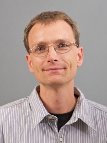 Olaf Ronneberger合作论文数Google DeepMind;Department of Computer Science, Faculty of Engineering, Albert-Ludwigs-Universität Freiburg54
 Marco Reisert合作论文数Chair of Pattern Recognition and Image Processing;Albert-Ludwigs-Universitat Freiburg;Institute for Computer Science20
 Lokesh Setia合作论文数Institute for Computer Science
Albert-Ludwigs-University Freiburg16Janis Fehr合作论文数15
Lokesh Setia合作论文数Institute for Computer Science
Albert-Ludwigs-University Freiburg16Janis Fehr合作论文数15 Alexandra Teynor合作论文数Albert-Ludwigs-University;Computer Science Department12
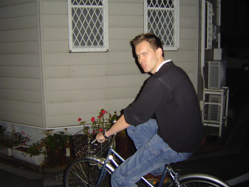 Henrik Skibbe合作论文数Integrated Systems Biology Laboratory,Kyoto11
 Alaa Halawani合作论文数Institute for Computer Science
Albert-Ludwigs-University11
Alaa Halawani合作论文数Institute for Computer Science
Albert-Ludwigs-University11 Jürgen Popp合作论文数Department of Spectroscopy and Imaging, Leibniz Institute of Photonic Technology;Jena Center for Soft Matter, Friedrich-Schiller-Universität Jena10
 Maja Temerinac-Ott合作论文数University of Freiburg, University of Freiburg10
 Zheming Lu (陆哲明)合作论文数Institute of Astronautic Electronic Engineering, School of Aeronautics and Astronautics, Zhejiang University9Qing Wang合作论文数9
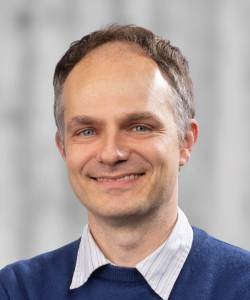 Thomas Brox合作论文数Department of Computer Science, Faculty of Engineering, University of Freiburg9An Luo合作论文数7
 Yonghua Xie (谢永华)合作论文数School of Computer Science, Nanjing University of Information Science & Technology;Nanjing University of Industry Technology7
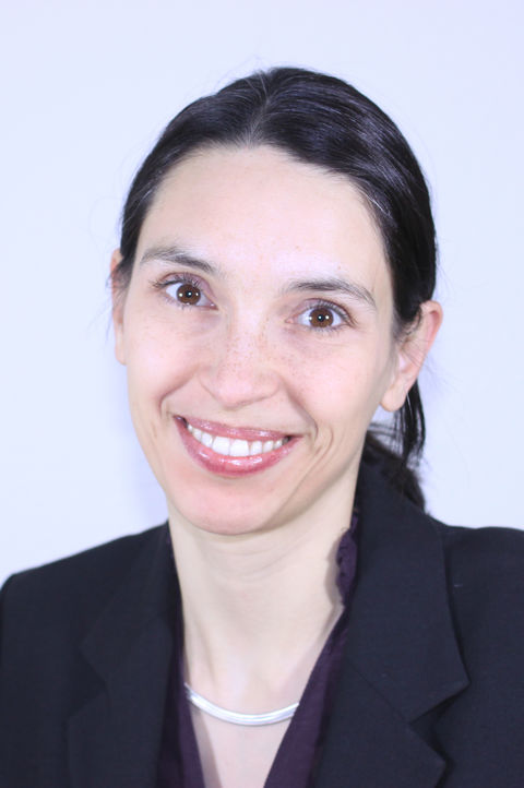 Margret Keuper合作论文数School of Business Informatics and Mathematics, University of Mannheim;Max Planck Institute for Informatics7
 Wolfgang Driever合作论文数Institute of Biology I, University of Freiburg6K.-D. Peschke合作论文数6
 Roland Nitschke合作论文数Life Imaging Center, University of Freiburg6
 Thorsten Schmidt合作论文数University of Frankfurt Cluster of Excellence Macromolecular Complexes, c/o Institute for Organic Chemistry and Chemical Biology6
 Michael Nolle合作论文数6
 Wan Nural Jawahir Hj Wan Yussof合作论文数6
 Da-Chuan Cheng合作论文数Department of Biomedical Imaging and Radiological Science, China Medical University5Michaela Harz合作论文数5Gerald Schreiber合作论文数5Petra Rösch合作论文数5Eckart Schultz合作论文数5Markus Lankers合作论文数Universität Würzburg Institut für Physikalische Chemie5
 Jürgen Hennig合作论文数Abteilung Röntgendiagnostik, Universität Freiburg;MR Development and application center, Universitätsklinikum Freiburg4
 Klaus Palme合作论文数Center of Biological Systems Analysis;Freiburg Institute For Advanced Studies, Head of Department, University of Freiburg4
 Wolfram Burgard合作论文数Department of Computer Science & Artificial Intelligence, University of Technology Nuremberg4Gerd Brunner合作论文数4Petra Rosch合作论文数4Hans Thiele合作论文数4Hanns Schulz-Mirbach合作论文数4
 Sven Siggelkow合作论文数Maurer Electronics GmbH4Andreas Bieniek合作论文数4
 Michael Markl合作论文数Department of Radiology, Feinberg School of Medicine, Northwestern University;Department of Biomedical Engineering, McCormick School of Engineering, Northwestern University4Jelena Bock合作论文数4Wenjing Tao合作论文数4Axel Fenske合作论文数3Hans-Walter Motzkus合作论文数3klausdieter peschke合作论文数3gunther schmauz合作论文数3Andreas Schüle合作论文数3s hofer合作论文数3
 Alex Frydrychowicz合作论文数3Susanne Schnell合作论文数Department of Radiology, Feinberg School of Medicine, Northwestern University3




