Odontogenic myxoma (OM) and odontogenic myxofibroma (OMF) are rare benign odontogenic tumors characterized by heterogeneous stromal composition, for which objective and reproducible pathological evaluation remains challenging. This multicenter study aimed to quantitatively assess fibrous tissue proportion (FTP) using AI-assisted digital pathology and to explore its clinicopathological relevance across odontogenic myxoma spectrum lesions. A total of 143 surgical specimens were collected from 34 institutions, and 100 cases were included after centralized pathological review. FTP was independently estimated by two board-certified oral pathologists to generate expert reference data. Whole-slide images of Masson's trichrome-stained sections were analyzed using a multi-stage deep learning pipeline implemented within a unified digital pathology platform. Agreement between expert assessment and quantitative measurements was evaluated, and associations between FTP and clinical variables were explored. Quantitative FTP measurements showed good agreement with expert pathological evaluation and revealed substantial inter-institutional variability in pathological diagnoses. In clinicopathological analyses, higher FTP was independently associated with unilocular radiological morphology. These findings demonstrate that AI-assisted quantitative pathology provides a reproducible framework for visualizing stromal heterogeneity in odontogenic myxoma spectrum lesions and may support more consistent clinicopathological interpretation across institutions.
Le Fort I osteotomy (LFI) can cause undesirable facial changes, including nasal widening. Alar cinch sutures are commonly used to reduce these effects, with varying efficacy. Herein, a modified cinch suture technique is introduced that integrates cartilage, bone, and soft-tissue sling components through bone anchorage. Existing literature on the topic is reviewed. This study included 31 patients (mean age, 30.6 y) who underwent LFI with modified cinch suture placement at our hospital between 2022 and 2024. The suture passed through the nasal septum and soft-tissue sling and was anchored to the bone at point A. The effectiveness of the technique was evaluated using three-dimensional analysis. Key nasal parameters before and 1-y postoperatively were compared using computed tomography. The mean increases in the nasal and alar-base widths were 2.15% and 3.32%, respectively. Nasal tip projection decreased by 6.03%, whereas the nasolabial angle increased by 5.54%. Patient satisfaction was assessed using a survey; 100% and 95% of participants expressed satisfaction with their facial and nasal aesthetics, respectively. Of the 22 studies retrieved from the PubMed database, 14 were included in the analysis. Compared with previously-reported studies, our technique resulted in lesser nasal widening and offered better morphological control. The method represents a novel combination of Rauso Types I–III that provides more comprehensive anchorage and stability. The modified alar cinch suture technique reduces postoperative nasal widening and offers a reproducible, integrative approach to nasal soft-tissue management in orthognathic surgery.
Le Fort I osteotomy (LFI) is commonly performed to correct maxillary deformities; however, it often results in undesirable changes to nasal morphology, particularly widening of the nasal width. The short-term course of nasal width stabilization remains poorly defined, and no indicators have been identified for changes in nasal width during the first postoperative year following LFI. This study examined temporal changes in nasal width following LFI with a modified cinch suture technique, applied by a single surgeon. Twenty-six patients were analyzed using standardized anthropometric measurements and CT imaging. Intra-examiner repeatability was excellent (intraclass correlation coefficient =0.96, 95% CI: 0.87–0.99), with Bland–Altman analysis showing a small mean bias (0.49 mm) and clinically acceptable limits of agreement (–1.44 to 2.42 mm). The cinch suture reduced nasal width by an average of 6.46 mm (16.33%) intraoperatively. Postoperatively, the nasal width gradually increased before stabilizing, with a mean stabilization period of 3.78 months (median, 3 months). At 1 year postoperatively, nasal width increased by 0.74 mm (2.01%) compared with the preoperative measurement. No correlation was found between stabilization period and patients’ demographic or clinical factors. Compared with previous reports, the technique achieved a smaller increase in nasal width at 1 year. The consistent stabilization around 3 months suggests its potential as a clinical indicator for postoperative monitoring. Limitations include the single-surgeon design and variability in individual healing and swelling. Future research should classify cinch suture techniques by type and systematically evaluate nasal- and soft-tissue changes following LFI.
Syphilis, a sexually transmitted disease induced by the bacterium Treponema pallidum, presents with diverse progression-based symptoms, and its incidence has exhibited a rising trend in recent years. Syphilis is frequently identified not only by genital or skin lesions but also by lesions on the oral mucosa. However, the diagnosis of oral syphilis remains challenging because of the diverse array of oral symptoms. Although neurosyphilis manifests at any stage, unfamiliarity with neurological symptoms among oral surgeons can lead to overlooking the potential presence of neurosyphilis. To effectively diagnose the increasing number of syphilis cases, a precise understanding of diverse oral and systemic manifestations, including neurological symptoms, is crucial. In this report, we delineate the case of a 37-year-old female with oral syphilis who was diagnosed based on lesions on the tongue and soft palate, prompting the consideration of neurosyphilis.
Neurofibromatosis type 1 (NF1) is an autosomal dominant nevus disease characterized by multiple manifestations, primarily café-au-lait macules and neurofibromas. Here, we present the case of an NF1 patient with 47,XYY mosaicism whose diagnosis was prompted by café-au-lait macules on the skin and mandibular neurofibromas. Targeted next-generation sequencing of the patient’s blood sample revealed a novel frameshift mutation in NF1 (NM_000267.3:c.6832dupA:p.Thr2278Asnfs*8) that is considered a pathogenic variant.
Osteoblastoma is a benign bone tumor with predominance in those <20 years of age and rarely occurs in the maxillofacial region. The identification of FOS gene rearrangements in osteoblastomas has been reported, however, a small subset of osteoblastomas do not show FOS gene rearrangements. There are no reports discussing the relationship between age and FOS gene rearrangement in osteoblastomas. We report the case of a 66-year-old female patient with histological features of osteoblastoma in the mandible without FOS gene rearrangement. According to a review of previously reported cases of osteoblastoma, FOS gene rearrangement is significantly less common in patients >40 years of age (P=0.041). Osteoblastomas occurring in older adult patients may exhibit pathogeneses different from those of young patients. Further data, including the relationship between age and FOS gene rearrangement, is necessary to clarify the pathogenesis of osteoblastoma.
Introduction: Marsupialization is a dependable choice for mandibular unicystic ameloblastoma (UA) management. However, investigations regarding its speed of shrinkage (SS) and reduction rate (RR) are lacking. This case report highlights the treatment of a huge mandibular UA with high SS and RR using marsupialization before secondary surgery. Presentation of case: A 45-year-old male patient presented with severe swelling of the right side of the mandible, resulting in prominent facial asymmetry. Panoramic radiograph revealed a unilocular, radiolucent lesion extending from the mandibular midline to the right ramus. Computed tomography (CT) revealed a large radiolucent lesion that expanded in the buccolingual direction. Incisional biopsy showed that the lesion was UA. After 1.5 years of marsupialization, an SS of 0.183 % per day was reached, leading to an impressive RR of 98.7 %. Treatment was followed by enucleation and peripheral osteotomy. No recurrence was observed at 1 year postsurgery. Discussion: The treatment of mandibular UA remains controversial, ranging from conservative approaches to aggressive interventions. In the current case, marsupialization was highly effective in reducing the volume of the lesion, thereby facilitating a minimally invasive secondary surgery to preserve function. The intact periosteum, which has the potential to differentiate into various cell types, may be associated with the regeneration of new bone after marsupialization. Conclusion: Marsupialization remains a successful strategy for managing mandibular UA. Even the huge lesions causing facial deformity can be treated with marsupialization combined with secondary surgery, avoiding the aesthetic and functional disruptions associated with radical treatment.
In patients with bilateral cleft lip and palate (BCLP), they sometimes need reposition premaxilla which is severely protruded and twisted as functional and aesthetic treatment. We introduce a surgical method of premaxillary fixation during alveolar bone grafting (ABG) with premaxillary osteotomy (PO). We included 9 patients with BCLP and a patient with bilateral cleft lip and alveolus who underwent ABG with PO in our institution between 2002 and 2020. They usually underwent two-stage ABG from the iliac crest with PO. Contralateral ABG was performed approximately 1 year after initial surgery. It is important to divide the surgery into two stages to perform the surgery more safely and surely. Premaxilla fixation was performed at bone-level from the anterior nasal spine to the maxillary sinus anterior wall with an absorbable plate and on dentition by wiring teeth to the labial archwire. This method provides firm fixation and facilitates feeding and dental cleaning without serious complications.
The clinical differences between odontogenic myxoma (OM) and odontogenic myxofibroma (OMF), and the clinical significance of their classifications, remain unclear. This study reviewed the clinicopathological characteristics of patients with OM or OMF and evaluated the fibrous component of the specimens. Medical records of 21 patients with OM or OMF who underwent tumour resection were reviewed. The percentage of fibrous tissue on the representative sections was evaluated using haematoxylin and eosin- and Masson's trichrome-stained specimens. Histopathological diagnoses included 11 OMs and 10 OMFs with no tumour recurrence except for two cases in which the dredging method was applied. More cortical bone perforation was observed in OM than in OMF cases, without significant differences. Location-locularity and apparent diffusion coefficient value (ADC)-cortical bone perforation were significantly correlated in all OM and OMF cases. The percentage of fibrous tissue in specimens showed bimodal distribution bordered by 45%. There was a significant association between diagnosis based on 45% fibrous tissue criterion and the final pathological diagnosis. Our study showed a tendency for cortical bone perforation in OM compared to OMF and correlation between ADC and cortical bone perforation. According to the histopathological analyses, the fibrous component of each case was bimodal with 45%, which may be a criterion to distinguish between OM and OMF. Accumulating knowledge, such as significant differences in prognosis, may allow for minimal surgical treatment options based on the diagnosis according to this novel histopathological criterion.
There are a few reports that focus on radiotherapy (RT) and cetuximab (CET) therapy exclusively for oral cancer. This retrospective study aimed to investigate the efficacy and safety of RT and CET therapy for locally advanced (LA) or recurrent/metastatic (R/M) oral squamous cell carcinoma (OSCC). Seventy-nine patients from 13 hospitals who underwent RT and CET therapy for LA or R/M OSCC between January 2013 and May 2015 were enrolled in the study. Response, overall survival (OS), disease-specific survival (DSS), and adverse events were investigated. The completion rate was 62/79 (78.5%). The response rates in patients with LA and R/M OSCC were 69% and 37.8%, respectively. When only completed cases were examined, the response rates were 72.2% and 62.9%, respectively. The 1- and 2-year OS were 51.5% and 27.8%, respectively (median, 14 months), for patients with LA OSCC, and 41.5% and 11.9% (median, 10 months) for patients with R/M OSCC. The 1- and 2-year DSS were 61.8% and 33.4%, respectively (median, 17 months), for patients with LA OSCC, and 76.6% and 20.4% (median, 12 months) for patients with R/M OSCC. The most common adverse event was oral mucositis (60.8%), followed by dermatitis, acneiform rash, and paronychia. The completion rate was 85.7% in LA patients and 70.3% in R/M patients. The most common reason for noncompletion was an inadequate radiation dose due to worsening general conditions in R/M patients. Although the standard treatment for LA or R/M oral cancer is concomitant RT with high-dose cisplatin (CCRT) and the efficacy of RT and CET therapy for oral cancer is not considered to be as high as that for other head and neck cancers, it was thought that RT and CET therapy could be possible treatments for patients who cannot use high-dose cisplatin.
Materials and Methods for Tissue microarray, Immunofluorescence, Quantitative Real-Time PCR, Proteomics analysis, Co-immunoprecipitation, Co-culture.
Objective: The goal of this study was to evaluate the performance of automatic machine learning (ML)-based classification of the impacted status of the mandibular third molar.Methods: The dataset consisted of 1864 mandibular third molar images, and the impaction pattern for each mandibular third molar was annotated based on the Pell and Gregory classification and the Winter classification. To improve performance, data augmentation techniques were applied, including rotation, flip, and pseudoimages that mimic prostheses, and ML was performed using the VGG16 convolutional neural network.Results: In the Pell and Gregory Class classification, the performance of the model trained on the augmented dataset exhibited good classification performance in three metrics, obtaining accuracy of 0.8609, macro-average F1-score of 0.7624, and an area under the receiver operating characteristic curve (AUC) of macro-average receiver operating characteristic (ROC) of 0.9334. For Pell and Gregory Position classification, the model obtained an accuracy value of 0.8432, a macro-average F1-score of 0.8156, and an AUC of macro-average ROC value of 0.9395. For Winter classification, the model obtained an accuracy value of 0.7959, a macro-average F1score of 0.6423, and an AUC of macro-average ROC value of 0.9549. Conclusions: The constructed ML classification model for mandibular third molar impaction status demonstrated good performance when data augmentation was applied.
Results of immunohistochemistry in breast and prostate cancer patients with metastasis.
 Avraham Raz合作论文数Karmanos Cancer Institute, Department of Pathology, School of Medicine, Wayne State University15
 Dhonghyo Kho合作论文数Department of Life Science, Sogang University13
 Nakajima Kosei合作论文数13Victor Hogan合作论文数13Tetsuya Yoda合作论文数12Takashi Yanagawa合作论文数Faculty of Science, Kyushu University10Chen Wei合作论文数9
 Rouba Ali-Fehmi合作论文数Department of Pathology, School of Medicine, Wayne State University;Department of Pathology, University of Michigan;Center of Global Health Equity, University of Michigan9Takahara Namiaki合作论文数7Keiichi Morita合作论文数7Kayamori Kou合作论文数5Hiroyuki Yoshitake合作论文数5Erina Tonouchi合作论文数4Hiroyuki Harada (原田浩之)合作论文数Department of Oral and Maxillofacial Surgical Oncology, Graduate School of Medical and Dental Sciences Medical, Institute of Science Tokyo4Eri Anzai合作论文数3Yasuhiro Kurasawa合作论文数3Yasuyuki Michi合作论文数3
 Tohru Ikeda合作论文数Division of Oral Pathology and Bone Metabolism, Nagasaki University Graduate School of Biomedical Sciences3
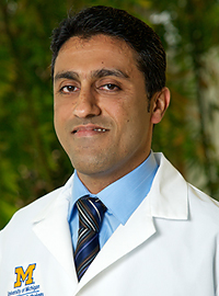 Rohit Mehra合作论文数Michigan Center for Translational Pathology;Department of Pathology, University of Michigan Medical School;Rogel Cancer Center, Michigan Medicine3Yu Akaike合作论文数2Eri Shibata合作论文数2Yuki Fukawa合作论文数2
 Samira Bernardino Ramos do Prado合作论文数2
 Tianpeng Zhang合作论文数Department of Genetics, Cell Biology and Development, University of Minnesota2Kiyoshi Harada合作论文数2Yoshiyuki Sasaki合作论文数2
 Tomoki Muramatsu合作论文数2Teruo Amagasa合作论文数2
 Vitaly Balan合作论文数Karmanos Cancer Institute, Wayne State University2
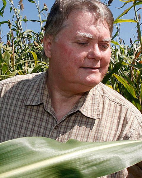 Nick Carpita合作论文数Department of Botany & Plant Pathology, College of Agriculture, Purdue University;National Renewable Energy Laboratory2Tânia Misuzu Shiga合作论文数2
 Issei Imoto (井本逸勢)合作论文数Aichi Cancer Center, Research Institute2
 João Paulo Fabi合作论文数Department of Food Science and Experimental Nutrition, School of Pharmaceutical Sciences, Universidade de São Paulo2
 Johji Inazawa (稲澤譲治)合作论文数Research Core Center, Tokyo Medical and Dental University;Bioresource Research Center, Tokyo Medical and Dental University2
 Ken-ichi Kozaki合作论文数2
 Narikazu Uzawa合作论文数2Yuko Katsuki合作论文数1Hirokazu Kachi合作论文数1Viet Vu Duc合作论文数1Jun Sumino合作论文数1Daisuke Miyajima合作论文数1Yuko Tsuji合作论文数1Masashi Yamashiro合作论文数1Hideo Miyamoto合作论文数1Yutaka Sato合作论文数1Takuya Iwasaki合作论文数1T. Sakuma合作论文数1Miho Suzuki合作论文数1




