Atomic resolution in cryo-electron microscopy was first demonstrated six years ago. This was accomplished using 300 kV electron microscopes equipped with new hardware that provided narrower energy spread, aberration correction and energy filtering. Here, we report the achievement of 1.24 Å atomic resolution on an upgraded 200 kV electron microscope featuring a cold field emission gun, a high-resolution objective lens polepiece and an energy filter. These components transform the instrument into a cost-effective single-particle cryo-EM platform with performance comparable to that of significantly more expensive 300 kV systems. The microscope can also be operated at 100 kV and by using a high-speed hybrid-pixel detector we were able to reach sub-2 Å resolution.
Cryo electron tomography (cryo-ET) enables three-dimensional visualization of biological macromolecules in their near-native environments, bridging the gap between structural and cellular biology. In this study, we evaluated high-resolution subtomogram averaging (STA) using a cryo-TEM (CRYO ARM (TM) 300 II, JEOL) and PACEtomo software system for both single-particle analysis (SPA) type grids and lamellae prepared by cryo-focused ion beam (cryo-FIB) milling (JIB-4700F, JEOL). Using mouse apoferritin and E. coli ribosomes in thin vitreous ice film, resolutions of 2.04 & Aring; and 2.86 & Aring; were achieved, respectively. Using cryo-FIB lamellae of Salmonella cells, in situ ribosome structures were resolved to 8.2 & Aring; resolution.
Tauopathies are characterized by the accumulation of abnormally phosphorylated tau filaments, with disease-specific folds revealed by cryo-electron microscopy (cryo-EM). Here, we delivered a recombinant human tau carrying the P301L/S320F double mutation into AppNL−G−F/MAPT double knock-in mice using a blood–brain-barrier-permeable AAV (AAV-PHP.eB), enabling rapid induction of tau aggregation in vivo. The mutant tau was systemically administered via retro-orbital injection, providing a minimally invasive approach for widespread neuronal transduction. Phosphorylated tau aggregation, as well as seeding activity, were observed without the need for exogenous seeds. Our cryo-EM analysis resolved a novel filament fold incorporating residues 279–329, in which the P301L and S320F mutations introduced stabilizing interactions that reinforced filament assembly. This conformation was distinct from previously reported folds, including Alzheimer and Pick tau filaments. Moreover, the filaments adopted a more compact architecture than those observed in patient-derived samples or other model mice. This finding demonstrates that P301L/S320F double-mutant tau adopts a structurally unique, highly aggregation-prone fold, and that this system provides a rapid platform for modeling tau filament formation.
Assembly of adaptor protein complex 2 (AP-2), the primary cargo adaptor in clathrin-mediated endocytosis, is regulated by the chaperones alpha- and gamma-adaptin binding protein (AAGAB) and coiled-coil domain-containing protein 32 (CCDC32), whose deletion causes loss of all AP-2 subunits in vivo. Here, we describe the molecular mechanism of CCDC32-mediated AP-2 assembly. CCDC32 interacts with the appendage domain of the AP-2 α subunit with high affinity, using the same binding site as canonical endocytic regulators in addition to a previously unidentified, yet highly conserved pocket on α. CCDC32 contains cargo sorting motifs normally found in transmembrane cargo and binds to AP-2 heterodimers using canonical cargo-binding sites. In addition, two amphipathic helices in CCDC32 bind to the α/σ2 heterodimer. Unexpectedly, in solution, CCDC32 prevents complex assembly and actively disassembles AP-2 tetramers. Inhibition requires the amphipathic helices of CCDC32, which also mediate binding to phosphatidylinositol 4,5-bisphosphate (PIP2)-containing membranes. The presence of PIP2-containing membrane stabilizes the final stages of assembly. We propose that the membrane acts as a molecular switch to release inhibitory interactions, allowing for full complex assembly to proceed.
Kinesin-1 walks along microtubules by alternating ATP hydrolysis and movement of its two motor domains ("head"). The detached head preferentially binds to the forward tubulin-binding site after ATP binds to the microtubule-bound head, but the mechanism preventing premature microtubule binding while the partner head awaits ATP remains unknown. Here, we examined the role of the neck linker, the segment connecting two heads, in this mechanism. Structural analyses of the nucleotide-free head revealed a bulge just ahead of the neck linker's base, creating an asymmetric constraint on its mobility. While the neck linker can stretch freely backward, it must navigate around this bulge to extend forward. We hypothesized that increased neck linker tension suppresses premature binding of the tethered head, which was supported by molecular dynamics simulations and single-molecule fluorescence assays. These findings demonstrate a tension-dependent allosteric mechanism that coordinates the movement of two heads, where neck linker tension modulates the allosteric conformational changes rather than directly affecting the nucleotide state.
Intracellular transport relies on motor proteins like kinesins to deliver essential cargoes along microtubules, yet the mechanisms of cargo recognition remain unclear. Here, we present high-resolution cryo-electron microscopy structures of the heterotrimeric kinesin-2 complex (KIF3A/KIF3B/KAP3) bound to the adenomatous polyposis coli (APC) cargo. Our findings reveal a previously uncharacterized KIF3 tail motif, termed the "Hitchdock domain," which plays a pivotal role in mediating interactions with both the KAP3 adaptor and the APC cargo. In this domain, the KIF3A helical regions facilitate specific cargo binding, while the β-hairpin region and KIF3B provide structural support. Mutagenesis and molecular dynamics simulations confirm the domain's functional importance. Interestingly, the Hitchdock/KAP3 structure suggests a conserved structural basis for cargo recognition across molecular motors, including kinesin-1 and dynein, which utilize similar hook-like architectures, highlighting the potential universality of this mechanism. Furthermore, our findings provide insights into kinesin-2 cargo specificity and offer a molecular framework for understanding related diseases. ### Competing Interest Statement The authors have declared no competing interest.
Cells have evolved a variety of assembly chaperones to aid in the difficult process of forming macromolecular complexes in a crowded cytoplasm. Assembly of adaptor protein complex 2 (AP-2), the primary cargo adaptor in clathrin-mediated endocytosis, is regulated by the chaperones AAGAB and CCDC32, whose deletion causes loss of all AP-2 subunits in vivo. AAGAB and CCDC32 are thought to act sequentially to assemble the AP-2 tetramer from its constituent heterodimers. However, the molecular requirements and structural consequences of CCDC32 interaction with AP-2 are not yet understood. Here, using in vitro reconstitution and integrative structural analysis, we describe the molecular mechanism of CCDC32-mediated AP-2 assembly. First, CCDC32 interacts with the appendage domain of the AP-2 α subunit, using the same binding site as canonical endocytic regulators in addition to a novel, yet highly conserved pocket on α. CCDC32 contains cargo sorting motifs normally found in trans-membrane cargo and binds to AP-2 heterodimers using canonical cargo-binding sites. Additionally, two amphipathic helices in CCDC32 bind to the α/σ2 heterodimer. Surprisingly, in solution, we find that CCDC32 prevents complex assembly and actively disassembles AP-2 tetramers. Inhibition requires the amphipathic helices of CCDC32, which also mediate binding to PIP2-containing membranes. The presence of PIP2-containing membrane stabilizes the final stages of assembly. We propose that the membrane acts as a molecular switch to release inhibitory interactions, allowing for full complex assembly to proceed. Using cryo-EM, we visualize an assembly intermediate that mimics the conformation of AP-2 found in vesicles, with CCDC32 bound at both cargo binding sites and both membrane-binding sites, suggesting that assembly leads to deposition of active complexes on the plasma membrane.
PMEL amyloids serve as essential scaffolds for melanin deposition in melanosomes, playing a crucial role in pigmentation. Despite their importance, the high-resolution structure of PMEL amyloids has remained unresolved. Using cryo-electron microscopy, we determine near-atomic resolution structures of wild-type PMEL amyloid core, revealing two distinct polymorphic forms with structural features. We further investigate the pathogenic G175S mutation associated with pigment dispersion syndrome (PDS). Structural analysis reveales that G175S introduces an additional hydrogen bond, stabilizing an alternative fibril conformation. In vitro, the G175S mutant exhibits a fourfold increase in polymerization efficiency compared to the wild type. In cells, G175S expression resultes in a twofold increase in intracellular amyloid content and a ~70% increase in extracellular amyloids, without altering melanosome morphology or number. These results indicate that the G175S mutation enhances amyloidogenesis within melanosomes, elevating amyloid load and potentially contributing to PDS pathophysiology. This study provides molecular insights into PMEL amyloid formation, highlighting its structural diversity and dysregulation in pigmentation disorders.
Convergent evolution of proteins provides insights into repeatability of genetic adaptation. While local convergence of proteins at residue or domain level has been characterized, global structural convergence by inter-domain/molecular interactions remains largely unknown. Here we present structural convergent evolution on fusion enzymes of aldehyde dehydrogenases (ALDHs) and alcohol dehydrogenases (ADHs). We discover BdhE (bifunctional dehydrogenase E), an enzyme clade that emerged independently from the previously known AdhE family through distinct gene fusion events. AdhE and BdhE show shared enzymatic activities and non-overlapping phylogenetic distribution, suggesting common functions in different species. Cryo-electron microscopy reveals BdhEs form donut-like homotetramers, contrasting AdhE's helical homopolymers. Intriguingly, despite distinct quaternary structures and < 30% amino acid sequence identity, both enzymes forms resemble dimeric structure units by ALDH-ADH interactions via convergently elongated loop structures. These findings suggest convergent gene fusions recurrently led to substrate channeling evolution to enhance two-step reaction efficiency. Our study unveils structural convergence at inter-domain/molecular level, expanding our knowledges on patterns behind molecular evolution exploring protein structural universe.
We report the first bioconjugation of Au25 nanocluster to a monoclonal antibody without protein engineering, in a step toward the development of high-resolution probes for cryogenic electron microscopy (cryo-EM) and tomography (cryo-ET). To achieve this, we improved the tryptophan (Trp)-selective bioconjugation step by using easy-to-analyze hydroxylamine (ABNOH) reagents in a pH-neutral buffer, instead of using N-oxyl radicals (ABNO) under acidic conditions as previously developed. This new protocol allowed for the application of Trp-selective bioconjugation to acid-sensitive proteins such as antibodies. We found that a two-step procedure, utilizing first Trp-selective bioconjugation for homogeneous introduction of azide groups to the protein and then strain-promoted azide-alkyne cycloaddition (SPAAC) to attach bicyclononyne (BCN)-presenting, redox-sensitive Au25 nanocluster, was key to successful immunogold synthesis. This procedure is scalable. The covalent labeling of the antibody with gold nanoclusters was confirmed by various analytical methods, including cryo-EM analysis of the Au25 nanocluster conjugates. In comparison with a non-homogenous variant prepared by lysine-selective bio-conjugation, Trp-selective conjugates exhibited both satisfactory gold cluster modification and minimal loss of antigen-binding ability.
Intracellular transport relies on motor proteins such as kinesins to deliver cargo along microtubules, yet how they recognize cargo remains unclear. Here, we present high-resolution cryo-electron microscopy structures of the heterotrimeric kinesin-2 complex (KIF3A/KIF3B/KAP3) bound to the cargo protein APC. Our findings reveal a previously uncharacterized KIF3 tail hook-like motif, termed the "HAC" domain, which mediates binding to both KAP3 adaptor and APC cargo. Within this domain, the KIF3A helical regions ensure cargo specificity, while a β-hairpin and KIF3B provide structural support. Biochemical and neuronal experiments confirm its functional importance. Notably, the HAC/KAP3 structure resembles hook-like architectures seen in kinesin-1 and dynein, suggesting a shared cargo recognition framework. These findings also shed light on kinesin-2 cargo specificity and offer a structural framework for understanding related neuronal transport mechanisms.
To identify proteins specific to the proximal ciliary axoneme, we used iTRAQ to compare short (~2 μm) and full-length (~11 μm) axonemes of Chlamydomonas. Known components of the proximal axoneme such as minor dynein heavy chains and LF5 kinase as well as the ciliary tip proteins FAP256 (CEP104) and EB1 were enriched in short axonemes whereas proteins present along the length of the axoneme were of similar abundance in both samples. The iTRAQ analysis revealed that FAP93, a protein of unknown function, and protein phosphatase 2A (PP2A) are enriched in the short axonemes. Consistently, immunoblots show enrichment of FAP93 and PP2A in short axonemes and immunofluorescence confirms the localization of FAP93 and enrichment of PP2A at the proximal axoneme. Ciliary regeneration reveals that FAP93 assembles continuously but more slowly than other axonemal structures and terminates at 1.03 μm in steady-state axonemes. The length of FAP93 assembly correlates with ciliary length, demonstrating ciliary length-dependent assembly of FAP93. Dikaryon rescue experiments show that FAP93 can assemble independently of IFT transport. In addition, FRAP analysis of GFP-tagged FAP93 demonstrates that FAP93 is stably anchored in the axoneme. FAP93 may function as a scaffold for assembly of other specific proteins at the proximal axoneme.
PMEL amyloids provide a vital scaffold for melanin deposition in melanosomes, playing a central role in pigmentation. Despite their importance, the high-resolution structure of PMEL amyloids has remained elusive. Here, we determined near-atomic resolution structures of wild-type PMEL amyloids using cryo-electron microscopy, revealing two distinct polymorphic forms with unique structural features. We further examined the pathogenic G175S mutation linked to pigment dispersion syndrome (PDS). Structural analysis showed that the G175S mutation introduces an additional hydrogen bond, stabilizing a novel fibril conformation. In vitro assays demonstrated a fourfold increase in polymerization efficiency for the G175S mutant compared to the wild-type. This enhanced polymerization correlated with a ~70% increase in secreted amyloids in G175S-expressing cells without detectable changes in melanosome morphology or number. These findings suggest that the G175S mutation promotes amyloidogenesis within melanosomes, increasing amyloid load and contributing to PDS pathophysiology. This study provides insights into the molecular basis of PMEL amyloid formation in both physiological and pathological contexts, offering new perspectives on their structural diversity and dysregulation in pigmentation disorders. ### Competing Interest Statement The authors have declared no competing interest.
Motile cilia have a so-called "9 + 2" structure, which consists of nine doublet microtubules and a central pair apparatus. The central pair apparatus (CA) is thought to interact mechanically with radial spokes and to control the flagellar beating. Recently, the components of the CA have been identified by proteomic and genomic analyses. Still, the mechanism of how the CA contributes to ciliary motility has much to be revealed. Here, we focused on one CA component with a large molecular weight: FAP47, and its relationship with two other CA components with large molecular weight: HYDIN, and CPC1. The analyses of motility of the Chlamydomonas mutants revealed that in contrast to cpc1 or hydin, which swam more slowly than the wild type, fap47 cells displayed wild-type swimming velocity and flagellar beat frequency, yet interestingly, fap47 cells have phototaxis defects and swim straighter than the wild-type cells. Furthermore, the double mutant fap47cpc1 and fap47hydin showed significantly slower swimming than cpc1 and hydin cells, and the motility defect of fap47cpc1 was rescued to the cpc1 level with GFP-tagged FAP47, indicating that the lack of FAP47 makes the motility defect of cpc1 worse. Cryo-electron tomography demonstrated that the fap47 lacks a part of the C1-C2 bridge of CA. Taken together, these observations indicate that FAP47 maintains the structural stiffness of the CA, which is important for flagellar regulation.
α- and β-tubulin have an unstructured glutamate-rich region at their C-terminal tails (CTT). The function of this region in cilia/flagella is still unclear, except that glutamates in CTT act as the sites for posttranslational modifications that affect ciliary motility. A unicellular alga Chlamydomonas possesses only two α-tubulin genes and two β-tubulin genes, each pair encoding an identical protein. This simple gene organization may enable a complete replacement of the wild-type tubulin with its mutated version. Here, using CRISPR/Cas9, we generated mutants expressing tubulins with modified CTTs. We found that the mutant whose four glutamate residues in the α-tubulin CTT have been replaced by alanine almost completely lacked polyglutamylated tubulin and displayed paralyzed cilia. In contrast, the mutant lacking the glutamate-rich region of the β-tubulin CTT assembled short cilia without the central apparatus. This phenotype is similar to the mutants harboring a mutation in a subunit of katanin, whose function has been shown to depend on the β-tubulin CTT. Therefore, our study reveals distinct and important roles of α- and β-tubulin CTT in the formation and function of cilia.Summary statement Chlamydomonas mutants were produced by CRISPR/Cas9 mediated gene editing to investigate ciliary function of tubulin C-terminal tails (CTTs). We found that α- and β-tubulin CTTs are essential for ciliary motility and assembly.### Competing Interest StatementThe authors have declared no competing interest.
We report the first bioconjugation of Au25 nanocluster to a monoclonal antibody at scarcely exposed tryptophan (Trp) residues toward the development of high-resolution probes for cryogenic electron microscopy (cryo-EM) and tomography (cryo-ET). To achieve this, we improved the Trp-selective bioconjugation using hydroxylamine (ABNOH) reagents instead of previously developed N-oxyl radicals (ABNO). This new protocol allowed for the application of Trp-selective bioconjugation to acid-sensitive proteins such as antibodies. We found that a two-step procedure utilizing first Trp-selective bioconjugation for the introduction of azide groups to the protein and then strain-promoted azide-alkyne cycloaddition (SPAAC) to attach a bicyclononyne (BCN)-presenting redox-sensitive Au25 nanocluster was essential for a scalable procedure. Covalent labeling of the antibody with gold nanoclusters was confirmed by various analytical methods, including cryo-EM analysis of the Au25 nanocluster conjugates.
Abstract α- and β-tubulin have an unstructured glutamate-rich region at their C-terminal tails (CTT). The function of this region in cilia/flagella is still unclear, except that glutamates in CTT act as the sites for posttranslational modifications that affect ciliary motility. A unicellular alga Chlamydomonas possesses only two α-tubulin genes and two β-tubulin genes, each pair encoding an identical protein. This simple gene organization may enable a complete replacement of the wild-type tubulin with its mutated version. Here, using CRISPR/Cas9, we generated mutants expressing tubulins with modified CTTs. We found that the mutant whose four glutamate residues in the α-tubulin CTT have been replaced by alanine almost completely lacked polyglutamylated tubulin and displayed paralyzed cilia. In contrast, the mutant lacking the glutamate-rich region of the β-tubulin CTT assembled short cilia without the central apparatus. This phenotype is similar to the mutants harboring a mutation in a subunit of katanin, whose function has been shown to depend on the β-tubulin CTT. Therefore, our study reveals distinct and important roles of α- and β-tubulin CTT in the formation and function of cilia. Summary statement Chlamydomonas mutants were produced by CRISPR/Cas9 mediated gene editing to investigate ciliary function of tubulin C-terminal tails (CTTs). We found that α- and β-tubulin CTTs are essential for ciliary motility and assembly.
ABSTRACT α- and β-tubulin have an unstructured glutamate-rich region at their C-terminal tails (CTTs). The function of this region in cilia and flagella is still unclear, except that glutamates in CTTs act as the sites for post-translational modifications that affect ciliary motility. The unicellular alga Chlamydomonas possesses only two α-tubulin and two β-tubulin genes, each pair encoding an identical protein. This simple gene organization might enable a complete replacement of the wild-type tubulin with its mutated version. Here, using CRISPR/Cas9, we generated mutant strains expressing tubulins with modified CTTs. We found that the mutant strain in which four glutamate residues in the α-tubulin CTT had been replaced by alanine almost completely lacked polyglutamylated tubulin and displayed paralyzed cilia. In contrast, the mutant strain lacking the glutamate-rich region of the β-tubulin CTT assembled short cilia without the central apparatus. This phenotype is similar to mutant strains harboring a mutation in a subunit of katanin, the function of which has been shown to depend on the β-tubulin CTT. Therefore, our study reveals distinct and important roles of α- and β-tubulin CTTs in the formation and function of cilia.
The urothelium, a distinct epithelial tissue lining the urinary tract, serves as an essential component in preserving urinary tract integrity and thwarting infections. The asymmetric unit membrane (AUM), primarily composed of the uroplakin complex, constitutes a critical permeability barrier in fulfilling this role. However, the molecular architectures of both the AUM and the uroplakin complex have remained enigmatic due to the paucity of high-resolution structural data. In this study, we utilized cryo-electron microscopy to elucidate the three-dimensional structure of the uroplakin complex within the porcine AUM. While the global resolution achieved was 3.5 Å, we acknowledge that due to orientation bias, the resolution in the vertical direction was determined to be 6.3 Å. Our findings unveiled that the uroplakin complexes are situated within hexagonally arranged crystalline lipid membrane domains, rich in hexosylceramides. Moreover, our research rectifies a misconception in a previous model by confirming the existence of a domain initially believed to be absent, and pinpointing the accurate location of a crucial Escherichia coli binding site implicated in urinary tract infections. These discoveries offer valuable insights into the molecular underpinnings governing the permeability barrier function of the urothelium and the orchestrated lipid phase formation within the plasma membrane.
 Masahide Kikkawa合作论文数Department of Cell Biology;Southwestern Medical Center;University of;Department of Life Sciences;School of Arts and Sciences;University of Tokyo;University of ;, University of Tokyo43
 Ritsu Kamiya合作论文数Dept Biol Sci, Univ Tokyo24
 Toshiyuki Oda合作论文数Graduate School of Medicine, The University of Tokyo21Toshiki Yagi合作论文数13
 Takuo Yasunaga合作论文数Faculty of Computer Science and Systems Engineering, Kyushu Institute of Technology13Mingyue Jin合作论文数8
 Osamu Nureki (濡木理)合作论文数Department of Biophysics and Biochemistry, Graduate School of Science, The University of Tokyo;Department of Biological Sciences, Graduate School of Science, The University of Tokyo7
 Keitaro Yamashita合作论文数Graduate School of Life Science, Hokkaido University;Graduate School of Life Science, Hokkaido University7
 Tomohiro Kubo合作论文数University of Tokyo, Gakushuin University7
 Eiichi Nakamura (中村荣一)合作论文数College of Chemistry, Nankai University;Department of Chemistry, Graduate School of Science, University of Tokyo7Masafumi Hirono合作论文数Institute of Biological Sciences, University of Tsukuba7Nakamuro Takayuki合作论文数6Koyasako Kotaro合作论文数6
 Radostin Danev合作论文数Graduate School of Medicine, University of Tokyo, S402, 7-3-1 Hongo, Bunkyo-ku, Japan 6Yoko Y. Toyoshima合作论文数4Ryosuke Yamamoto合作论文数4Yuma Tani合作论文数3reiko chijimatsu合作论文数3
 Laura A Fox合作论文数3Sale Winfield S合作论文数3Saito Kei合作论文数3Hirasaka Yuka合作论文数3Yamanaka Ryutaro合作论文数3
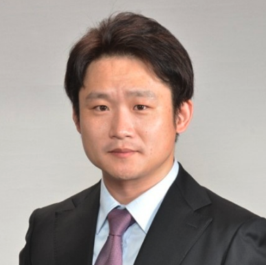 Rui Shang (尚睿)合作论文数School of Science, Westlake University3Yasuharu Kushida合作论文数2migyue jin合作论文数2Yoshihiro Kita合作论文数Life Sci Core Facil, Univ Tokyo2Hiroaki Ishikawa合作论文数Department of Biochemistry and Biophysics, University of California2
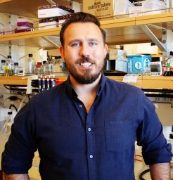 Richard W Baker合作论文数School of Medicine, University of North Carolina2
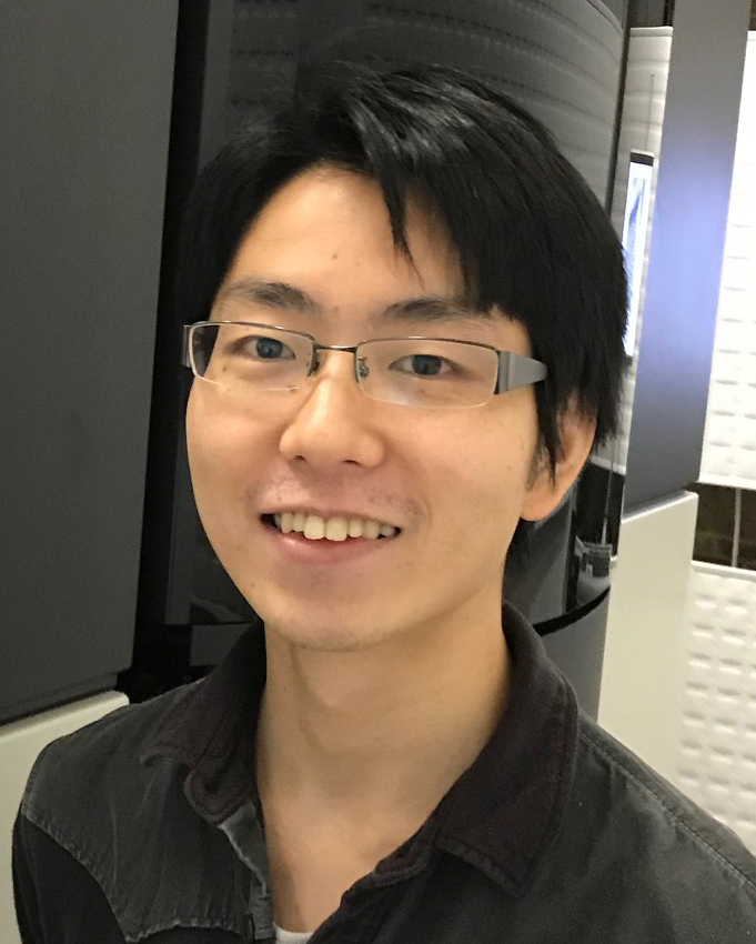 Muneyoshi Ichikawa (市川宗严)合作论文数State Key Laboratory of Genetic Engineering, Department of Biochemistry and Biophysics, School of Life Sciences, Fudan University2
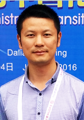 Jiangwei Tian (田蒋为)合作论文数中国药科大学研究生院2
 Wallace F. Marshall合作论文数School of Medicine, University of California, San Francisco2
 George B Witman合作论文数Department of Radiology, UMass Chan Medical School;Department of Radiology, T.H. Chan School of Medicine2
 Nobutaka Hirokawa (廣川信隆)合作论文数Department of Cell Biology and Anatomy, Graduate School of Medicine, The University of Tokyo2Takano Shinjiro合作论文数2
 Xuguang Jiang合作论文数2
 Takuzo Aida合作论文数Department of Chemistry and Biotechnology, School of Engineering, The University of Tokyo2Osamu Numata合作论文数2
 Motomu Kanai (金井求)合作论文数Graduate School of Pharmaceutical Sciences, The University of Tokyo2
 Tsukuda Tatsuya (佃達哉)合作论文数Department of Chemistry, Graduate School of Science, The University of Tokyo2
 Mary E. Porter合作论文数Department of Cell Biology and Neuroanatomy, University of Minnesota Medical School2
 Maureen Wirschell合作论文数Department of Cell Biology, Emory University School of Medicine2
 Kounosuke Oisaki合作论文数Graduate School of Pharmaceutical Sciences, The University of Tokyo2
 Koji Harano合作论文数Dept Chem, Univ Tokyo2
 Takeyuki Wakabayashi合作论文数2
 Hongmin Qin合作论文数Xuzhou Medical College, Xuzhou Medical College2
 Kentaro Nakano合作论文数2Ken‐ichi Miyazono合作论文数1Ken-ichi Wakabayashi合作论文数1




