Domain shift across imaging modalities and acquisition sites remains a significant barrier to the clinical deployment of segmentation models. Source-free unsupervised domain adaptation (SFUDA) addresses this by adapting a pretrained model to an unlabeled target domain without requiring access to sensitive source data. We introduce a novel SFUDA framework built on Symmetrical Flow Matching, a unified generative model that segments an input image and synthesizes a source-like image from a mask within the same learned flow. By initializing inference from a domain-agnostic Gaussian origin, the model preserves structural consistency across domains and grounds predictions in learned anatomy rather than shifted texture statistics. Our pipeline leverages this symmetry to generate reliable pseudo-labels and corresponding source-like synthetic images from unlabeled target data, creating a generative replay buffer that anchors source knowledge during a generative self-training stage that fine-tunes on a joint set of real target and synthetic source-like images. We evaluate on abdominal multi-organ and cardiac segmentation, covering cross-modality MRI<->CT shifts, and multi-site prostate segmentation. Our approach outperforms SFUDA baselines and is competitive with conventional UDA methods.
Segmentation models such as Segment Anything Model (SAM) and SAM2 achieve strong prompt-driven zero-shot performance. However, their training on natural images limits domain transfer to medical data. Consequently, accurate segmentation typically requires extensive fine-tuning and expert-designed prompts. We propose DiffuSAM, a diffusion-based adaptation of SAM2 for prompt-free medical image segmentation. Our framework synthesizes SAM2-compatible segmentation mask-like embeddings via a lightweight diffusion-prior from off-the-shelf frozen SAM2 image features. The generated embeddings are integrated into SAM2's mask decoder to produce accurate segmentations, thereby eliminating the need for user prompts. The diffusion prior is further conditioned on previously segmented slices, enforcing spatial consistency across volumes. Evaluated on the BTCV and CHAOS datasets for CT and MRI under Source-Free Unsupervised Domain Adaptation (SF-UDA) and Few-Shot settings, DiffuSAM achieves competitive performance with efficient training and inference. Code is available upon request from the corresponding author.
Full data acquisition in MRI is inherently slow, which limits clinical throughput and increases patient discomfort. Compressed Sensing MRI (CS-MRI) seeks to accelerate acquisition by reconstructing images from under-sampled k-space data, requiring both an optimal sampling trajectory and a high-fidelity reconstruction model. In this work, we propose a novel active sampling framework that leverages the inherent discrete structure of a pretrained medical image tokenizer and a latent transformer. By representing anatomy through a dictionary of quantized visual tokens, the model provides a well-defined probability distribution over the latent space. We utilize this distribution to derive a principled uncertainty measure via token entropy, which guides the active sampling process. We introduce two strategies to exploit this latent uncertainty: (1) Latent Entropy Selection (LES), projecting patch-wise token entropy into the k-space domain to identify informative sampling lines, and (2) Gradient-based Entropy Optimization (GEO), which identifies regions of maximum uncertainty reduction via the k-space gradient of a total latent entropy loss. We evaluate our framework on the fastMRI singlecoil Knee and Brain datasets at × 8 and × 16 acceleration. Our results demonstrate that our active policies outperform state-of-the-art baselines in perceptual metrics, and feature-based distances. Our code is available at https://github.com/levayz/TRUST-MRI.
Rationale: Obstructive sleep apnea (OSA) is associated with an increased risk of cardiovascular disease (CVD), but the effectiveness of continuous positive airway pressure (CPAP) therapy in improving cardiovascular outcomes has been inconsistent. Visceral obesity has emerged as a potential mediator in this relationship and is a known risk factor for CVD. This study aimed to explore the relationship between OSA severity and abdominal obesity metrics, including visceral and subcutaneous adipose tissue (VAT and SAT) volumes and VAT metabolic activity, and to assess changes in these metrics following CPAP intervention, using [18F]-Fluoro-2-deoxy-D-glucose (FDG) positron emission tomography (PET) combined with magnetic resonance imaging (MRI). Methods: We retrospectively analyzed PET/MRI scans from 115 adults with newly diagnosed OSA, both before and after three months of CPAP therapy. OSA severity was determined using portable sleep testing, defined by the respiratory disturbance index (pRDI). A deep learning model, using a transfer learning approach, segmented regions of interest (ROIs) within the subcutaneous and visceral adipose tissue compartments on MRI. Adipose volume and SUVmean values were calculated for the ROIs at each time point. Log-transformed linear regression and linear mixed-effects models were used to evaluate associations between OSA severity, adipose tissue metrics, and the effects of CPAP, adjusted for age, sex, hypertension, diabetes, diet, and body mass index (BMI). Results: Participants had an average age of 47.0 years (standard deviation [SD] 11.97), were predominantly male (84.0%), and had an average BMI of 31.85 kg/m² (SD 5.08). The mean pRDI was 32.53 events/hour (SD 19.21). OSA severity was not significantly associated with baseline VAT SUVmean (0.02%, confidence interval [CI] [-0.21-0.25], p=0.87). CPAP therapy did not significantly alter VAT SUVmean (-2.17%, CI [-5.02-0.77], p=0.15), VAT volume (-0.98%, CI [-2.41-0.47], p=0.19), or weight (0.30%, CI [-0.83-1.43], p=0.61). However, there was a significant reduction in the VAT/SAT volume ratio (-1.70%, CI [-3.28-0.09], p=0.04). Conclusion: This study found no change in visceral adipose tissue metabolic activity after three months of CPAP. However, despite no significant changes in weight following CPAP, a notable reduction in the VAT/SAT volume ratio suggests that CPAP may differentially impact abdominal fat distribution. Future research should explore how alternative therapies for OSA, such as GLP-1 receptor agonists, may modulate abdominal fat distribution and metabolic activity in patients with obesity-related OSA.
Acute ischemic stroke (AIS) is a leading cause of death and long‐term disability worldwide, where rapid reperfusion remains critical for salvaging brain tissue. Although CT perfusion (CTP) imaging provides essential hemodynamic information, its limitations—including extended processing times, additional radiation exposure, and variable software outputs—can delay treatment. In contrast, non-contrast head CT (NCHCT) is ubiquitously available in acute stroke settings. This study explores a generative artificial intelligence approach to predict key perfusion parameters (relative cerebral blood flow [rCBF] and time-to-maximum [Tmax]) directly from NCHCT, potentially streamlining stroke imaging workflows and expanding access to critical perfusion data. We retrospectively identified patients evaluated for AIS who underwent NCHCT, CT angiography, and CTP. Ground truth perfusion maps (rCBF and Tmax) were extracted from VIZ.ai post-processed CTP studies. A modified pix2pix‐turbo generative adversarial network (GAN) was developed to translate co-registered NCHCT images into corresponding perfusion maps. The network was trained using paired NCHCT–CTP data, with training, validation, and testing splits of 80
We present CXR-DINO, an adaptation of the DINOv2 training method tailored to enhance medical imaging analysis, with a focus on chest X-ray interpretation. This work leverages the strengths of self-supervised learning within a teacher-student framework, employing contrastive learning to train Vision Transformers (ViTs) on manipulated medical images. The images undergo processes such as cropping, augmentation, and segmentation, focusing on clinically relevant features, enabling the model to discern essential details from noise. By doing so, CXR-DINO preserves DINOv2's capability to generate resilient features across broad and detailed visual tasks as well as addresses the unique challenges of medical imagery. This approach mitigates the need for extensive labeled datasets, which are costly and labor-intensive to produce, by allowing the model to learn directly from the data itself. We demonstrate increased performance using CXR-DINO in disease classification and segmentation tasks. The presented approach is general and can be extended to other modalities and tasks, thus supporting self-supervised learning for automated medical diagnostics.
Hepatocellular carcinoma (HCC) surveillance primarily relies on ultrasound (U/S), which often exhibits decreased sensitivity in high-risk populations, such as individuals with cirrhosis or obesity. Abbreviated magnetic resonance imaging (AMRI) offers a potential alternative by employing targeted MRI sequences to enhance HCC detection. AMRI encompasses three primary strategies: non-contrast, dynamic contrast-enhanced, and hepatobiliary phase imaging, showing potential for overcoming U/S limitations in these populations. This study investigates the application of deep learning (DL) techniques to automate HCC tumor detection and segmentation within dynamic contrast-enhanced (Dyn-AMRI) protocols. Specifically, we leverage the capabilities of Vision Transformers (ViTs) to analyze complex image data and extract relevant features. Additionally, a novel heuristic is introduced to enhance the segmentation performance of the MedNeXt architecture. Our aim is to develop a robust DL pipeline for accurate HCC detection and segmentation on Dyn-AMRI, ultimately improving diagnostic outcomes.
This work presents an advance in one-shot medical image segmentation, where a single image-label sample from a new site is used for finetuning the solution - particularly valuable in scenarios where labeled data is scarce or rapid adaptation to new classes and sites is required. We introduce ProtoSAM, a novel, fully automated framework, for one-shot medical image segmentation that combines Prototypical networks, known for few-shot segmentation, with the Segment Anything Model (SAM), a natural image foundation model for segmentation. The proposed method creates an initial coarse segmentation mask using the ALPnet prototypical network, augmented with a DINOv2 encoder. Following the extraction of an initial mask, prompts are extracted, such as points and bounding boxes, which are then input into SAM. We present extensive validation on multiple datasets including CT, MRI, and endoscopy images, demonstrating state-of-the-art results in many scenarios. Our results show that an untrained ProtoSAM can match or exceed the performance of existing one-shot trained methods, with further improvements possible through self-supervised finetuning of the encoder. Our code is available at: https://github.com/levayz/ProtoSAM/ .
Chest X-rays or chest radiography (CXR), commonly used for medical diagnostics, typically enables limited imaging compared to computed tomography (CT) scans, which offer more detailed and accurate three-dimensional data, particularly contrast-enhanced scans like CT Pulmonary Angiography (CTPA). However, CT scans entail higher costs, greater radiation exposure, and are less accessible than CXRs. In this work, we explore cross-modal translation from a 2D low contrast-resolution X-ray input to a 3D high contrast and spatial-resolution CTPA scan. Driven by recent advances in generative AI, we introduce a novel diffusion-based approach to this task. We employ the synthesized 3D images in a classification framework and show improved AUC in a Pulmonary Embolism (PE) categorization task, using the initial CXR input. Furthermore, we evaluate the model's performance using quantitative metrics, ensuring diagnostic relevance of the generated images. The proposed method is generalizable and capable of performing additional cross-modality translations in medical imaging. It may pave the way for more accessible and cost-effective advanced diagnostic tools. The code for this project is available: https://github.com/NoaCahan/X-ray2CTPA .
Retinal abnormalities impact millions of people globally. Early diagnosis and treatment can halt disease progression, preventing countless cases of avoidable blindness. However, manual detection is often time-consuming, labor-intensive, and lacks consistency. One frequent challenge when effectively deploying deep learning systems in practical scenarios is the decline in performance that occurs when a network trained on data is applied to another medical site. This study centers on the realistic scenario of Unsupervised Domain Adaptation (UDA). It proposes a self-training approach that enables a continuous shift from the source domain to the target domain. We implemented our method on publicly available retinal segmentation and glaucoma classification datasets collected from several medical sites and showed improved performance compared to previous methods.
Pulmonary Embolism (PE) is a life-threatening condition. Computed tomography pulmonary angiography (CTPA) is the gold standard for PE diagnosis, offering high-resolution soft tissue visualization and three-dimensional imaging. However, its high cost, increased radiation exposure, and limited accessibility restrict its widespread use. In this work, we aim to introduce faster diagnosis opportunities by using 2D chest X-ray (CXR) data. CXR provides only limited two-dimensional visualization and is not typically used for PE diagnosis due to its inability to capture soft tissue contrast effectively. Here, we develop a novel methodology that distills knowledge from a trained CTPA-based teacher classifier model embedding to a CXR-based student embedding, by feature alignment - leveraging paired CTPA and CXR features as supervision, which can be readily acquired. This enables us to train without requiring annotated data. Our approach utilizes a latent diffusion model to generate CTPA-based PE classifier embeddings from CXR embeddings. In addition, we show that incorporating cross-entropy loss together with the corresponding loss of the teacher-student embeddings increases performance, bringing it close to clinical-level performance. We show state-of-the-art AUC in a PE categorization task using only the initial CXR input. This approach broadens the diagnostic capabilities of CXRs by enabling their use in PE classification, thereby extending their applicability beyond traditional imaging roles. The code for this project is available: https://github.com/meshims/Cross-Modal CXR- CTPA Knowledge Distillation.
Background: Accurate quantification of visceral (VAT) and subcutaneous adipose tissue (SAT) is critical for understanding the cardiometabolic consequences of obstructive sleep apnea (OSA) and other chronic diseases. This study validates a customization framework using pre-trained networks for the development of automated VAT/SAT segmentation models using hybrid positron emission tomography (PET)/magnetic resonance imaging (MRI) data from OSA patients. While the widespread adoption of deep learning models continues to accelerate the automation of repetitive tasks, establishing a customization framework is essential for developing models tailored to specific research questions. Methods: A UNet-ResNet50 model, pre-trained on RadImageNet, was iteratively trained on 59, 157, and 328 annotated scans within a closed-loop system on the Discovery Viewer platform. Model performance was evaluated against manual expert annotations in 10 independent test cases (with 80-100 MR slices per scan) using Dice similarity coefficients, segmentation time, intraclass correlation coefficients (ICC) for volumetric and metabolic agreement (VAT/SAT volume and standardized uptake values [SUVmean]), and Bland-Altman analysis to evaluate the bias. Results: The proposed deep learning pipeline substantially improved segmentation efficiency. Average annotation time per scan was 121.8 min (manual segmentation), 31.8 min (AI-assisted segmentation), and only 1.2 min (fully automated AI segmentation). Segmentation performance, assessed on 10 independent scans, demonstrated high Dice similarity coefficients for masks (0.98 for VAT and SAT), though lower for contours/boundary delineation (0.43 and 0.54). Agreement between AI-derived and manual volumetric and metabolic VAT/SAT measures was excellent, with all ICCs exceeding 0.98 for the best model and with minimal bias. Conclusions: This scalable and accurate pipeline enables efficient abdominal fat quantification using hybrid PET/MRI for simultaneous volumetric and metabolic fat analysis. Our framework streamlines research workflows and supports clinical studies in obesity, OSA, and cardiometabolic diseases through multi-modal imaging integration and AI-based segmentation. This facilitates the quantification of depot-specific adipose metrics that may strongly influence clinical outcomes.
Aging is associated with structural brain changes, cognitive decline, and neurodegenerative diseases. Brain age, an imaging biomarker sensitive to deviations from healthy aging, offers insights into structural aging variations and is a potential prognostic biomarker in neurodegenerative conditions. This study introduces BrainAgeNeXt, a novel convolutional neural network inspired by the MedNeXt framework, designed to predict brain age from T1-weighted magnetic resonance imaging (MRI) scans. BrainAgeNeXt was trained and validated on 11,574 MRI scans from 33 private and publicly available datasets of healthy volunteers, aged 5 to 95 years, imaged with 3T and 7T MRI. Performance was compared against three state-of-the-art brain age prediction methods. BrainAgeNeXt achieved a mean absolute error (MAE) of 2.78 ± 3.64 years, lower than the compared methods (MAE range 3.55-4.16 years). We also tested all methods across different levels of image quality, and BrainAgeNeXt performed well even with motion artifacts and less common 7T MRI data. In three longitudinal multiple sclerosis (MS) cohorts (273 individuals), brain age was, on average, 4.21 ± 6.51 years greater than chronological age. Longitudinal analysis indicated that brain age increased by 1.15 years per chronological year in individuals with MS (95% CI = [1.05, 1.26]). Moreover, in early MS, individuals with worsening disability had a higher annual increase in brain age compared to those with stable clinical assessments (1.24 vs 0.75,p< 0.01). These findings suggest that brain age is a promising prognostic biomarker for MS progression and potentially a valuable endpoint for clinical trials.
Rationale: Central abdominal obesity, particularly visceral adiposity may be a key player in mediating obstructive sleep apnea (OSA)-related cardiovascular disease (CVD) risk. Accurately measuring changes in visceral (VAT) and subcutaneous adipose tissue (SAT) volumes and metabolic activity could be crucial for evaluating the effectiveness of OSA therapies such as continuous positive airway pressure (CPAP) and novel weight-loss drugs. Manual analysis of abdominal adipose tissue on MRI can be time-intensive. We developed a dynamic training approach leveraging pre-trained AI models for abdominal fat segmentation in patients with OSA who underwent 18F-FDG positron emission tomography (PET) / magnetic resonance imaging (MRI), before and after CPAP. Methods: We utilized the AI Discovery Viewer (DV) platform, a web application for developing and deploying Medical AI models. In total, 328 abdominal PET/MRI scans from OSA patients were annotated within DV, with contours delineated for external (EXT) and internal (INT) SAT, as well as exclusionary (EXC) regions (i.e. kidneys, bone marrow). Initial training was conducted on a RadImageNet (RIN) UNet-ResNet50 model with 40 manually annotated cases, allowing for rapid model learning and facilitating AI-assisted annotation. This closed-loop system within DV enabled continuous fine-tuning of models with new annotations (Figure 1). Three versions of the models were assessed against manual segmentations in Osirix/Horos for segmentation speed, contour accuracy (Dice score), and VAT/SAT volumes and SUV. Performance was analyzed using the Wilcoxon Signed-Rank test. Results: The models achieved an average processing time of 1.14±0.19 minutes per scan, while expert-corrected AI segmentations took 31.8±17 minutes in DV, versus 134.5±27 minutes (Horos) and 96.8±7.8 minutes (Osirix) manually. Fat mask Dice scores showed high reliability, exceeding 0.98 for INT and EXT and reaching 0.83 for EXC. Contour Dice scores improved with each model iteration: INT from 0.39 to 0.45, EXT from 0.52 to 0.55, and EXC from 0.34 to 0.38. For VAT/SAT metrics, no significant differences were found between AI and manual annotations, except for VAT SUV mean (p=0.039), although the mean difference of 0.01 was not clinically significant. Conclusion: In summary, the abdominal adipose volumes and metabolic activity values derived using our AI models demonstrate a reasonable correlation to manual segmentation values. This novel approach has accelerated the time-for-annotation process by a factor of four and promises continued improvements with further model refinement. It demonstrates promise for abdominal fat quantification measures in OSA, to explore how therapies such as GLP-1 receptor agonists may modulate abdominal fat distribution and metabolic activity.
Background Right ventricular ejection fraction (RVEF) and end‐diastolic volume (RVEDV) are not readily assessed through traditional modalities. Deep learning–enabled ECG analysis for estimation of right ventricular (RV) size or function is unexplored. Methods and Results We trained a deep learning–ECG model to predict RV dilation (RVEDV >120 mL/m2), RV dysfunction (RVEF ≤40%), and numerical RVEDV and RVEF from a 12‐lead ECG paired with reference‐standard cardiac magnetic resonance imaging volumetric measurements in UK Biobank (UKBB; n=42 938). We fine‐tuned in a multicenter health system (MSHoriginal [Mount Sinai Hospital]; n=3019) with prospective validation over 4 months (MSHvalidation; n=115). We evaluated performance with area under the receiver operating characteristic curve for categorical and mean absolute error for continuous measures overall and in key subgroups. We assessed the association of RVEF prediction with transplant‐free survival with Cox proportional hazards models. The prevalence of RV dysfunction for UKBB/MSHoriginal/MSHvalidation cohorts was 1.0%/18.0%/15.7%, respectively. RV dysfunction model area under the receiver operating characteristic curve for UKBB/MSHoriginal/MSHvalidation cohorts was 0.86/0.81/0.77, respectively. The prevalence of RV dilation for UKBB/MSHoriginal/MSHvalidation cohorts was 1.6%/10.6%/4.3%. RV dilation model area under the receiver operating characteristic curve for UKBB/MSHoriginal/MSHvalidation cohorts was 0.91/0.81/0.92, respectively. MSHoriginal mean absolute error was RVEF=7.8% and RVEDV=17.6 mL/m2. The performance of the RVEF model was similar in key subgroups including with and without left ventricular dysfunction. Over a median follow‐up of 2.3 years, predicted RVEF was associated with adjusted transplant‐free survival (hazard ratio, 1.40 for each 10% decrease; P=0.031). Conclusions Deep learning–ECG analysis can identify significant cardiac magnetic resonance imaging RV dysfunction and dilation with good performance. Predicted RVEF is associated with clinical outcome.
This work introduces a new framework, ProtoSAM, for one-shot medical image segmentation. It combines the use of prototypical networks, known for few-shot segmentation, with SAM - a natural image foundation model. The method proposed creates an initial coarse segmentation mask using the ALPnet prototypical network, augmented with a DINOv2 encoder. Following the extraction of an initial mask, prompts are extracted, such as points and bounding boxes, which are then input into the Segment Anything Model (SAM). State-of-the-art results are shown on several medical image datasets and demonstrate automated segmentation capabilities using a single image example (one shot) with no need for fine-tuning of the foundation model. Our code is available at: https://github.com/levayz/ProtoSAM
Catheter based radiofrequency ablation for pulmonary vein isolation has become the first line of treatment for atrial fibrillation in recent years. This requires a rather accurate map of the left atrial sub-endocardial surface including the ostia of the pulmonary veins, which requires dense sampling of the surface and takes more than 10 minutes. The focus of this work is to provide left atrial visualization early in the procedure to ease procedure complexity and enable further workflows, such as using catheters that have difficulty sampling the surface. We propose a dense encoder-decoder network with a novel regularization term to reconstruct the shape of the left atrium from partial data which is derived from simple catheter maneuvers. To train the network, we acquire a large dataset of 3D atria shapes and generate corresponding catheter trajectories. Once trained, we show that the suggested network can sufficiently approximate the atrium shape based on a given trajectory. We compare several network solutions for the 3D atrium reconstruction. We demonstrate that the solution proposed produces realistic visualization using partial acquisition within a 3-minute time interval. Synthetic and human clinical cases are shown.
The adoption of artificial intelligence (AI) tools in medicine poses challenges to existing clinical workflows. This commentary discusses the necessity of context-specific quality assurance (QA), emphasizing the need for robust QA measures with quality control (QC) procedures that encompass (1) acceptance testing (AT) before clinical use, (2) continuous QC monitoring, and (3) adequate user training. The discussion also covers essential components of AT and QA, illustrated with real-world examples. We also highlight what we see as the shared responsibility of manufacturers or vendors, regulators, healthcare systems, medical physicists, and clinicians to enact appropriate testing and oversight to ensure a safe and equitable transformation of medicine through AI.
Magnetic resonance imaging (MRI) is a ubiquitous medical imaging technology with applications in disease diagnostics, intervention, and treatment planning. Accurate MRI segmentation is critical for diagnosing abnormalities, monitoring diseases, and deciding on a course of treatment. With the advent of advanced deep learning frameworks, fully automated and accurate MRI segmentation is advancing. Traditional supervised deep learning techniques have advanced tremendously, reaching clinical-level accuracy in the field of segmentation. However, these algorithms still require a large amount of annotated data, which is oftentimes unavailable or impractical. One way to circumvent this issue is to utilize algorithms that exploit a limited amount of labeled data. This paper aims to review such state-of-the-art algorithms that use a limited number of annotated samples. We explain the fundamental principles of self-supervised learning, generative models, few-shot learning, and semi-supervised learning and summarize their applications in cardiac, abdomen, and brain MRI segmentation. Throughout this review, we highlight algorithms that can be employed based on the quantity of annotated data available. We also present a comprehensive list of notable publicly available MRI segmentation datasets. To conclude, we discuss possible future directions of the field—including emerging algorithms, such as contrastive language-image pretraining, and potential combinations across the methods discussed—that can further increase the efficacy of image segmentation with limited labels.
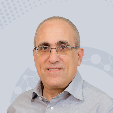 Jacob Goldberger合作论文数Faculty of Engineering, Bar Ilan University62
 Klang Eyal合作论文数22Eli Konen合作论文数21
 Idit Diamant合作论文数Sony Semiconductor Israel17
 Avi Ben-Cohen合作论文数17
 Shiri Gordon合作论文数Tel Aviv University16
 Gali Zimmerman Moreno合作论文数15
 Arnaldo Mayer合作论文数Tel-Aviv University15
 Serge Belongie合作论文数Department of Computer Science, University of Copenhagen;European Laboratory for Learning and Intelligent Systems;Det Nationale Center for AI i Samfundet13
 Tanveer Syeda-Mahmood合作论文数IBM Almaden Research Center;Stanford University12Uri Avni合作论文数Medical Engineering, Tel-Aviv University11Michal M Amitai合作论文数10
 Maayan Frid-Adar合作论文数RADLogics Inc10
 Jitendra Malik合作论文数Department of Electrical Engineering and Computer Sciences, University of California, Berkeley10Noa Cahan合作论文数Faculty of Engineering, Tel-Aviv University9Rula Amer合作论文数Dept Biomed Engn, Tel Aviv Univ9
 Michal Marianne Amitai合作论文数Wolfson Medical Center;Division of Internal Medicine, Sheba Medical Center8
 Raja Giryes合作论文数Department of EE - Systems, School of Electrical Engineering, Faculty of Engineering, Tel Aviv University8
 Daniel Rubin合作论文数Department of Biomedical Data Science, School of Medicine, Stanford University7
 V. Fauveau合作论文数6
 Ophir Gozes合作论文数RADLogics Ltd.6
 Nimrod Sagie合作论文数6Guy Alexandroni合作论文数Tel Aviv Univ, Dept Elect Engn, IL-69978 Tel Aviv, Israel6Shelly Soffer合作论文数DeepVision Lab, Sheba Medical Center6
 Nir Sochen合作论文数Department of Applied Mathematics, School of Mathematical Sciences, University of Tel Aviv6
 Zahi A. Fayad合作论文数BioMedical Engineering and Imaging Institute, Icahn School of Medicine, Mount Sinai;Department of Diagnostic, Molecular and Interventional Radiology, Icahn School of Medicine6
 Anant Madabhushi合作论文数Wallace H. Coulter Department of Biomedical Engineering, Emory University;Winship Cancer Institute, Emory University6Michal Sharon合作论文数6
 Sigal Trattner合作论文数Faculty of Engineering, Tel-Aviv University6C Carson合作论文数5
 Jannette Nassar合作论文数5Yiftach Barash合作论文数DeepVis Lab, Sheba Med Ctr5Dana Cohen合作论文数Tel Aviv University, Israel5
 Ofer Geva合作论文数5Lieberman Sivan合作论文数5Philip M Robson合作论文数5
 Fei Wang合作论文数IBM Almaden Research Center5
 David James Beymer合作论文数Almaden Research Center, IBM5Chad Carson合作论文数5
 Sameer Antani合作论文数Lister Hill National Center for Biomedical Communications, National Library of Medicine, National Institutes of Health5R GOODMAN合作论文数4itzhak wilf合作论文数4
 Michal Heker合作论文数Dept Biomed Engn, Tel Aviv Univ4Xueyan Mei合作论文数Icahn Sch Med Mt Sinai, Biomed Engn & Imaging Inst4Vaishnavi Kundel合作论文数4
 Natan Tzvi Shaked合作论文数Department of Biomedical Engineering, Tel Aviv University4
 Edith M. Marom合作论文数Sackler Faculty of Medicine, Tel Aviv University;Thoracic Imaging Section, Sheba Medical Center4
 Yaniv Bar合作论文数4
 Ronald M. Summers合作论文数Imaging Biomarkers and Computer-Aided Diagnosis Laboratory, Clinical Center, National Institutes of Health, U.S. Department of Health and Human Services;Radiology and Imaging Sciences Department, Clinical Center, National Institutes of Health, U.S. Department of Health and Human Services4




