Background Continuous positive airway pressure (CPAP) remains the cornerstone of therapy for obstructive sleep apnea, yet its impact on preventing cardiovascular disease remains uncertain. Despite widespread clinical use, randomized controlled trials have not shown cardiovascular benefits with CPAP. Emerging evidence suggests that obstructive sleep apnea is a heterogeneous disease, and a uniform approach to treatment may obscure potential benefits or harm for individuals. Methods To address this, we applied causal survival forest analysis to data from the SAVE trial (n = 2,687), the largest clinical trial evaluating CPAP for cardiovascular disease prevention, to estimate individualized treatment effect scores for each participant. Results Our model reveals significant heterogeneity in treatment response across the cohort (area under the target operator characteristic curve 2.6; 95% confidence interval 2.03-4.55; p < 0.001). Survival analysis demonstrates that participants in the tertile predicted to benefit from CPAP experienced a 100-fold improvement in event-free survival when randomized to CPAP (p < 0.001), whereas those in the tertile predicted to be harmed experienced a > 100-fold increase in major adverse cardiovascular outcomes (p < 0.001). Conclusions To our knowledge, these findings provide the first evidence of individualized treatment effect estimates for CPAP therapy in obstructive sleep apnea. These results also highlight the potential for precision medicine approaches to guide treatment decisions, reduce cardiovascular disease risk, and avoid harm in susceptible individuals.
BACKGROUND:Electronic cigarette use, vaping, is common among young adults. The cardiovascular (CV) risk remains unknown. OBJECTIVES:This study evaluated the association of long-term vaping with markers of CV risk in young adults. METHODS:We recruited 372 CV disease-free participants aged 18 to 49 years into 4 groups: vaping/never smoking, vaping/former smoking, vaping/current smoking (dual use), or never vaping/never smoking and measured systolic and diastolic blood pressure (SBP/DBP), endothelial function (Framingham reactive hyperemia index), and coronary artery calcification (CAC) (spatially weighted CAC score). We used multivariable-adjusted linear and log-linear regression to model associations of vaping with markers of CV risk. RESULTS:The median (Q1-Q3) age was 26 (21-33) years, and 50.5% were males. The median (Q1-Q3) years of vaping was 4 (2-6) years. After adjustment for age, gender, education, and body mass index, least squares mean differences (95% CI) for SBP/DBP in mm Hg were 4.79 (1.83-7.75)/2.88 (0.54-5.21), 1.60 (-1.65 to 4.85)/1.44 (-1.12 to 4.00), 1.10 (-1.86 to 4.06)/1.48 (-0.86 to 3.82), respectively, comparing vaping/never smoking, vaping/former smoking, and dual use to never vaping/never smoking. The corresponding least squares mean differences (95% CI) of SBP/DBP for greater than 100 vs fewer than 36 e-cigarette puffs/day were 4.84 (0.94-8.74)/3.57 (0.74-6.41) mm Hg. There was no association between vaping categories and Framingham reactive hyperemia index. For spatially weighted CAC score, there was an inverse association for the 3 vaping categories vs never vaping/never smoking. CONCLUSIONS:Long-term vaping in young adults was associated with higher blood pressure, with evidence of a dose-response by vaping intensity. Vaping alone or vaping plus smoking provided no risk reduction for hypertension compared to never vaping or smoking.
There is a critical gap in our understanding of how treatment with continuous positive airway pressure (CPAP) modulates vascular inflammation in patients with obstructive sleep apnea (OSA). We have shown that the effects of CPAP treatment on cardiovascular outcomes in OSA vary between individuals (heterogeneity of treatment effects [HTE]). In this study, we evaluate HTE on vascular inflammation in OSA patients undergoing CPAP therapy. We recruited adults with moderate-to-severe OSA (respiratory disturbance index, RDI ≥15) who underwent 18F-FDG Positron Emission Tomography/Magnetic Resonance Imaging (PET/MRI) at baseline and after 3 months of CPAP. We measured vascular inflammation in the carotid arteries and aorta, using standardized uptake values (SUV). Carotid SUVmax was the primary outcome. We used multivariable linear regression and linear mixed-effects models to evaluate associations between OSA metrics and vascular inflammation at baseline, and after CPAP. We explored HTE using k-means clustering to identify distinct clusters based on changes in carotid vascular inflammation after CPAP treatment. 180 patients completed baseline imaging, and 135 returned for follow-up. OSA severity and hypoxic burden were significantly associated with vascular inflammation, though this association was attenuated after adjusting for cardiovascular risk factors. There were no significant changes in vascular inflammation after CPAP. However, significant HTE was observed, with three distinct clusters of patients: those showing a decrease in carotid vascular inflammation (-16.6% [CI -19.8%,-13.3%]), an increase (+24.2% [CI 19.3%, 29.2%]), or no significant change (-1.18% [CI -3.7%, 1.4%]) in inflammation post-CPAP. Patients with no change or an increase in vascular inflammation post-CPAP were more likely to have a history of smoking (P = 0.009) and a higher baseline delta heart rate (P = 0.012) compared to those with a decrease in vascular inflammation post-CPAP. Although CPAP therapy did not uniformly reduce vascular inflammation in OSA patients, there was substantial heterogeneity in treatment responses across participant clusters. Differences in baseline lifestyle and polysomnographic features between these clusters may provide insight into factors driving variability in subclinical cardiovascular disease and treatment outcomes in OSA.
RATIONALE:There is a critical gap in our understanding of how treatment with continuous positive airway pressure (CPAP) modulates vascular inflammation in patients with obstructive sleep apnea (OSA). We have shown that the effects of CPAP treatment on cardiovascular outcomes in OSA vary between individuals (heterogeneity of treatment effects [HTE]). In this study, we evaluate HTE on vascular inflammation in OSA patients undergoing CPAP therapy. METHODS:We recruited adults with moderate-to-severe OSA (respiratory disturbance index, RDI ≥15) who underwent 18F-fluorodeoxyglucose positron emission tomography/magnetic resonance imaging at baseline and after 3 months of CPAP. We measured vascular inflammation in the carotid arteries and aorta, using standardized uptake values (SUVs). Carotid SUVmax was the primary outcome. We used multivariable linear regression and linear mixed-effects models to evaluate associations between OSA metrics and vascular inflammation at baseline and after CPAP. We explored HTE using K-means clustering to identify distinct clusters based on changes in carotid vascular inflammation after CPAP treatment. RESULTS:One hundred eighty patients completed baseline imaging, and 135 returned for follow-up. OSA severity and hypoxic burden were significantly associated with vascular inflammation, though this association was attenuated after adjusting for cardiovascular risk factors. There were no significant changes in vascular inflammation after CPAP. However, significant HTE was observed, with 3 distinct clusters of patients: those showing a decrease in carotid vascular inflammation (-16.6% [95% CI, -19.8% to -13.3%]), an increase (+24.2% [95% CI, 19.3%-29.2%]), or no significant change (-1.18% [95% CI, -3.7% to 1.4%]) in inflammation post-CPAP. Patients with no change or an increase in vascular inflammation post-CPAP were more likely to have a history of smoking (P = .009) and a higher baseline delta heart rate (P = .012) compared to those with a decrease in vascular inflammation post-CPAP. CONCLUSIONS:Although CPAP therapy did not uniformly reduce vascular inflammation in OSA patients, there was substantial heterogeneity in treatment responses across participant clusters. Differences in baseline lifestyle and polysomnographic features between these clusters may provide insight into factors driving variability in subclinical cardiovascular disease and treatment outcomes in OSA.
BACKGROUND:Obesity is a major risk factor for OSA, and visceral adiposity may mediate its cardiometabolic consequences. However, studies evaluating the impact of CPAP on visceral obesity have yielded conflicting results. RESEARCH QUESTION:What is the relationship between OSA and visceral adipose tissue (VAT) volume and metabolic activity, and how does short-term CPAP therapy modify these measures? STUDY DESIGN AND METHODS:Adults with newly diagnosed, moderate to severe OSA underwent combined [18F]-fluoro-2-deoxy-D-glucose (FDG) PET imaging and MRI before and after short-term CPAP therapy. Using a novel deep learning approach to segment abdominal adipose compartments, we quantified adipose volumes and metabolic activity using mean standardized uptake values (SUVmean). The primary outcome was VAT SUVmean. Secondary outcomes included VAT and subcutaneous adipose tissue (SAT) volume, and VAT to SAT volume ratio. Associations between OSA severity and adipose metrics and CPAP effects were assessed using multivariable regression and linear mixed-effects models. RESULTS:Among 134 participants, OSA severity was not associated with increased VAT SUVmean in adjusted analyses (P = .70) and CPAP did not alter VAT metabolic activity significantly after 3 months (P = .66). A modest reduction in VAT to SAT volume ratio (P = .03) was observed after CPAP therapy, despite no changes in weight or total abdominal adipose tissue volume. Although CPAP did not affect VAT volume (P = .09) overall, we observed a significant reduction in VAT volume in patients with obesity (-2.08%; 95% CI, -3.9 to -0.24; P = .03). INTERPRETATION:In this study, short-term CPAP therapy was not associated with a reduction in VAT metabolic activity, though a modest shift in fat distribution from the visceral to subcutaneous compartment was observed. Notably, patients with obesity had a significant reduction in VAT volume, highlighting potential heterogeneity of treatment effects. Our findings underscore the need for longer-term studies to evaluate how CPAP therapy and, importantly, adjunct weight loss pharmacotherapies may alter fat distribution and visceral adiposity, potentially modifying OSA-related cardiometabolic risk.
Rationale: There is mounting evidence that short sleep and irregular sleep patterns are associated with impaired cardiovascular health. However, few studies have explored this link independent of obstructive sleep apnea (OSA), an important confounder in this relationship. Here, we investigate the link between short sleep and imaging-based surrogate measures of cardiovascular health in a cohort of patients with mild to no OSA. Methods: We recruited adults from sleep and internal medicine clinics, with ≥1 cardiovascular risk factor and without significant OSA (respiratory disturbance index ≤20). Participants’ sleep patterns were monitored for 10-14 days using actigraphy for measures of total sleep time (TST) and sleep irregularity (sleep efficiency, variability of TST, and variability of time of sleep onset). Hybrid positron emission tomography/magnetic resonance imaging (PET/MRI) with 18F-fluorodeoxyglucose (FDG) radiotracer was conducted to characterize vascular inflammation of the carotid arteries and ascending aorta, and visceral and subcutaneous adipose tissue (VAT and SAT respectively) volume and metabolic activity. Image tracings were performed with OsiriX and Matlab. FDG-uptake was measured using standardized uptake values (SUVmean and SUVmax). Patients who averaged <6 hours TST were classified as short sleepers. Comparisons were performed using unpaired t-tests or Mann-Whitney U tests as appropriate. We report descriptive baseline characteristics, sleep-wake patterns, and imaging metrics. Results: 37 participants completed actigraphy and PET/MRI. Baseline characteristics are noted in Figure 1. Nine participants met criteria for short sleep duration (TST 5.4±0.57 hours), and 28 were categorized as normal sleepers (TST 6.98±0.61 hours). There were no statistically significant differences in vascular inflammation or adipose tissue metrics between the two groups, though participants with short sleep had higher VAT SUVmean (0.645 versus 0.593), higher VAT/SAT volume ratios (0.561 versus 0.481), and lower carotid SUVmax (2.02 versus 2.27) and aortic SUVmax (1.97 versus 2.14). Short sleepers tended to be younger, with a significantly lower sleep efficiency (83.3% versus 89.4%, p=0.04) and significantly greater variability in sleep-onset time (85.1 minutes versus 41.2 minutes, p=0.02). Conclusion: In preliminary analysis, we found no significant differences in vascular inflammation or adipose tissue volume and metabolic activity between short and normal sleepers. Patients with short sleep duration had reduced sleep efficiency and higher sleep-onset variability, suggesting increased sleep irregularity. Our results are limited by sample size. In this ongoing study, future analyses with a larger cohort will allow us to better characterize the effects of short sleep and sleep irregularity as modifiable risk factors for cardiometabolic health, independent of OSA.
The objective of this study is to describe the prevalence of inflammatory cardiopulmonary findings in a prospective cohort of long coronavirus disease (LC) patients. Methods: Subjects with a history of coronavirus disease 2019 infection, persistent cardiopulmonary symptoms 9-12 mo after initial infection, and a clinical assessment compatible with LC underwent cardiopulmonary 18F-FDG PET/MRI, dual-energy CT (DECT) of the lungs, and plasma protein analysis (subgroup). A control group that included subjects with a history of acute severe acute respiratory syndrome coronavirus 2 infection but without cardiopulmonary symptoms at recruitment was also characterized. Results: Ninety-eight patients (median age, 48.5 y; 47% men) were enrolled. The most common LC symptom was shortness of breath (80%), and 27% of participants were hospitalized. Of the subjects, 90% presented abnormalities in DECT, with 67% and 59% of participants demonstrating pulmonary infiltrates and abnormal perfusion, respectively. PET/MRI was abnormal for 57% of subjects: 24% showed cardiac involvement suggestive of myocarditis, 22% presented uptake reminiscent of pericarditis, 11% showed periannular uptake, and 30% showed vascular uptake (aortic or pulmonary). There was no myocardial, pericardial, periannular, or pulmonary uptake on the PET/MRI scans of the control group (n = 9). Analysis of plasma protein concentrations showed significant differences between the LC and the control groups. Lastly, the plasma protein profile was significantly different among LC patients with abnormal and normal PET/MRI. Conclusion: In LC subjects evaluated up to a year after coronavirus disease 2019 infection, our results indicate a high prevalence of abnormalities on PET/MRI and DECT, as well as significant differences in the peripheral biomarker profile, which might warrant further monitoring to exclude the development of complications such as pulmonary hypertension and valvular disease.
Rationale: Obstructive sleep apnea (OSA) is associated with atherosclerosis and cardiovascular disease (CVD). Yet randomized trials have not demonstrated unanimous CVD benefit with continuous positive airway pressure (CPAP) therapy. Our work has demonstrated heterogeneity of treatment effects (HTE) with respect to cardiovascular outcomes. As such, we aimed to evaluate HTE in OSA at the vascular level to better understand biologic mechanisms. We employed hybrid PET/MRI with 18F-FDG to characterize vascular inflammation and assess HTE in OSA patients before and after CPAP. Methods: We recruited adults with moderate-to-severe OSA (respiratory disturbance index, RDI≥15) who underwent PET/MRI at baseline and after 3 months of CPAP. 180 patients completed baseline imaging, and 135 returned for follow-up. We conducted a quantitative analysis of FDG-uptake in the carotid arteries and aorta, using standardized uptake values (SUVmean and SUVmax) to assess arterial wall inflammation. Carotid SUVmax was the primary outcome. We used multivariable linear regression and linear mixed-effects models to evaluate associations between OSA metrics and vascular inflammation at baseline, and after CPAP. We performed hierarchical clustering and principal component analysis to identify differential clusters on the basis of delta vascular inflammation post-CPAP. Results: Baseline vascular inflammation in OSA patients was higher than historical healthy controls, and similar to patients with increased CVD risk (Figure-1). Age, BMI, RDI, and hypoxic burden were significantly associated with vascular inflammation, but the association between OSA and hypoxic burden attenuated after adjusting for covariates. There were no significant changes in vascular inflammation following short-term CPAP. To explore HTE, we defined 3 clusters by delta carotid SUVmax post-CPAP, with a decrease (-16.6%), increase (+24.15%), or no significant changes (-1.18%) in inflammation, respectively. Table-1 shows baseline characteristics by cluster. Compared to patients with a decrease in inflammation, those with increased vascular inflammation following CPAP had lower baseline vascular inflammation, were more likely to be smokers, with a trend toward more severe OSA/hypoxia, and a higher delta heart-rate. Conclusion: In baseline unadjusted models, OSA severity and hypoxic burden were associated with vascular inflammation. While there were no changes in vascular inflammation following CPAP, we expanded on our previous findings showing heterogeneity of treatment effects with CPAP. We identified three clusters by vascular inflammatory response to CPAP, highlighting the variability in CPAP's impact on subclinical cardiovascular disease. Future research should go beyond examining average treatment effects in OSA with a focus on identifying differential factors that contribute to this variability in treatment outcomes.
Chronic stress is a recognized risk factor for atherosclerotic cardiovascular disease (CVD), yet the underlying biological mechanisms remain incompletely understood. Dysregulation of cortico-limbic brain circuits, leading to heightened systemic inflammation and accelerated progression of CVD risk factors, has been proposed as a central pathway. Prior research has primarily focused on altered connectivity between the amygdala and the prefrontal cortex (PFC); however, the contributions of additional cortical and subcortical regions have not been fully delineated. In this study, we utilized a multimodal imaging approach, integrating brain magnetic resonance imaging (MRI) and vascular imaging, to assess both functional and structural connectivity between the amygdala and broader brain networks. Consistent with previous findings demonstrating increased inflammation, greater atherosclerotic burden, and impaired amygdala- PFC connectivity in chronically stressed individuals, we show that task-based functional and structural connectivity measures independently distinguish participants with higher versus lower atherosclerotic burden. Importantly, while limbic and prefrontal regions remain critical, our findings also highlight brain regions involved in sensorimotor and autonomic processes, including the sensorimotor cortices and cerebellum, in the stress-atherosclerosis pathway. By expanding the scope beyond the amygdala-PFC axis, these results offer a more comprehensive framework for understanding the neural mechanisms linking chronic stress to CVD and may guide the development of novel therapeutic strategies aimed at neuroimmune modulation.
There is mounting evidence that irregular sleep patterns are associated with impaired cardiometabolic health. However, few studies have explored this link independent of obstructive sleep apnea (OSA), an important confounder in this relationship. Here, we report a descriptive analysis of our study, investigating the link between irregular sleep, vascular inflammation, and abdominal adiposity in a cohort of patients without significant OSA. We recruited adults from ambulatory clinics with ≥1 cardiovascular risk factor and without significant OSA (Respiratory Disturbance Index ≤20). Sleep patterns were monitored for 10-14 days using actigraphy. Hybrid PET/MRI with 18F-fluorodeoxyglucose (FDG) radiotracer was conducted to characterize vascular inflammation of the carotid arteries and ascending aorta, and visceral and subcutaneous adipose tissue (VAT and SAT) volume and metabolic activity. Image analysis was performed using OsiriX. FDG-uptake was measured using standardized uptake values (SUVmean and SUVmax). Patients with standard deviations in sleep-onset timing >60 minutes were classified as irregular sleepers. We report descriptive baseline characteristics, sleep-wake patterns, and imaging metrics. 39 participants completed actigraphy and PET/MRI thus far. Baseline characteristics are noted in Table 1. Twelve (31%) participants met criteria for irregular sleep. Irregular sleepers had higher subjective symptoms of daytime sleepiness (p< 0.05), shorter sleep (TST 5.9 vs 6.9 hours, p< 0.05), and more irregular total sleep time (TST standard deviation 80.7 versus 54.5 min, p< 0.01). Participants with irregular sleep were also significantly younger in age (p< 0.01). There were no significant differences in adiposity measures or vascular inflammation between the two groups. In exploratory analysis, both age (β=0.007 p=0.07) and BMI (β=0.034, p< 0.05) were positively associated with carotid vascular inflammation. In preliminary analysis, we found that irregular sleepers were younger in age, with a lower TST, and greater complaints of daytime sleepiness. There were no significant differences in vascular inflammation and abdominal adiposity measures between the two groups. Our results are limited by sample size. In this ongoing study, future analyses with a larger cohort and use of novel metrics for sleep regularity will allow us to better characterize its effects as a modifiable risk factor for cardiometabolic biomarkers, independent of OSA. NIH/NHLBI-K23HL161324, AASM-274-BS-22
Background: Accurate quantification of visceral (VAT) and subcutaneous adipose tissue (SAT) is critical for understanding the cardiometabolic consequences of obstructive sleep apnea (OSA) and other chronic diseases. This study validates a customization framework using pre-trained networks for the development of automated VAT/SAT segmentation models using hybrid positron emission tomography (PET)/magnetic resonance imaging (MRI) data from OSA patients. While the widespread adoption of deep learning models continues to accelerate the automation of repetitive tasks, establishing a customization framework is essential for developing models tailored to specific research questions. Methods: A UNet-ResNet50 model, pre-trained on RadImageNet, was iteratively trained on 59, 157, and 328 annotated scans within a closed-loop system on the Discovery Viewer platform. Model performance was evaluated against manual expert annotations in 10 independent test cases (with 80-100 MR slices per scan) using Dice similarity coefficients, segmentation time, intraclass correlation coefficients (ICC) for volumetric and metabolic agreement (VAT/SAT volume and standardized uptake values [SUVmean]), and Bland-Altman analysis to evaluate the bias. Results: The proposed deep learning pipeline substantially improved segmentation efficiency. Average annotation time per scan was 121.8 min (manual segmentation), 31.8 min (AI-assisted segmentation), and only 1.2 min (fully automated AI segmentation). Segmentation performance, assessed on 10 independent scans, demonstrated high Dice similarity coefficients for masks (0.98 for VAT and SAT), though lower for contours/boundary delineation (0.43 and 0.54). Agreement between AI-derived and manual volumetric and metabolic VAT/SAT measures was excellent, with all ICCs exceeding 0.98 for the best model and with minimal bias. Conclusions: This scalable and accurate pipeline enables efficient abdominal fat quantification using hybrid PET/MRI for simultaneous volumetric and metabolic fat analysis. Our framework streamlines research workflows and supports clinical studies in obesity, OSA, and cardiometabolic diseases through multi-modal imaging integration and AI-based segmentation. This facilitates the quantification of depot-specific adipose metrics that may strongly influence clinical outcomes.
Rationale: Central abdominal obesity, particularly visceral adiposity may be a key player in mediating obstructive sleep apnea (OSA)-related cardiovascular disease (CVD) risk. Accurately measuring changes in visceral (VAT) and subcutaneous adipose tissue (SAT) volumes and metabolic activity could be crucial for evaluating the effectiveness of OSA therapies such as continuous positive airway pressure (CPAP) and novel weight-loss drugs. Manual analysis of abdominal adipose tissue on MRI can be time-intensive. We developed a dynamic training approach leveraging pre-trained AI models for abdominal fat segmentation in patients with OSA who underwent 18F-FDG positron emission tomography (PET) / magnetic resonance imaging (MRI), before and after CPAP. Methods: We utilized the AI Discovery Viewer (DV) platform, a web application for developing and deploying Medical AI models. In total, 328 abdominal PET/MRI scans from OSA patients were annotated within DV, with contours delineated for external (EXT) and internal (INT) SAT, as well as exclusionary (EXC) regions (i.e. kidneys, bone marrow). Initial training was conducted on a RadImageNet (RIN) UNet-ResNet50 model with 40 manually annotated cases, allowing for rapid model learning and facilitating AI-assisted annotation. This closed-loop system within DV enabled continuous fine-tuning of models with new annotations (Figure 1). Three versions of the models were assessed against manual segmentations in Osirix/Horos for segmentation speed, contour accuracy (Dice score), and VAT/SAT volumes and SUV. Performance was analyzed using the Wilcoxon Signed-Rank test. Results: The models achieved an average processing time of 1.14±0.19 minutes per scan, while expert-corrected AI segmentations took 31.8±17 minutes in DV, versus 134.5±27 minutes (Horos) and 96.8±7.8 minutes (Osirix) manually. Fat mask Dice scores showed high reliability, exceeding 0.98 for INT and EXT and reaching 0.83 for EXC. Contour Dice scores improved with each model iteration: INT from 0.39 to 0.45, EXT from 0.52 to 0.55, and EXC from 0.34 to 0.38. For VAT/SAT metrics, no significant differences were found between AI and manual annotations, except for VAT SUV mean (p=0.039), although the mean difference of 0.01 was not clinically significant. Conclusion: In summary, the abdominal adipose volumes and metabolic activity values derived using our AI models demonstrate a reasonable correlation to manual segmentation values. This novel approach has accelerated the time-for-annotation process by a factor of four and promises continued improvements with further model refinement. It demonstrates promise for abdominal fat quantification measures in OSA, to explore how therapies such as GLP-1 receptor agonists may modulate abdominal fat distribution and metabolic activity.
Introduction: High resolution vessel wall MRI (VWMRI) has been increasingly used in stroke patients to evaluate the arterial pathologies and underlying cause of stroke. We aimed to explore VWMRI features of intracranial vasculopathies in ischemic stroke patients and assess their associations with stroke mechanism and with clinical outcome. Methods: Ischemic stroke or TIA patients with intracranial vasculopathy as the potential cause of stroke were included if they underwent VWMRI (3D T1-weighted, pre- & post- contrast, resolution isotropic 0.625 mm). The VWMRI features including the number of arterial lesions with thickened wall, presence of T1 hyperintensity, degree of enhancement [absent (0), mild (1), significant (2 =similar to infundibulum)], pattern of enhancement [eccentric, concentric, and mixed (both)] were evaluated by board certified neuroradiologists. Primary outcome was defined as recurrent stroke or other cardiovascular events. The association between clinical characteristic VWMRI features & clinical outcome were examined. Results: A total of 46 patients [29 (63%) males; mean age (SD) 55.7 (15) years] were included. Among 41 patients (89%) had at least one lesion with thickened wall, 21 patients (51.2%) had multiple lesions, 28 (68.3%) had T1 hyperintensity, 39 (95%) had contrast enhancement (eccentric n=24, concentric n=10, mixed n=5 ) and 27 (66%) had significant enhancement. Overall, VWMRI suggested a different pathological mechanism of stroke in 16 patients comparing with the original impression. Higher creatinine and lower HDL levels were significantly (p<0.05) associated with presence of multiple vs single lesion on VWMRI. LDL was significantly (p=0.04) higher in those with eccentric vs concentric enhancement. The median follow-up was 28.4 (IQR 13- 47) months, and primary outcome occurred in 6 (13.3%) patients. VWMRI features were not associated with the primary outcome. Conclusions: Presence of some degree of enhancement is common (95%) in patients with ischemic stroke attributed to intracranial vasculopathies. The VWMRI features were not associated with the primary clinical outcome. VWMRI may help to clarify the etiology of the stroke mechanisms. These findings warrant further evaluation in larger studies.
Background Visual interpretation of PET and CMR may fail to identify cardiac sarcoidosis (CS) with high specificity. This study aimed to evaluate the role of [ 18 F]FDG PET and late gadolinium enhancement (LGE)-CMR radiomic features in differentiating CS from another cause of myocardial inflammation, in this case patients with cardiac-related clinical symptoms following COVID-19. Methods [ 18 F]FDG PET and LGE-CMR were treated separately in this work. There were 35 post-COVID-19 (PC) and 40 CS datasets. Regions of interest were delineated manually around the entire left ventricle for the PET and LGE-CMR datasets. Radiomic features were then extracted. The ability of individual features to correctly identify image data as CS or PC was tested to predict the clinical classification of CS vs. PC using Mann–Whitney U -tests and logistic regression. Features were retained if the P -value was <0.00053, the AUC was >0.5, and the accuracy was >0.7. After applying the correlation test, uncorrelated features were used as a signature (joint features) to train machine learning classifiers. For LGE-CMR analysis, to further improve the results, different classifiers were used for individual features besides logistic regression, and the results of individual features of each classifier were screened to create a signature that included all features that followed the previously mentioned criteria and used it them as input for machine learning classifiers. Results The Mann–Whitney U -tests and logistic regression were trained on individual features to build a collection of features. For [ 18 F]FDG PET analysis, the maximum target-to-background ratio ( TBR max ) showed a high area under the curve (AUC) and accuracy with small P -values (<0.00053), but the signature performed better (AUC 0.98 and accuracy 0.91). For LGE-CMR analysis, the Gray Level Dependence Matrix (gldm)-Dependence Non-Uniformity showed good results with small error bars (accuracy 0.75 and AUC 0.87). However, by applying a Support Vector Machine classifier to individual LGE-CMR features and creating a signature, a Random Forest classifier displayed better AUC and accuracy (0.91 and 0.84, respectively). Conclusion Using radiomic features may prove useful in identifying individuals with CS. Some features showed promising results in differentiating between PC and CS. By automating the analysis, the patient management process can be accelerated and improved.
 Zahi A. Fayad合作论文数BioMedical Engineering and Imaging Institute, Icahn School of Medicine, Mount Sinai;Department of Diagnostic, Molecular and Interventional Radiology, Icahn School of Medicine111
 Claudia Mani合作论文数Tunnell Government Services, Inc.40
 Maria Giovanna Trivieri合作论文数Icahn Sch Med Mt Sinai, Cardiovasc Inst39
 Venkatesh Mani合作论文数the Zena and Michael A. Wiener Cardiovascular Institute, Mount Sinai School of Medicine33
 Marc Dweck合作论文数Centre for Cardiovascular Science, University of Edinburgh27
 Abgral Ronan合作论文数18Ramachandran Sarayu合作论文数17
 Willem Mulder合作论文数Department of Biomedical Engineering, Eindhoven University of Technology;Institute for Complex Molecular Systems, Eindhoven University of Technology;Radboud University Medical Center17
 Nikolaos Karakatsanis合作论文数Department of Radiology, Weill Cornell Medicine, Cornell University16Mark E. Lobatto合作论文数14Adam Jacobi合作论文数13
 Valentin Fuster合作论文数Centro Nacional de Investigaciones Cardiovasculares;Zena and Michael A. Wiener Cardiovascular Institute, Icahn School of Medicine at Mount Sinai;The Mount Sinai Hospital13Johanna P Contreras合作论文数12Nicolas A Karakatsanis合作论文数12Vaishnavi Kundel合作论文数12
 Charalampos Tsoumpas合作论文数St. Thomas’ Hospital, King’s College London12
 Max L. Senders合作论文数Translational and Molecular Imaging Institute, Icahn School of Medicine at Mount Sinai;Translational and Molecular Imaging Institute, Icahn School of Medicine at Mount Sinai11
 Carlos Perez-Medina合作论文数Spain Translational and Molecular Imaging Institute, Icahn School of Medicine at Mount Sinai11Mootaz Eldib合作论文数Department of Physics and Astronomy;University of Texas at Brownsville11Audrey E Kaufman合作论文数10Thomas Reiner合作论文数10Ana Devesa合作论文数10Jason Bini合作论文数Bioimaging Sciences, Radiology & Biomedical Imaging, Yale School of Medicine, Yale University10Marc A. Miller合作论文数10
 David C. Alsop合作论文数Beth Israel Deaconess Medical Center, Harvard Medical School10Renata Pyzik合作论文数9
 Jagat P Narula合作论文数Icahn School of Medicine, Mount Sinai9
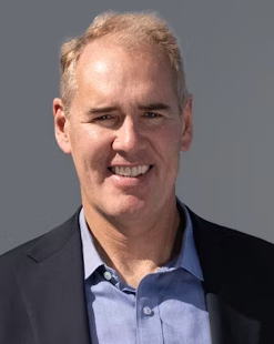 Jason Kovacic合作论文数Victor Chang Cardiac Research Institute;Graduate School of Biomedical Science, Icahn School of Medicine at Mount Sinai;The Mount Sinai Hospital;St Vincent's Health Australia;University of New South Wales9
 Francois Fay合作论文数Translational and Molecular Imaging Institute, Icahn School of Medicine at Mount Sinai;Translational and Molecular Imaging Institute, Icahn School of Medicine at Mount Sinai8
 Tina Binderup合作论文数Faculty of Health Sciences, University of Copenhagen8
 Neomi Shah合作论文数Montefiore Medical Center, Albert Einstein College of Medicine8
 Maria L Padilla合作论文数7
 Mandy M T van Leent合作论文数BioMedical Engineering and Imaging Institute, Icahn School of Medicine at Mount Sinai7
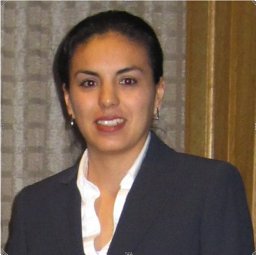 Sanchez-Gaytan Brenda L合作论文数Department of Chemistry, University of Pennsylvania7
 David Newby合作论文数Centre for Cardiovascular Science, The University of Edinburgh7
 Erik Stroes合作论文数Department of Vascular Medicine, Amsterdam University Medical Center7
 Weiying Dai合作论文数Beth Israel Deaconess Medical Center, Harvard Medical School7
 Anuradha Lala-Trindade合作论文数Icahn School of Medicine at Mount Sinai7Steve Liao合作论文数6Samantha Baxter合作论文数6
 Suárez-Fariñas Mayte合作论文数Laboratory for Investigative Dermatology, The Rockefeller University6Antoine Millon合作论文数6
 Mhairi Doris合作论文数6Seigo Ishino合作论文数6Olivier Lairez合作论文数5Alison Pruzan合作论文数5
 V. Fauveau合作论文数5Munir Ghesani合作论文数5
 Dimostenis Pandis合作论文数5




