In this study, we present a Transformer-based encoder model to predict Alzheimer's Disease (AD) progression from longitudinal multi-modal patient data. Our model, Longitudinal Survival Model for AD (LSM-AD), leverages rich temporal patterns present in sequences of patient visits, integrating multi-modal data, such as cognitive assessments and Magnetic Resonance Imaging (MRI) biomarkers to compute accurate diagnostic predictions. We conduct an empirical evaluation across two patient groups-Cognitively Normal (CN) individuals and those with Mild Cognitive Impairment (MCI)-tracking their progression for up to five follow-up years. Our results indicate that incorporating longer patient histories can yield superior performance compared to relying solely on a single visit, emphasizing the importance of historical context in improving predictive accuracy. Additionally, we show that the choice of the prediction head, training loss function and method for handling input missingness can significantly impact the quality of predictions. Notably, LSM-AD can improve Area Under the Receiver Operating Characteristic (AUROC) curve by up to 15% over previous state-of-the-art, when MRI biomarkers serve as the sole longitudinal feature. Our findings reinforce the value of multi-modal longitudinal data in evaluating patients, demonstrating its potential to improve early detection and monitoring of AD progression. Our code is available at https://github.com/batuhankmkaraman/LSM-AD.
Purpose: To investigate whether an AI-based method can detect subtle inter-fraction changes in MR-Linac images acquired during radiotherapy and explore the broader potential of MRLinac imaging. Methods: This retrospective study included longitudinal 0.35T MR-Linac images from 761 patients. To identify temporal changes, we employed a deep learning model using temporal ordering via pairwise comparison, previously shown effective for longitudinal imaging studies. The model was trained using first-to-last fraction pairs (F1-FL) and all pairs (All-pairs). Performance was assessed using quantitative metrics (accuracy and AUC) and compared against a radiologist's performance. Qualitative evaluation was performed using saliency maps, which identify anatomical regions associated with temporal imaging changes. Results: The F1-FL model demonstrated high performance (AUC=0.99, accuracy=0.95) and outperformed the radiologist in temporal ordering task. The All-pairs model also showed high performance (AUC=0.97, accuracy=0.91). Regions contributing to predictions included the prostate, bladder, and pubic symphysis. The performance was correlated to fractional intervals and was reduced for non-radiation-exposed timepoints (Sim and F1), suggesting that observed changes may reflect both temporal variation and radiation exposure. Conclusion: MR-Linac imaging appears capable of capturing subtle changes during prostate radiotherapy that can be detected by AI models, even over approximately two-day intervals. The model's high performance, together with quantitative and qualitative analyses, supports a potential role for MR-Linac in clinical applications beyond image guidance.
Clinical MRI frequently acquires anisotropic volumes with high in-plane resolution and low through-plane resolution to reduce acquisition time. Multiple orientations are therefore acquired to provide complementary anatomical information. Conventional integration of these views relies on registration followed by interpolation, which can degrade fine structural details. Recent deep learning-based super-resolution (SR) approaches have demonstrated strong performance in enhancing single-view images. However, their clinical reliability is often limited by the need for large-scale training datasets, resulting in increased dependence on cohort-level priors. Self-supervised strategies offer an alternative by learning directly from the target scans. Prior work either neglects the existence of multi-view information or assumes that in-plane information can supervise through-plane reconstruction under the assumption of pre-alignment between images. However, this assumption is rarely satisfied in clinical settings. In this work, we introduce Single-Subject Implicit Multi-View Super-Resolution for MRI (SIMS-MRI), a framework that operates solely on anisotropic multi-view scans from a single patient without requiring pre- or post-processing. Our method combines a multi-resolution hash-encoded implicit representation with learned inter-view alignment to generate a spatially consistent isotropic reconstruction. We validate the SIMS-MRI pipeline on both simulated brain and clinical prostate MRI datasets. Code will be made publicly available for reproducibility: https://github.com/abhshkt/SIMS-MRI
Multi-objective optimization (MOO) underlies many machine learning problems, yet MOO solvers across the loss-balancing, gradient-balancing, and Pareto-based families almost universally hand their reconciled directions to Adam . We show this coupling introduces two systematic gaps between the solver's intent and the optimizer's execution. The first is a weighting mismatch: Adam's second-moment denominator entangles the time-varying preference vector with gradient statistics, marginalizing the preference into a history average and collapsing distinct Pareto trade-offs toward a near-uniform mixture. The second is a geometric mismatch: Adam's adaptive metric distorts the Euclidean geometry MOO solvers assume, turning aligned objectives into apparent conflicts. To resolve both jointly, we introduce MAdam (Metric-Aware Multi-Objective Adam), a drop-in wrapper that leaves both solver and optimizer unchanged. MAdam preconditions the reconciled direction by the preference-conditioned curvature of the scalarized objective; on this whitened input, Adam's second moment collapses to identity, so the realized update is governed by the preference-conditioned metric. Across multi-task learning, Pareto-front recovery, physics-informed neural networks, and medical imaging, MAdam consistently improves over Adam for every solver family.
Deep learning models benefit from rich (e.g., multi-modal) input features. However, multimodal models might be challenging to deploy, because some inputs may be missing at inference. Current popular solutions include marginalization, imputation, and training multiple models. Marginalization achieves calibrated predictions, but it is computationally expensive and only feasible for low dimensional inputs. Imputation may result in inaccurate predictions, particularly when high-dimensional data, such as images, are missing. Training multiple models, where each model is designed to handle different subsets of inputs, can work well but requires prior knowledge of missing input patterns. Furthermore, training and retaining multiple models can be costly. We propose an efficient method to learn both the conditional distribution using full inputs and the marginal distributions. Our method, Knockout, randomly replaces input features with appropriate placeholder values during training. We provide a theoretical justification for Knockout and show that it can be interpreted as an implicit marginalization strategy. We evaluate Knockout across a wide range of simulations and real-world datasets and show that it offers strong empirical performance.
Longitudinal imaging data are routinely acquired for health studies and patient monitoring. A central goal in longitudinal studies is tracking relevant change over time. Traditional methods remove nuisance variation with custom pipelines to focus on significant changes. In this work, we present a machine learning–based method that automatically ignores irrelevant changes and extracts the time-varying signal of interest. Our method, called Learning-based Inference of Longitudinal imAge Changes (LILAC), performs a pairwise comparison of longitudinal images in order to make a temporal difference prediction. LILAC employs a convolutional Siamese architecture to extract feature pairs, followed by subtraction and a bias-free fully connected layer to learn meaningful temporal image differences. We first showcase LILAC’s ability to capture key longitudinal changes by simply training it to predict the temporal ordering of images. In our experiments, temporal ordering accuracy exceeded 0.98, and predicted time differences were strongly correlated with actual changes in relevant variables (Pearson Correlation Coefficient r = 0.911 with embryo phase change, and r = 0.875 with time interval in wound healing). Next, we trained LILAC to explicitly predict specific targets, such as the change in clinical scores in patients with mild cognitive impairment. LILAC models achieved over a 40% reduction in root mean square error compared to baseline methods. Our empirical results demonstrate that LILAC effectively localizes and quantifies relevant individual-level changes in longitudinal imaging data, offering valuable insights for studying temporal mechanisms or guiding clinical decisions.
Healthcare data often come from multiple sites in which the correlations between confounding variables can vary widely. If deep learning models exploit these unstable correlations, they might fail catastrophically in unseen sites. Although many methods have been proposed to tackle unstable correlations, each has its limitations. For example, adversarial training forces models to completely ignore unstable correlations, but doing so may lead to poor predictive performance. Other methods (e.g. Invariant risk minimization [4]) try to learn domain-invariant representations that rely only on stable associations by assuming a causal data-generating process (input X causes class label Y ). Thus, they may be ineffective for anti-causal tasks (Y causes X), which are common in computer vision. We propose a method called CoPA (Conditional Prevalence-Adjustment) for anti-causal tasks. CoPA assumes that (1) generation mechanism is stable, i.e. label Y and confounding variable(s) Z generate X, and (2) the unstable conditional prevalence in each site E fully accounts for the unstable correlations between X and Y . Our crucial observation is that confounding variables are routinely recorded in healthcare settings and the prevalence can be readily estimated, for example, from a set of (Y, Z) samples (no need for corresponding samples of X). CoPA can work even if there is a single training site, a scenario which is often overlooked by existing methods. Our experiments on synthetic and real data show CoPA beating competitive baselines.
Parkinson's disease patients with motor complications are often considered for deep brain stimulation (DBS) surgery. DBS candidate selection involves an assessment known as the levodopa challenge test (LCT). The LCT aims to predict DBS outcomes by measuring symptom improvement accompanying changes in levodopa dosage. While used in the patient selection process, inconsistent LCT predictions have been widely documented, verified here with Pearson's correlation r=0.12. Estimating symptom improvement to separate DBS responders and non-responders remains an unmet need. Quantitative susceptibility mapping (QSM) is routinely acquired for pre-surgical planning and depicts the iron distribution in substantia nigra and subthalamic nuclei. Iron deposition in these nuclei has correlated with disease progression and motor symptom severity. A novel QSM radiomic approach is presented using a regression model and features extracted from the pre-surgical targeting acquisition. Noise compensation in training labels improves outcome prediction in regression and classification models. The model predicts improvement in the unified Parkinson's disease rating scale (UPDRS-III) (r=0.75). Predictive feature maps in deep gray nuclei offer contrast between responders and non-responders. The QSM radiomic approach has potential to improve DBS candidate selection by accurately estimating symptom improvement, eliminating difficult medication manipulation, and avoiding time-consuming evaluations to reduce patient and clinician burden. ### Competing Interest Statement Pascal Spincemaille and Yi Wang are co-inventors on QSM-related patents owned by Cornell University and have ownership shares in MedImageMetric LLC. The remaining authors have no conflicts of interest to report. ### Funding Statement This research was supported by grants from the National Institutes of Health R01 NS095562 and National Multiple Sclerosis Society TA-2204-39428 and FG-2008-36976. ### Author Declarations I confirm all relevant ethical guidelines have been followed, and any necessary IRB and/or ethics committee approvals have been obtained. Yes The details of the IRB/oversight body that provided approval or exemption for the research described are given below: Institutional Review Boards of Weill Cornell Medicine and Mount Sinai and Changhai Hospitals gave ethical approval for this work. I confirm that all necessary patient/participant consent has been obtained and the appropriate institutional forms have been archived, and that any patient/participant/sample identifiers included were not known to anyone (e.g., hospital staff, patients or participants themselves) outside the research group so cannot be used to identify individuals. Yes I understand that all clinical trials and any other prospective interventional studies must be registered with an ICMJE-approved registry, such as ClinicalTrials.gov. I confirm that any such study reported in the manuscript has been registered and the trial registration ID is provided (note: if posting a prospective study registered retrospectively, please provide a statement in the trial ID field explaining why the study was not registered in advance). Yes I have followed all appropriate research reporting guidelines, such as any relevant EQUATOR Network research reporting checklist(s) and other pertinent material, if applicable. Yes Code and data will be available upon publication.
Prostate MRI plays an important role in imaging the prostate gland and surrounding tissues, particularly in the diagnosis and management of prostate cancer. With the widespread adoption of multiparametric magnetic resonance imaging in recent years, the concerns surrounding the variability of imaging quality have garnered increased attention. Several factors contribute to the inconsistency of image quality, such as acquisition parameters, scanner differences and interobserver variabilities. While efforts have been made to standardize image acquisition and interpretation via the development of systems, such as PI-RADS and PI-QUAL, the scoring systems still depend on the subjective experience and acumen of humans. Artificial intelligence (AI) has been increasingly used in many applications, including medical imaging, due to its ability to automate tasks and lower human error rates. These advantages have the potential to standardize the tasks of image interpretation and quality control of prostate MRI. Despite its potential, thorough validation is required before the implementation of AI in clinical practice. In this article, we explore the opportunities and challenges of AI, with a focus on the interpretation and quality of prostate MRI.
Segmentation is a crucial task in the medical imaging field and is often an important primary step or even a prerequisite to the analysis of medical volumes. Yet treatments such as surgery complicate the accurate delineation of regions of interest. The BraTS Post-Treatment 2024 Challenge published the first public dataset for post-surgery glioma segmentation and addresses the aforementioned issue by fostering the development of automated segmentation tools for glioma in MRI data. In this effort, we propose two straightforward approaches to enhance the segmentation performances of deep learning-based methodologies. First, we incorporate an additional input based on a simple linear combination of the available MRI sequences input, which highlights enhancing tumors. Second, we employ various ensembling methods to weigh the contribution of a battery of models. Our results demonstrate that these approaches significantly improve segmentation performance compared to baseline models, underscoring the effectiveness of these simple approaches in improving medical image segmentation tasks.
Distribution shifts between sites can seriously degrade model performance since models are prone to exploiting unstable correlations. Thus, many methods try to find features that are stable across sites and discard unstable features. However, unstable features might have complementary information that, if used appropriately, could increase accuracy. More recent methods try to adapt to unstable features at the new sites to achieve higher accuracy. However, they make unrealistic assumptions or fail to scale to multiple confounding features. We propose Generalized Prevalence Adjustment (GPA for short), a flexible method that adjusts model predictions to the shifting correlations between prediction target and confounders to safely exploit unstable features. GPA can infer the interaction between target and confounders in new sites using unlabeled samples from those sites. We evaluate GPA on several real and synthetic datasets, and show that it outperforms competitive baselines.
We present a keypoint-based foundation model for general purpose brain MRI registration, based on the recently-proposed KeyMorph framework. Our model, called BrainMorph, serves as a tool that supports multi-modal, pairwise, and scalable groupwise registration. BrainMorph is trained on a massive dataset of over 100,000 3D volumes, skull-stripped and non-skull-stripped, from nearly 16,000 unique healthy and diseased subjects. BrainMorph is robust to large misalignments, interpretable via interrogating automatically-extracted keypoints, and enables rapid and controllable generation of many plausible transformations with different alignment types and different degrees of nonlinearity at test-time. We demonstrate the superiority of BrainMorph in solving 3D rigid, affine, and nonlinear registration on a variety of multi-modal brain MRI scans of healthy and diseased subjects, in both the pairwise and groupwise setting. In particular, we show registration accuracy and speeds that surpass many classical and learning-based methods, especially in the context of large initial misalignments and large group settings. All code and models are available at https://github.com/alanqrwang/brainmorph.
Interpretability for machine learning models in medical imaging (MLMI) is an important direction of research. However, there is a general sense of murkiness in what interpretability means. Why does the need for interpretability in MLMI arise? What goals does one actually seek to address when interpretability is needed? To answer these questions, we identify a need to formalize the goals and elements of interpretability in MLMI. By reasoning about real-world tasks and goals common in both medical image analysis and its intersection with machine learning, we identify five core elements of interpretability: localization, visual recognizability, physical attribution, model transparency, and actionability. From this, we arrive at a framework for interpretability in MLMI, which serves as a step-by-step guide to approaching interpretability in this context. Overall, this paper formalizes interpretability needs in the context of medical imaging, and our applied perspective clarifies concrete MLMI-specific goals and considerations in order to guide method design and improve real-world usage. Our goal is to provide practical and didactic information for model designers and practitioners, inspire developers of models in the medical imaging field to reason more deeply about what interpretability is achieving, and suggest future directions of interpretability research.
Most state-of-the-art techniques for medical image segmentation rely on deep-learning models. These models, however, are often trained on narrowly-defined tasks in a supervised fashion, which requires expensive labeled datasets. Recent advances in several machine learning domains, such as natural language generation have demonstrated the feasibility and utility of building foundation models that can be customized for various downstream tasks with little to no labeled data. This likely represents a paradigm shift for medical imaging, where we expect that foundation models may shape the future of the field. In this paper, we consider a recently developed foundation model for medical image segmentation, UniverSeg. We conduct an empirical evaluation study in the context of prostate imaging and compare it against the conventional approach of training a task-specific segmentation model. Our results and discussion highlight several important factors that will likely be important in the development and adoption of foundation models for medical image segmentation.
Longitudinal studies, where a series of images from the same set of individuals are acquired at different time-points, represent a popular technique for studying and characterizing temporal dynamics in biomedical applications. The classical approach for longitudinal comparison involves normalizing for nuisance variations, such as image orientation or contrast differences, via pre-processing. Statistical analysis is, in turn, conducted to detect changes of interest, either at the individual or population level. This classical approach can suffer from pre-processing issues and limitations of the statistical modeling. For example, normalizing for nuisance variation might be hard in settings where there are a lot of idiosyncratic changes. In this paper, we present a simple machine learning-based approach that can alleviate these issues. In our approach, we train a deep learning model (called PaIRNet, for Pairwise Image Ranking Network) to compare pairs of longitudinal images, with or without supervision. In the self-supervised setup, for instance, the model is trained to temporally order the images, which requires learning to recognize time-irreversible changes. Our results from four datasets demonstrate that PaIRNet can be very effective in localizing and quantifying meaningful longitudinal changes while discounting nuisance variation. Our code is available at \url{https://github.com/heejong-kim/learning-to-compare-longitudinal-images.git}
 Mert Sabuncu合作论文数School of Electrical and Computer Engineering, Cornell University;Cornell Tech, Cornell University;Department of Radiology, Weill Cornell Medicine17
 Do-Young Yoon合作论文数College of Sciences, Konkuk University10
 illhee yoon合作论文数8Jung-Hee Kim合作论文数8Lee Sojung合作论文数8
 Jin Tae Hong合作论文数Chungbuk National University8Eunjin Kim合作论文数7
 Margolis Daniel J合作论文数Department of Radiology, University of California, Los Angeles7Alan Q. Wang合作论文数6Jeong-Woo Kang合作论文数6Kim Man Sub合作论文数6
 Himanshu Nagar合作论文数Radiation Oncology, Weill Cornell Medicine, Cornell University6Byung Hyuk Kwon合作论文数6
 Jeongwoo Kang合作论文数Res Inst Vet Sci, Seoul Natl Univ5
 Young Yang合作论文数5Young Yoon合作论文数4
 Minh Tho Nguyen合作论文数4
 Heesook Choi合作论文数4Yun Sun Park合作论文数3
 Batuhan K Karaman合作论文数3
 Rachit Saluja合作论文数Department of Radiology and Biomedical Imaging, University of California, San Francisco;Hospital, the University of Pennsylvania;University of California, San Francisco, Hospital of the University of Pennsylvania3Soohyun Kim合作论文数Lab Cytokine Immunol, Konkuk Univ3Bak Yesol合作论文数3
 Fengbei Liu合作论文数Cornell Tech, Cornell University3
 Dong-hun Lee合作论文数College of Veterinary Medicine, Konkuk University;Konkuk Wildlife Health Laboratory, Konkuk University3
 Joseph Piven合作论文数Department of Psychiatry, School of Medicine, The University of North Carolina at Chapel Hill3
 Guido Gerig合作论文数Department of Computer Science and Engineering, Tandon School of Engineering, New York University;Department of Biomedical Engineering, Tandon School of Engineering, New York University3Min-Chul Cho合作论文数2Chang Han Kim合作论文数2
 Min Chul Cho合作论文数2Hyun-Jeong Park合作论文数2
 dae-yeul yu合作论文数2Hee Jin Kim合作论文数2
 Jin-Man Kim合作论文数2Eun-Hee Seo合作论文数2
 Dominick Romano合作论文数Dept Radiol, Weill Cornell Med2R. Pennell合作论文数Sch Med, NYU2Man Jin Kim合作论文数2
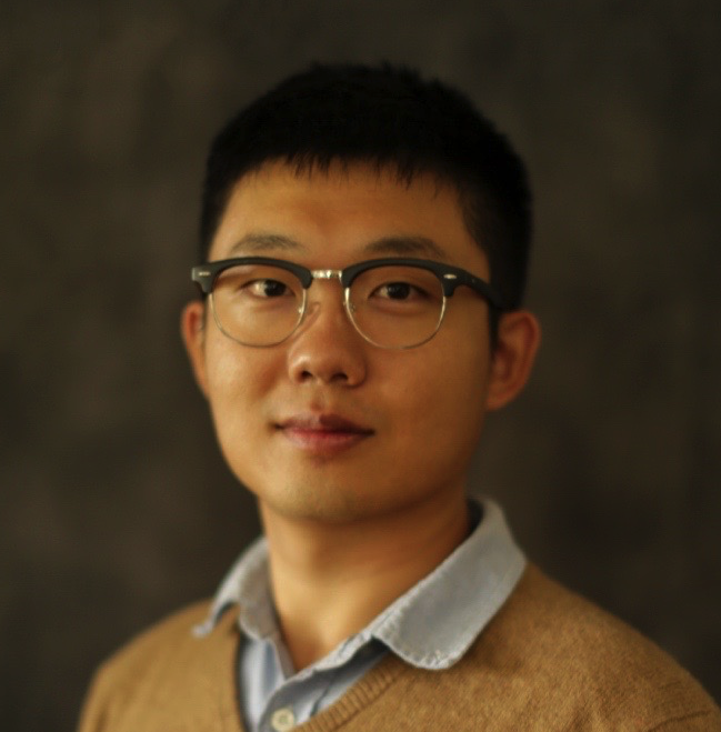 Qingyu Zhao合作论文数Department of Psychiatry and Behavioral Sciences, Stanford University2
 Pascal Spincemaille合作论文数Weill Cornell Medicine, Cornell University2
 Yi Wang合作论文数Meinig School of Biomedical Engineering, Cornell University;MRI Research Institute Radiology, Cornell University2
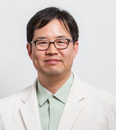 Chan Kyo Kim合作论文数Dept Radiol, Sungkyunkwan Univ2
 Sang Won Seo合作论文数Department of Neurology, Samsung Medical Center;Department of Clinical Research Design and Evaluation, Sungkyunkwan University;Samsung Advanced Institute for Health Sciences & Technology, Sungkyunkwan University2
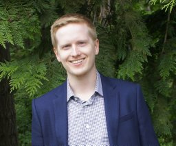 Michael Eggleston合作论文数2
 Dongsu Kim合作论文数dankook university2
 Bain Daniel J合作论文数Department of Geology and Planetary Science, University of Pittsburgh2
 Shah Shreyas合作论文数2
 Jianqi Li (李建奇)合作论文数Institute of Magnetic Resonance and Molecular Imaging in Medicine, East China Normal University;School of Physics, East China Normal University2
 Neel Dey合作论文数Computer Science and Artificial Intelligence Laboratory, Massachusetts Institute of Technology2




