Vulnerable atherosclerotic plaques, especially hemorrhaged lesions, are the major cause of mortalities related to vascular pathologies. The early identification of vulnerable plaques helps to stratify patients at risk of developing acute vascular events. In this study, proteomics analyses of human carotid artery samples collected from patients with atheromatous plaques and complicated lesions, respectively, as well as from healthy controls were performed. The proteins isolated from the carotid artery samples were analyzed by a bottom-up shotgun approach that relied on nanoflow liquid chromatography–tandem mass spectrometry analyses (LC–MS/MS) using both data-dependent (DDA) and data-independent (DIA) acquisitions. The data obtained by high-resolution DIA analyses displayed a stronger distinction among groups compared to DDA analyses. Differentially expressed proteins were further examined using Ingenuity Pathway Analysis® with focus on pathological and molecular processes driving atherosclerosis. From the more than 150 significantly regulated canonical pathways, atherosclerosis signaling and neutrophil extracellular trap signaling were verified by protein-targeted data extraction. The results of our study are expected to facilitate a better understanding of the disease progression’s molecular drivers and provide inspiration for further multiomics and hypothesis-driven studies.
Hydrogen sulfide (H2S) was previously revealed to inhibit osteoblastic differentiation of valvular interstitial cells (VICs), a pathological feature in calcific aortic valve disease (CAVD). This study aimed to explore the metabolic control of H2S levels in human aortic valves.Lower levels of bioavailable H2S and higher levels of interleukin-1β (IL-1β) and tumor necrosis factor-α (TNF-α) were detected in aortic valves of CAVD patients compared to healthy individuals, accompanied by higher expression of cystathionine γ-lyase (CSE) and same expression of cystathionine β-synthase (CBS). Increased biogenesis of H2S by CSE was found in the aortic valves of CAVD patients which is supported by increased production of lanthionine. In accordance, healthy human aortic VICs mimic human pathology under calcifying conditions, as elevated CSE expression is associated with low levels of H2S. The expression of mitochondrial enzymes involved in H2S catabolism including sulfide quinone oxidoreductase (SQR), the key enzyme in mitochondrial H2S oxidation, persulfide dioxygenase (ETHE1), sulfite oxidase (SO) and thiosulfate sulfurtransferase (TST) were up-regulated in calcific aortic valve tissues, and a similar expression pattern was observed in response to high phosphate levels in VICs. AP39, a mitochondria-targeting H2S donor, rescued VICs from an osteoblastic phenotype switch and reduced the expression of IL-1β and TNF-α in VICs. Both pro-inflammatory cytokines aggravated calcification and osteoblastic differentiation of VICs derived from the calcific aortic valves. In contrast, IL-1β and TNF-α provided an early and transient inhibition of VICs calcification and osteoblastic differentiation in healthy cells and that effect was lost as H2S levels decreased. The benefit was mediated via CSE induction and H2S generation.We conclude that decreased levels of bioavailable H2S in human calcific aortic valves result from an increased H2S metabolism that facilitates the development of CAVD. CSE/H2S represent a pathway that reverses the action of calcifying stimuli.
Previous investigations have demonstrated that treatment of animals with rapamycin increases levels of autophagy, which is a process by which cells degrade intracellular detritus, thus suppressing the emergence of senescent cells, whose pro‐inflammatory properties, are primary drivers of age‐associated physical decline. A hypothesis is tested here that rapamycin treatment of mice approaching the end of their normal lifespan exhibits increased survival, enhanced expression of autophagic proteins; and klotho protein—a biomarker of aging that affects whole organism senescence, and systemic suppression of inflammatory mediator production. Test groups of 24‐month‐old C57BL mice were injected intraperitoneally with either 1.5 mg/kg/week rapamycin or vehicle. All mice administered rapamycin survived the 12‐week course, whereas 43% of the controls died. Relative to controls, rapamycin‐treated mice experienced minor but significant weight loss; moreover, nonsignificant trends toward decreased levels of leptin, IL‐6, IL‐1β, TNF‐α, IL‐1α, and IGF‐1, along with slight elevations in VEGF, MCP‐1 were observed in the blood serum of rapamycin‐treated mice. Rapamycin‐treated mice exhibited significantly enhanced autophagy and elevated expression of klotho protein, particularly in the kidney. Rapamycin treatment also increased cardiomyocyte Ca2+‐sensitivity and enhanced the rate constant of force re‐development, which may also contribute to the enhanced survival rate in elderly mice.
Hidradenitis suppurativa (HS) is a Th1/17-driven inflammatory skin disease of the apocrine gland-rich (AGR) skin regions, where keratinocytes seem to be the crucial drivers of the initial pathogenic steps. However, the possible role of permeability barrier alteration in activating keratinocytes during HS development has not been clarified. We compared the major permeability barrier elements of non-lesional HS (HS-NL; n = 10) and lesional HS (HS-L; n = 10) skin with healthy AGR regions (n = 10) via RT-qPCR and immunohistochemistry. Stratum corneum components related to cornified envelope formation, corneocyte desquamation and (corneo)desmosome organization were analyzed along with tight junction molecules and barrier alarmins. The permeability barrier function was also investigated with transepidermal water loss (TEWL) measurements (n = 16). Junction structures were also visualized using confocal microscopy. At the gene level, none of the investigated molecules were significantly altered in HS-NL skin, while 11 molecules changed significantly in HS-L skin versus control. At the protein level, the investigated molecules were similarly expressed in HS-NL and AGR skin. In HS-L skin, only slight changes were detected; however, differences did not show a unidirectional alteration, as KRT1 and KLK5 were detected in decreased levels, and KLK7, KRT6 and DSG1 in increased levels. No significant differences in TEWL or the expression of junction structures were assessed. Our findings suggest that the permeability barrier is not significantly damaged in HS skin and permeability barrier alterations are not the driver factors of keratinocyte activation in this disease.
Background: Acne vulgaris provides a unique disease setting in which a prominent skin inflammation is coupled with the overproduction of lipid rich sebum.Objectives: Our goal was to evaluate the expression of barrier molecules in papular acne skin samples obtained from untreated patients and compare those to the results of healthy and of papulopustular rosacea-involved ones at the mRNA and protein levels. In addition, we aimed to assess the effects of various sebum composing lipids on the expression of proteins involved in barrier formation in keratinocytes.Methods: Available microarray data sets of papular acne and papulopustular rosacea-affected skin samples were re-analysed with a focus on epidermal barrier related pathways. Immunohistochemistry was performed to detect barrier molecules in the interfollicular regions of human acne and healthy skin samples. Protein levels of barrier-related genes were measured by western blot in samples of HaCaT keratinocytes treated with selected lipids.Results: Meta-analysis of whole transcriptome data sets revealed that barrier-related pathways are significantly affected in acne vulgaris skin samples. While an altered expression of key molecules in maintaining barrier functions such as filaggrin, keratin 1, involucrin, desmoglein 1, kallikrein 5 and 7, was also observed at the protein levels, our data demonstrated that sebum composing lipids may selectively modify the levels of epidermal barrier-related molecules.Conclusions: Our results suggest that although not as prominently as in the dry papulopustular rosacea skin, the epidermal barrier in the interfollicular region may be damaged also in the lipid rich skin samples of papular acne. Furthermore, our findings indicating diverse regulatory effects of various sebum lipids on the expression of barrier molecules in keratinocytes suggest, that they may influence the moisturization of the skin as well. Altogether, our findings could have implications in the development of sebum-modulating anti acne therapies and even in the care of symptom free skin.
Atopic dermatitis is an inflammatory skin disease characterized by significant permeability barrier damage. Regulation and maintenance of permeability and antimicrobial skin barriers are strongly connected. There is a lack of comprehensive studies of the expression of all 5 major antimicrobial peptide functional groups in atopic dermatitis. The aim of this study was to investigate the major antimicrobial peptide functional groups in lesional atopic dermatitis, non-lesional atopic dermatitis, and healthy control samples, using real-time quantitative PCR and immunohistochemistry. Lesional psoriatic skin was also examined as a diseased control. No differences in mRNA levels were detected between non-lesional atopic dermatitis and healthy control skin, and, at the protein level, the only change was the significantly decreased LL-37 in non-lesional atopic dermatitis. In lesional atopic dermatitis, several antimicrobial peptides were significantly altered at the mRNA level, while, at the protein level, all antimicrobial peptides were significantly upregulated or unchanged, except for LL-37, which decreased, compared with healthy controls. Antimicrobial peptides were similarly elevated in lesional atopic dermatitis and lesional psoriatic skin, with somewhat higher expression in lesional psoriatic skin, except for LL-37. In conclusion, LL-37 was the only antimicrobial peptide that was impaired in both non-lesional and lesional atopic dermatitis, highlighting its potential pathogenetic or exacerbating role in the initial stages of the disease.
Recent data indicate that distinct skin areas show different microbial/chemical milieu. Keratinocytes (KC) respond to these stimuli by producing cytokine mediators. Therefore, we aimed to determine KC-derived cytokine expression in distinct healthy skin regions (gland-poor [GP], sebaceous gland-rich [SGR] and apocrine gland-rich [AGR]), and their changes in skin diseases of the given regions (atopic dermatitis [AD], papulopustular rosacea [PPR] and psoriasis). Cytokines were analysed at the mRNA and protein levels, and literature analysis was performed for functional categorization. The three regions showed characteristically different cytokine patterns. GP was featured by an IL-25/IL-33/IL-36RA/IL-38/IL-18 cytokine milieu, SGR was characterized by IL-23/IL-17C/IL-18, and AGR skin exhibited a mixed IL-25/IL-33/IL-23/IL-18 profile. Literature analyses revealed different homeostatic and proinflammatory roles of these cytokine patterns (Th2 related in GP, Th17 related in SGR and mixed Th2/Th17 in AGR). In skin diseases which are primarily epidermal cytokine-driven (AD, PPR), the level of the regionally characteristic cytokines were further elevated, in contrast to the autoantigen-driven psoriasis, where the cytokine pattern was independent from the localization. Healthy skin regions are equipped with different KC-derived cytokine profiles, which may influence each region's capability of mediator production in certain types of dermatoses.
Aim: The aim of our study was to explore the pathophysiologic role of oxidation of hemoglobin (Hb) to ferrylHb in human atherosclerosis. Results: We observed a severe oxidation of Hb to ferrylHb in complicated atherosclerotic lesions of carotid arteries with oxidative changes of the globin moieties, detected previously described oxidation hotspots in Hb (β1Cys93; β1Cys112; β2Cys112) and identified a novel oxidation hotspot (α1Cys104). After producing a monoclonal anti-ferrylHb antibody, ferrylHb was revealed to be localized extracellularly and also internalized by macrophages in the human hemorrhagic complicated lesions. We demonstrated that ferrylHb is taken up via phagocytosis as well as CD163 receptor-mediated endocytosis and then transported to lysosomes involving actin polymerization. Internalization of ferrylHb was accompanied by upregulation of heme oxygenase-1 and H-ferritin and accumulation of iron within lysosomes as a result of heme/iron uptake. Importantly, macrophages exposed to ferrylHb in atherosclerotic plaques exhibited a proinflammatory phenotype, as reflected by elevated levels of IL-1β and TNF-α. To find further signatures of ferrylHb in complicated lesions, we performed RNA-seq analysis on biopsies from patients who underwent endarterectomies. RNA-seq analysis demonstrated that human complicated lesions had a unique transcriptomic profile different from arteries and atheromatous plaques. Pathways affected in complicated lesions included gene changes associated with phosphoinositide 3-kinase (PI3K) signaling, lipid transport, tissue remodeling, and vascularization. Targeted analysis of gene expression associated with calcification, apoptosis, and hemolytic-specific clusters indicated an increase in the severity of complicated lesions compared with atheroma. A 39% overlap in the differential gene expression profiles of human macrophages exposed to ferrylHb and the complicated lesion profiles was uncovered. Among these 547 genes, we found inflammatory, angiogenesis, and iron metabolism gene clusters regulated in macrophages. Innovation and Conclusion: We conclude that oxidation of Hb to ferrylHb contributes to the progression of atherosclerosis via polarizing macrophages into a proatherogenic phenotype. Antioxid. Redox Signal. 35, 917–950.
Hemorrhage and hemolysis with subsequent heme release are implicated in many pathologies. Endothelial cells (ECs) encounter large amount of free heme after hemolysis and are at risk of damage from exogenous heme. Here we show that hemorrhage aggravates endoplasmic reticulum (ER) stress in human carotid artery plaques compared to healthy controls or atheromas without hemorrhage as demonstrated by RNA sequencing and immunohistochemistry. In EC cultures, heme also induces ER stress. In contrast, if cultured ECs are pulsed with heme arginate, cells become resistant to heme-induced ER (HIER) stress that is associated with heme oxygenase-1 (HO-1) and ferritin induction. Knocking down HO-1, HO-2, biliverdin reductase, and ferritin show that HO-1 is the ultimate cytoprotectant in acute HIER stress. Carbon monoxide-releasing molecules (CORMs) but not bilirubin protects cultured ECs from HIER stress via HO-1 induction, at least in part. Knocking down HO-1 aggravates heme-induced cell death that cannot be counterbalanced with any known cell death inhibitors. We conclude that endothelium and perhaps other cell types can be protected from HIER stress by induction of HO-1, and heme-induced cell death occurs via HIER stress that is potentially involved in the pathogenesis of diverse pathologies with hemolysis and hemorrhage including atherosclerosis.
Infiltration of red blood cells into atheromatous plaques and oxidation of hemoglobin (Hb) and lipoproteins are implicated in the pathogenesis of atherosclerosis. α1-microglobulin (A1M) is a radical-scavenging and heme-binding protein. In this work, we examined the origin and role of A1M in human atherosclerotic lesions. Using immunohistochemistry, we observed a significant A1M immunoreactivity in atheromas and hemorrhaged plaques of carotid arteries in smooth muscle cells (SMCs) and macrophages. The most prominent expression was detected in macrophages of organized hemorrhage. To reveal a possible inducer of A1M expression in ruptured lesions, we exposed aortic endothelial cells (ECs), SMCs and macrophages to heme, Oxy- and FerrylHb. Both heme and FerrylHb, but not OxyHb, upregulated A1M mRNA expression in all cell types. Importantly, only FerrylHb induced A1M protein secretion in aortic ECs, SMCs and macrophages. To assess the possible function of A1M in ruptured lesions, we analyzed Hb oxidation and heme-catalyzed lipid peroxidation in the presence of A1M. We showed that recombinant A1M markedly inhibited Hb oxidation and heme-driven oxidative modification of low-density lipoproteins as well plaque lipids derived from atheromas. These results demonstrate the presence of A1M in atherosclerotic plaques and suggest its induction by heme and FerrylHb in the resident cells.
The chemical milieu, microbiota composition, and immune activity show prominent differences in distinct healthy skin areas. The objective of the current study was to compare the major permeability barrier components (stratum corneum and tight junction (TJ)), investigate the distribution of (corneo)desmosomes and TJs, and measure barrier function in healthy sebaceous gland-rich (SGR), apocrine gland-rich (AGR), and gland-poor (GP) skin regions. Molecules involved in cornified envelope (CE) formation, desquamation, and (corneo)desmosome and TJ organization were investigated at the mRNA and protein levels using qRT-PCR and immunohistochemistry. The distribution of junction structures was visualized using confocal microscopy. Transepidermal water loss (TEWL) functional measurements were also performed. CE intracellular structural components were similarly expressed in gland-rich (SGR and AGR) and GP areas. In contrast, significantly lower extracellular protein levels of (corneo)desmosomes (DSG1 and CDSN) and TJs (OCLN and CLDN1) were detected in SGR/AGR areas compared to GP areas. In parallel, kallikrein proteases were significantly higher in gland-rich regions. Moreover, gland-rich areas were characterized by prominently disorganized junction structures ((corneo)desmosomes and TJs) and significantly higher TEWL levels compared to GP skin, which exhibited a regular distribution of junction structures. According to our findings, the permeability barrier of our skin is not uniform. Gland-rich areas are characterized by weaker permeability barrier features compared with GP regions. These findings have important clinical relevance and may explain the preferred localization of acantholytic skin diseases on gland-rich skin regions (e.g., Pemphigus foliaceus, Darier’s disease, and Hailey–Hailey disease).
Background Wolcott-Rallison Syndrome (WRS) is a rare autosomal recessive disease that is the most common cause of neonatal diabetes in consanguineous families. WRS is caused by various genetic alterations of the Eukaryotic Translation Initiation Factor 2-Alpha Kinase 3 (EIF2AK3) gene. Methods Genetic analysis of a consanguineous family where two children were diagnosed with WRS was performed by Sanger sequencing. The altered protein was investigated by in vitro cloning, expression and immunohistochemistry. Results The first cases in Hungary, - two patients in one family, where the parents were fourth-degree cousins - showed the typical clinical features of WRS: early onset diabetes mellitus with hyperglycemia, growth retardation, infection-induced multiple organ failure. The genetic background of the disease was a novel alteration in the EIF2AK3 gene involving the splice site of exon 11- intron 11-12 boundary: g.53051_53062delinsTG. According to cDNA sequencing this created a new splice site and resulted in a frameshift and the development of an early termination codon at amino acid position 633 (p.Pro627AspfsTer7). Based on in vitro cloning and expression studies, the truncated protein was functionally inactive. Immunohistochemistry revealed that the intact protein was absent in the islets of pancreas, furthermore insulin expressing cells were also dramatically diminished. Elevated GRP78 and reduced CHOP protein expression were observed in the liver. Conclusions The novel genetic alteration causing the absence of the EIF2AK3 protein resulted in insufficient handling of severe endoplasmic reticulum stress, leading to liver failure and demise of the patients.
Abstract is missing (Short communication)
Intraplaque hemorrhage frequently occurs in atherosclerotic plaques resulting in cell-free hemoglobin, which is oxidized to ferryl hemoglobin (FHb) in the highly oxidative environment. Osteoclast-like cells (OLCs) derived from macrophages signify a counterbalance mechanism for calcium deposition in atherosclerosis. Our aim was to investigate whether oxidized hemoglobin alters osteoclast formation, thereby affecting calcium removal from mineralized atherosclerotic lesions. RANKL- (receptor activator of nuclear factor kappa-Βligand-) induced osteoclastogenic differentiation and osteoclast activity of RAW264.7 cells were studied in response to oxidized hemoglobin via assessing bone resorption activity, expression of osteoclast-specific genes, and the activation of signalization pathways. OLCs in diseased human carotid arteries were assessed by immunohistochemistry. FHb, but not ferrohemoglobin, decreased bone resorption activity and inhibited osteoclast-specific gene expression (tartrate-resistant acid phosphatase, calcitonin receptor, and dendritic cell-specific transmembrane protein) induced by RANKL. In addition, FHb inhibited osteoclastogenic signaling pathways downstream of RANK (receptor activator of nuclear factor kappa-Β). It prevented the induction of TRAF6 (tumor necrosis factor (TNF) receptor-associated factor 6) and c-Fos, phosphorylation of p-38 and JNK (c-Jun N-terminal kinase), and nuclear translocation of NFκB (nuclear factor kappa-Β) and NFATc1 (nuclear factor of activated T-cells, cytoplasmic 1). These effects were independent of heme oxygenase-1 demonstrated by knocking down HO-1 gene in RAW264.7 cells and in mice. Importantly, FHb competed with RANK for RANKL binding suggesting possible mechanisms by which FHb impairs osteoclastic differentiation. In diseased human carotid arteries, OLCs were abundantly present in calcified plaques and colocalized with regions of calcium deposition, while the number of these cells were lower in hemorrhagic lesions exhibiting accumulation of FHb despite calcium deposition. We conclude that FHb inhibits RANKL-induced osteoclastic differentiation of macrophages and suggest that accumulation of FHb in a calcified area of atherosclerotic lesion with hemorrhage retards the formation of OLCs potentially impairing calcium resorption.
The lysis of red blood cells was shown to occur in human ruptured atherosclerotic lesions and intraventricular hemorrhage (IVH) of the brain. Liberated cell-free hemoglobin was found to undergo oxidation in both pathologies. We hypothesize that hemoglobin-derived peptides are generated during hemoglobin oxidation both in complicated atherosclerotic lesions and IVH of the brain, triggering endothelial cell dysfunction. Oxidized hemoglobin and its products were followed with spectrophotometry, LC–MS/MS analysis and detection of the cross-linking of globin chains in complicated atherosclerotic lesions of the human carotid artery and the hemorrhaged cerebrospinal liquid of preterm infants. The vascular pathophysiologic role of oxidized hemoglobin and the resultant peptides was assessed by measuring endothelial integrity, the activation of endothelial cells and the induction of proinflammatory genes. Peptide fragments of hemoglobin (VNVDEVGGEALGRLLVVYPWTQR, LLVVYPWTQR, MFLSFPTTK, VGAHAGEYGAELERMFLSFPTTK, and FLASVSTVLTSKYR) were identified in ruptured atherosclerotic lesions and in IVH of the human brain. Fragments resulting from the oxidation of hemoglobin were accompanied by the accumulation of ferryl hemoglobin. Similar to complicated atherosclerotic lesions of the human carotid artery, a high level of oxidized and cross-linked hemoglobin was observed in the cerebrospinal fluid after IVH. Haptoglobin inhibited hemoglobin fragmentation provoked by peroxide. The resultant peptides failed to bind haptoglobin or albumin. Peptides derived from hemoglobin oxidation and ferryl hemoglobin induced intercellular gap formation, decreased junctional resistance in the endothelium, and enhanced monocyte adhesion to endothelial cells. Enhanced expression of TNF and the activation of NLRP3 and CASP1 followed by the increased generation of IL-1β and nuclear translocation of the NF-κβ transcription factor occurred in response to hemoglobin-derived peptides, and ferryl hemoglobin in endothelium was upregulated in both pathologies. We conclude that the oxidation of hemoglobin in complicated atherosclerotic lesions and intraventricular hemorrhage of the brain generates peptide fragments and ferryl hemoglobin with the potential to trigger endothelial cell dysfunction.
Rosacea is a common chronic inflammation of sebaceous gland-rich facial skin characterized by severe skin dryness, elevated pH, transepidermal water loss, and decreased hydration levels. Until now, there has been no thorough molecular analysis of permeability barrier alterations in the skin of patients with rosacea. Thus, we aimed to investigate the barrier alterations in papulopustular rosacea samples compared with healthy sebaceous gland-rich skin, using RNA sequencing analysis (n = 8). Pathway analyses by Cytoscape ClueGO revealed 15 significantly enriched pathways related to skin barrier formation. RT-PCR and immunohistochemistry were used to validate the pathway analyses. The results showed significant alterations in barrier components in papulopustular rosacea samples compared with sebaceous gland-rich skin, including the cornified envelope and intercellular lipid lamellae formation, desmosome and tight junction organizations, barrier alarmins, and antimicrobial peptides. Moreover, the barrier damage in papulopustular rosacea was unexpectedly similar to atopic dermatitis; this similarity was confirmed by immunofluorescent staining. In summary, besides the well-known dysregulation of immunological, vascular, and neurological functions, we demonstrated prominent permeability barrier alterations in papulopustular rosacea at the molecular level, which highlight the importance of barrier repair therapies for rosacea.
Objective- Calcific aortic valve disease is a prominent finding in elderly and in patients with chronic kidney disease. We investigated the potential role of iron metabolism in the pathogenesis of calcific aortic valve disease. Approach and Results- Cultured valvular interstitial cells of stenotic aortic valve with calcification from patients undergoing valve replacement exhibited significant susceptibility to mineralization/osteoblastic transdifferentiation in response to phosphate. This process was abrogated by iron via induction of H-ferritin as reflected by lowering ALP and osteocalcin secretion and preventing extracellular calcium deposition. Cellular phosphate uptake and accumulation of lysosomal phosphate were decreased. Accordingly, expression of phosphate transporters Pit1 and Pit2 were repressed. Translocation of ferritin into lysosomes occurred with high phosphate-binding capacity. Importantly, ferritin reduced nuclear accumulation of RUNX2 (Runt-related transcription factor 2), and as a reciprocal effect, it enhanced nuclear localization of transcription factor Sox9 (SRY [sex-determining region Y]-box 9). Pyrophosphate generation was also increased via upregulation of ENPP2 (ectonucleotide pyrophosphatase/phosphodiesterase-2). 3H-1, 2-dithiole-3-thione mimicked these beneficial effects in valvular interstitial cell via induction of H-ferritin. Ferroxidase activity of H-ferritin was essential for this function, as ceruloplasmin exhibited similar inhibitory functions. Histological analysis of stenotic aortic valve revealed high expression of H-ferritin without iron accumulation and its relative dominance over ALP in noncalcified regions. Increased expression of H-ferritin accompanied by elevation of TNF-α (tumor necrosis factor-α) and IL-1β (interleukin-1β) levels, inducers of H-ferritin, corroborates the essential role of ferritin/ferroxidase via attenuating inflammation in calcific aortic valve disease. Conclusions- Our results indicate that H-ferritin is a stratagem in mitigating valvular mineralization/osteoblastic differentiation. Utilization of 3H-1, 2-dithiole-3-thione to induce ferritin expression may prove a novel therapeutic potential in valvular mineralization.
Calcification of heart valves is a frequent pathological finding in chronic kidney disease and in elderly patients. Hydrogen sulfide (H2S) may exert anti‐calcific actions. Here we investigated H2S as an inhibitor of valvular calcification and to identify its targets in the pathogenesis.
The infiltration of red blood cells into atheromatous plaques is implicated in atherogenesis. Inside the lesion, hemoglobin (Hb) is oxidized to ferri- and ferrylHb which exhibit prooxidant and proinflammatory activities. Cystathione gamma-lyase- (CSE-) derived H2S has been suggested to possess various antiatherogenic actions. Expression of CSE was upregulated predominantly in macrophages, foam cells, and myofibroblasts of human atherosclerotic lesions derived from carotid artery specimens of patients. A similar pattern was observed in aortic lesions of apolipoprotein E-deficient mice on high-fat diet. We identified several triggers for inducing CSE expression in macrophages and vascular smooth muscle cells including heme, ferrylHb, plaque lipids, oxidized low-density lipoprotein, tumor necrosis factor-α, and interleukin-1β. In the interplay between hemoglobin and atheroma lipids, H2S significantly mitigated oxidation of Hb preventing the formation of ferrylHb derivatives, therefore providing a novel function as a heme-redox-intermediate-scavenging antioxidant. By inhibiting Hb-lipid interactions, sulfide lowered oxidized Hb-mediated induction of adhesion molecules in endothelium and disruption of endothelial integrity. Exogenous H2S inhibited heme and Hb-mediated lipid oxidation of human atheroma-derived lipid and human complicated lesion. Our study suggests that the CSE/H2S system represents an atheroprotective pathway for removing or limiting the formation of oxidized Hb and lipid derivatives in the atherosclerotic plaque.
 Méhes Gábor合作论文数Department of Pathology, Faculty of Medicine, University of Debrecen, Debrecen, 4032,
Hungary16
Méhes Gábor合作论文数Department of Pathology, Faculty of Medicine, University of Debrecen, Debrecen, 4032,
Hungary16 György Balla合作论文数Faculty of Medicine, University of Debrecen;H-4012 Debrecen, Hungary;Faculty of Medicine, University of Debrecen14
 Jozsef Balla合作论文数Div. of Nephrology, Faculty of Medicine, University of Debrecen;Department of Internal Medicine, University of Debrecen;Department of Internal Medicine, Internal Medicine Clinic, University of Debrecen Clinical Centre14
 Zsolt Dajnoki合作论文数10
 Andrea Szegedi合作论文数Department of Dermatology, Faculty of Medicine, University of Debrecen;University of Debrecen Clinical Centre10
 Krisztian Gaspar合作论文数9
 Laszlo Potor合作论文数Thrombosis and Hemostasis Research Group, Hungarian Academy of Sciences9
 Anikó Kapitány合作论文数Institute of Medicine, University of Debrecen Medical and Health Science Center9
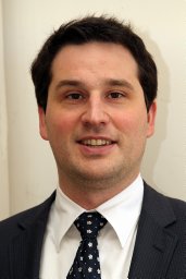 Törőcsik Dániel合作论文数8
 David Petho合作论文数Kalman Laki Doctoral Sch Biomed & Clin Sci, Univ Debrecen6Beke Lívia合作论文数6Orsolya Somogyi合作论文数5
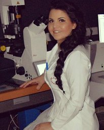 Katalin Éva Sikura合作论文数ELKH UD Vasc Biol & Myocardial Pathophysiol Res G, Hungarian Acad Sci5Lilla Szabó合作论文数5Gáll Tamás合作论文数Medical and Health Science Centre, University of Debrecen5
 Szilard Poliska合作论文数Department of Biochemistry and Molecular Biology, Faculty of Medicine, University of Debrecen5
 Csaba D. Toth合作论文数Department of Mathematics, California State University Northridge;Department of Computer Science, Tufts University5Niké Posta合作论文数4Barbara Medgyesi合作论文数4Melinda Oros合作论文数Laboratory of Neurobehavior Genetics, The Nathan S. Kline Institute for Psychiatric Research4
 Peter Gergely合作论文数Second Department of Medicine, Semmelweis University4Roberta Torregrossa合作论文数3
 Katalin Dull合作论文数Faculty of Medicine, University of Debrecen3
 Enikő Balogh合作论文数Faculty of Medicine, University of Debrecen3
 Peter Szucs合作论文数Dept Anat Histol & Embryol, Univ Debrecen3
 Eva Csosz合作论文数Medical and Health Science Center, University of Debrecen3
 Tamas Szerafin合作论文数3Annamaria Nagy合作论文数3Matt Whiteman合作论文数Institute of Biomedical and Clinical Science, College of Medicine and Health, University of Exeter3
 Klaudia Docs合作论文数Faculty of Medicine, University of Debrecen3
 Gergo Kallo合作论文数3Béke Gabriella合作论文数3
 Peter Nagy合作论文数Department of Molecular Immunology and Toxicology, National Institute of Oncology, Hungary;Debrecen University;Department of Pathology, University of Otago3
 Viktória Jeney合作论文数Medical and Health Science Center, University of Debrecen3
 Tamás Sándor Biró合作论文数Institute for Particle and Nuclear Physics, Wigner Research Centre for Physics3
 Imre Szabo合作论文数the Fels Institute for Cancer Research and Molecular Biology, Temple University3
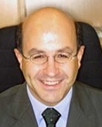 Christos C. Zouboulis合作论文数Klinik für Dermatologie, Venerologie und Allergologie;Forschungslaboratorien für Biogerontologie, Dermato-Pharmakologie und Dermato-Endokrinologie und für Immunologie, Städtisches Klinikum;Zentrums für Klinische Studien3Abolfazl Zarjou合作论文数Department of Medicine, University of Debrecen3Agnes Gyetvai合作论文数Medical and Health Science Center Laboratory of Immunology, University of Debrecen3Ágnes Papp合作论文数2
 Zsolt Combi合作论文数2
 Adrienn Jenei合作论文数Ctr Clin, Univ Debrecen2
 Andreas Patsalos合作论文数Johns Hopkins All Childrens Hosp, Johns Hopkins Univ2
 Paolo Arosio合作论文数Biochemical Engineering Laboratory, Institute for Chemical and Bioengineering, Department of Chemistry and Applied Biosciences, ETH Zurich2Janka Eszter合作论文数2
 Fagyas Miklós合作论文数Általános Orvostudományi Kar, Debreceni Egyetem;Kardiológiai Intézet, Debreceni Egyetem;Kardiológiai és Szívsebészeti Klinika, Debreceni Egyetem2
 Magnus Gram合作论文数2Bódi Beáta合作论文数2
 Laszlo Nagy合作论文数University of Debrecen;Institute for Fundamental Biomedical Research, Johns Hopkins All Children’s Hospital;Johns Hopkins Center for Metabolic Origins of Disease;Division of Endocrinology, Diabetes and Metabolism, Department of Medicine, School of MedicineJohns Hopkins University;Department of Biological Chemistry, School of Medicine, Johns Hopkins University2




