Abdominal aortic aneurysm (AAA) is a lethal vascular disease characterized by intramural hemorrhage. This study delineates the signatures of heme and its metabolic imbalance related to progression and inflammation in AAA. Clinical analyses of patients undergoing open AAA surgery show that AAA patients exhibit vascular inflammation, with elevated serum CRP, IL-6, and heme levels correlating with the expression of heme-regulated gene Hmox1/HO-1 (heme oxygenase-1) in the affected aortic wall. Oxidation of hemoglobin to ferri state leading to accumulation of methemoglobin readily releasing heme occurs in human AAA and in angiotensin II (AngII)-induced AAA in apolipoprotein E-deficient mice. Transcriptomic analysis for AngII-induced AAA identifies upregulated genes predominantly enriched in inflammatory signaling, extracellular matrix degradation, oxidative stress pathways, and altered expression of genes related to heme metabolism including Hmox1. Immunohistochemistry for IL1β and TNFα confirms inflammatory activation within AAA tissues. The signatures of heme-responsive gene inductions, enhanced expression of HO-1 and H-ferritin, are detected. Mechanistic studies employing endothelial cells and smooth muscle cells reveal that heme exposure of resident cells markedly enhances the expression of IL1β and ICAM1, as well as the inflammasome component NLRP3, and such inflammatory response is controlled by HO-1. Intervention with Normosang (heme arginate), an HO-1 inducer, attenuates aneurysm progression, whereas HO-1 inhibition by Tin protoporphyrin IX abolishes this protection. Induction of HO-1 accompanied by elevated H-ferritin level also mitigated aortic wall inflammation as reflected by lowering IL1β and TNFα. These findings highlight the heme-HO-1-H-ferritin axis as an element of AAA pathogenesis and a potential therapeutic target.
Macrophages, endowed with remarkable phenotypic plasticity, are essential for orchestrating injury responses and regulating iron homeostasis. Given the central role of ferritin heavy chain (FtH) as a molecular rheostat linking iron sequestration to redox-dependent signaling, we examined how myeloid FtH governs renal iron trafficking and ensuing oxidative stress pathways during acute kidney injury (AKI). Transcriptome analysis revealed coupling of FtH deficiency in monocytes and macrophages with activation of ferroptosis, a regulated cell death associated with iron accumulation. Moreover, myeloid FtH deletion worsened AKI, increasing leukocyte infiltration and iron deposition, together with ferroptosis-associated gene induction, oxidative stress, and lipid peroxidation. Notably, α-synuclein (SNCA), an iron-binding protein and the main pathological driver of Parkinson’s disease, was robustly induced both by FtH deficiency and following AKI. Mechanistic studies showed that monomeric SNCA exhibits ferrireductase activity, amplifying redox cycling and promoting ferroptotic cell death. Furthermore, SNCA expression was elevated in kidney pathologies characterized by leukocyte expansion in both mouse models and human cohorts, suggesting that inflammatory microenvironments promote SNCA accumulation and redox imbalance. These findings define a macrophage FtH/SNCA regulatory axis as a key driver of ferroptosis in AKI, implicating SNCA as a pathological nexus between iron dyshomeostasis and inflammatory kidney injury.
Abdominal aortic aneurysm (AAA) with potentially fatal outcomes affects cardiovascular health via inflammation-related abnormalities. We revealed the presence of the oxidized hemoglobin (Hb), ferrylHb in the circulation in patients diagnosed with ruptured AAA. The aim of our study was to identify the source of oxidation of Hb in human hemorrhaged AAA and in an angiotensin II provoked AAA model in mice leading to generation of ferrylHb and to describe its fate in the pathophysiology. We demonstrate that two electron oxidations of Hb leading to the formation of ferrylHb with the characteristics of oxidation of βCys93, αCys104, and βCys112 residues is the terminal product of the interaction of neutrophils and macrophages with Hb in the hemorrhaged AAA. This oxidized Hb, ferrylHb is taken up by neutrophils and macrophages via CD163 mediated endocytosis with subsequent activations including peptidylarginine deiminase 4 (PAD4) driven NETosis, liberation of elastase, myeloperoxidase and subsequent degradation of extracellular matrix. AAA exhibits increased expression of CD163 in macrophages and neutrophils revealed to be inducible by ferrylHb involving PAD4 signaling. RNA-seq analysis demonstrated that human ruptured AAA has a unique transcriptomic profile, different from healthy aorta, with a 43 % overlap in the differential gene expressions of human macrophages exposed to ferrylHb. Among these 884 genes, we found inflammatory-, angiogenesis-, and tissue remodeling gene clusters. These data suggest that oxidation of Hb to ferryl state and the interactions of neutrophils and macrophages with ferrylHb within aortic wall contribute to AAA progression.
Vulnerable atherosclerotic plaques, especially hemorrhaged lesions, are the major cause of mortalities related to vascular pathologies. The early identification of vulnerable plaques helps to stratify patients at risk of developing acute vascular events. In this study, proteomics analyses of human carotid artery samples collected from patients with atheromatous plaques and complicated lesions, respectively, as well as from healthy controls were performed. The proteins isolated from the carotid artery samples were analyzed by a bottom-up shotgun approach that relied on nanoflow liquid chromatography–tandem mass spectrometry analyses (LC–MS/MS) using both data-dependent (DDA) and data-independent (DIA) acquisitions. The data obtained by high-resolution DIA analyses displayed a stronger distinction among groups compared to DDA analyses. Differentially expressed proteins were further examined using Ingenuity Pathway Analysis® with focus on pathological and molecular processes driving atherosclerosis. From the more than 150 significantly regulated canonical pathways, atherosclerosis signaling and neutrophil extracellular trap signaling were verified by protein-targeted data extraction. The results of our study are expected to facilitate a better understanding of the disease progression’s molecular drivers and provide inspiration for further multiomics and hypothesis-driven studies.
Vulnerable atherosclerotic plaques with hemorrhage considerably contribute to cardiovascular morbidity and mortality. Calcification is the main characteristic of advanced atherosclerotic lesions and calcified aortic valve disease (CAVD). Lyses of red blood cells and hemoglobin (Hb) release occur in human hemorrhagic complicated lesions. During the interaction of cell-free Hb with plaque constituents, Hb is oxidized to ferric and ferryl states accompanied by oxidative changes of the globin moieties and heme release. Accumulation of both ferryl-Hb and metHb has been observed in atherosclerotic plaques. The oxidation hotspots in the globin chain are the cysteine and tyrosine amino acids associated with the generation of Hb dimers, tetramers and polymers. Moreover, fragmentation of Hb occurs leading to the formation of globin-derived peptides. A series of these pro-atherogenic cellular responses can be suppressed by hydrogen sulfide (H2S). Since H2S has been explored to exhibit a wide range of physiologic functions to maintain vascular homeostasis, it is not surprising that H2S may play beneficial effects in the progression of atherosclerosis. In the present review, we summarize the findings about the effects of H2S on atherosclerosis and CAVD with a special emphasis on the oxidation of Hb/heme in atherosclerotic plaque development and vascular calcification.
Aim: The aim of our study was to explore the pathophysiologic role of oxidation of hemoglobin (Hb) to ferrylHb in human atherosclerosis. Results: We observed a severe oxidation of Hb to ferrylHb in complicated atherosclerotic lesions of carotid arteries with oxidative changes of the globin moieties, detected previously described oxidation hotspots in Hb (β1Cys93; β1Cys112; β2Cys112) and identified a novel oxidation hotspot (α1Cys104). After producing a monoclonal anti-ferrylHb antibody, ferrylHb was revealed to be localized extracellularly and also internalized by macrophages in the human hemorrhagic complicated lesions. We demonstrated that ferrylHb is taken up via phagocytosis as well as CD163 receptor-mediated endocytosis and then transported to lysosomes involving actin polymerization. Internalization of ferrylHb was accompanied by upregulation of heme oxygenase-1 and H-ferritin and accumulation of iron within lysosomes as a result of heme/iron uptake. Importantly, macrophages exposed to ferrylHb in atherosclerotic plaques exhibited a proinflammatory phenotype, as reflected by elevated levels of IL-1β and TNF-α. To find further signatures of ferrylHb in complicated lesions, we performed RNA-seq analysis on biopsies from patients who underwent endarterectomies. RNA-seq analysis demonstrated that human complicated lesions had a unique transcriptomic profile different from arteries and atheromatous plaques. Pathways affected in complicated lesions included gene changes associated with phosphoinositide 3-kinase (PI3K) signaling, lipid transport, tissue remodeling, and vascularization. Targeted analysis of gene expression associated with calcification, apoptosis, and hemolytic-specific clusters indicated an increase in the severity of complicated lesions compared with atheroma. A 39% overlap in the differential gene expression profiles of human macrophages exposed to ferrylHb and the complicated lesion profiles was uncovered. Among these 547 genes, we found inflammatory, angiogenesis, and iron metabolism gene clusters regulated in macrophages. Innovation and Conclusion: We conclude that oxidation of Hb to ferrylHb contributes to the progression of atherosclerosis via polarizing macrophages into a proatherogenic phenotype. Antioxid. Redox Signal. 35, 917–950.
Hemorrhage and hemolysis with subsequent heme release are implicated in many pathologies. Endothelial cells (ECs) encounter large amount of free heme after hemolysis and are at risk of damage from exogenous heme. Here we show that hemorrhage aggravates endoplasmic reticulum (ER) stress in human carotid artery plaques compared to healthy controls or atheromas without hemorrhage as demonstrated by RNA sequencing and immunohistochemistry. In EC cultures, heme also induces ER stress. In contrast, if cultured ECs are pulsed with heme arginate, cells become resistant to heme-induced ER (HIER) stress that is associated with heme oxygenase-1 (HO-1) and ferritin induction. Knocking down HO-1, HO-2, biliverdin reductase, and ferritin show that HO-1 is the ultimate cytoprotectant in acute HIER stress. Carbon monoxide-releasing molecules (CORMs) but not bilirubin protects cultured ECs from HIER stress via HO-1 induction, at least in part. Knocking down HO-1 aggravates heme-induced cell death that cannot be counterbalanced with any known cell death inhibitors. We conclude that endothelium and perhaps other cell types can be protected from HIER stress by induction of HO-1, and heme-induced cell death occurs via HIER stress that is potentially involved in the pathogenesis of diverse pathologies with hemolysis and hemorrhage including atherosclerosis.
Purpose: Our aim was to analyse the clinical effect of intravitreal bevacizumab treatment for macular oedema due to central/branch retinal vein occlusion (CRVO/BRVO). The end points were final best-corrected visual acuity (BCVA), BCVA improvement, final central 1-mm macular subfield thickness (CST) and change in CST. Methods: Our study included 34 CRVO and 25 BRVO patients. Patients received intravitreal bevacizumab (IVB) treatment at our department. Our control group consisted of 50 CRVO and 30 BRVO patients, who had not received this treatment because their disease developed before the anti-VEGF treatment became available. For statistical analysis, two-samplet-test, Pearson's correlation, and ANOVA were used. The level of significance was defined atp < 0.05. Results: With the two-sample t-test we found significant improvement of BCVA in the IVB-treated group (CRVO: 0.171 +/- 0.270, p1 = 3.25x10⁻⁴; BRVO: 0.215 +/- 0.282, p2 = 5.52x10⁻⁴). The difference in BCVA improvement was also significant compared to the control group (CRVO: p1 = 3.46x10⁻⁴; BRVO: p2 = 0.003). Significant decrease was observed in the CST in the treated group (CRVO: -345.114 +/- 280.577, p1 = 6.94x10⁻⁹; BRVO: -151.875 +/- 174.341, p2 = 1.67x10⁻⁴). In case of BRVO patients the final BCVA was significantly better in the treated group (0.617 +/- 0.334) compared to the control group (0.406 +/- 0.357),p = 0.016. Conclusion: IVB treatment results in significantly better final visual acuity and leads to significantly increased BCVA improvement compared to patients with RVO-induced macular oedema receiving no treatment.
Hemoglobin, heme and iron are implicated in the progression of atherosclerosis. Therefore, we investigated whether the hydrophobic fungal iron chelator siderophore, desferricoprogen (DFC) inhibits atherosclerosis. DFC reduced atherosclerotic plaque formation in ApoE−/− mice on an atherogenic diet. It lowered the plasma level of oxidized LDL (oxLDL) and inhibited lipid peroxidation in aortic roots. The elevated collagen/elastin content and enhanced expression of adhesion molecule VCAM-1 were decreased. DFC diminished oxidation of Low-density Lipoprotein (LDL) and plaque lipids catalyzed by heme or hemoglobin. Formation of foam cells, uptake of oxLDL by macrophages, upregulation of CD36 and increased expression of TNF-α were reduced by DFC in macrophages. TNF-triggered endothelial cell activation (vascular cell adhesion molecule-1 (VCAM-1), intercellular adhesion molecules (ICAMs), E-selectin) and increased adhesion of monocytes to endothelium were attenuated. The increased endothelial permeability and intracellular gap formation provoked by TNF-α was also prevented by DFC. DFC acted as a cytoprotectant in endothelial cells and macrophages challenged with a lethal dose of oxLDL and lowered the expression of stress-responsive heme oxygenase-1 as sublethal dose was employed. Saturation of desferrisiderophore with iron led to the loss of the beneficial effects. We demonstrated that DFC accumulated within the atheromas of the aorta in ApoE−/− mice. DFC represents a novel therapeutic approach to control the progression of atherosclerosis.
Intraplaque hemorrhage frequently occurs in atherosclerotic plaques resulting in cell-free hemoglobin, which is oxidized to ferryl hemoglobin (FHb) in the highly oxidative environment. Osteoclast-like cells (OLCs) derived from macrophages signify a counterbalance mechanism for calcium deposition in atherosclerosis. Our aim was to investigate whether oxidized hemoglobin alters osteoclast formation, thereby affecting calcium removal from mineralized atherosclerotic lesions. RANKL- (receptor activator of nuclear factor kappa-Βligand-) induced osteoclastogenic differentiation and osteoclast activity of RAW264.7 cells were studied in response to oxidized hemoglobin via assessing bone resorption activity, expression of osteoclast-specific genes, and the activation of signalization pathways. OLCs in diseased human carotid arteries were assessed by immunohistochemistry. FHb, but not ferrohemoglobin, decreased bone resorption activity and inhibited osteoclast-specific gene expression (tartrate-resistant acid phosphatase, calcitonin receptor, and dendritic cell-specific transmembrane protein) induced by RANKL. In addition, FHb inhibited osteoclastogenic signaling pathways downstream of RANK (receptor activator of nuclear factor kappa-Β). It prevented the induction of TRAF6 (tumor necrosis factor (TNF) receptor-associated factor 6) and c-Fos, phosphorylation of p-38 and JNK (c-Jun N-terminal kinase), and nuclear translocation of NFκB (nuclear factor kappa-Β) and NFATc1 (nuclear factor of activated T-cells, cytoplasmic 1). These effects were independent of heme oxygenase-1 demonstrated by knocking down HO-1 gene in RAW264.7 cells and in mice. Importantly, FHb competed with RANK for RANKL binding suggesting possible mechanisms by which FHb impairs osteoclastic differentiation. In diseased human carotid arteries, OLCs were abundantly present in calcified plaques and colocalized with regions of calcium deposition, while the number of these cells were lower in hemorrhagic lesions exhibiting accumulation of FHb despite calcium deposition. We conclude that FHb inhibits RANKL-induced osteoclastic differentiation of macrophages and suggest that accumulation of FHb in a calcified area of atherosclerotic lesion with hemorrhage retards the formation of OLCs potentially impairing calcium resorption.
The lysis of red blood cells was shown to occur in human ruptured atherosclerotic lesions and intraventricular hemorrhage (IVH) of the brain. Liberated cell-free hemoglobin was found to undergo oxidation in both pathologies. We hypothesize that hemoglobin-derived peptides are generated during hemoglobin oxidation both in complicated atherosclerotic lesions and IVH of the brain, triggering endothelial cell dysfunction. Oxidized hemoglobin and its products were followed with spectrophotometry, LC–MS/MS analysis and detection of the cross-linking of globin chains in complicated atherosclerotic lesions of the human carotid artery and the hemorrhaged cerebrospinal liquid of preterm infants. The vascular pathophysiologic role of oxidized hemoglobin and the resultant peptides was assessed by measuring endothelial integrity, the activation of endothelial cells and the induction of proinflammatory genes. Peptide fragments of hemoglobin (VNVDEVGGEALGRLLVVYPWTQR, LLVVYPWTQR, MFLSFPTTK, VGAHAGEYGAELERMFLSFPTTK, and FLASVSTVLTSKYR) were identified in ruptured atherosclerotic lesions and in IVH of the human brain. Fragments resulting from the oxidation of hemoglobin were accompanied by the accumulation of ferryl hemoglobin. Similar to complicated atherosclerotic lesions of the human carotid artery, a high level of oxidized and cross-linked hemoglobin was observed in the cerebrospinal fluid after IVH. Haptoglobin inhibited hemoglobin fragmentation provoked by peroxide. The resultant peptides failed to bind haptoglobin or albumin. Peptides derived from hemoglobin oxidation and ferryl hemoglobin induced intercellular gap formation, decreased junctional resistance in the endothelium, and enhanced monocyte adhesion to endothelial cells. Enhanced expression of TNF and the activation of NLRP3 and CASP1 followed by the increased generation of IL-1β and nuclear translocation of the NF-κβ transcription factor occurred in response to hemoglobin-derived peptides, and ferryl hemoglobin in endothelium was upregulated in both pathologies. We conclude that the oxidation of hemoglobin in complicated atherosclerotic lesions and intraventricular hemorrhage of the brain generates peptide fragments and ferryl hemoglobin with the potential to trigger endothelial cell dysfunction.
Objective- Calcific aortic valve disease is a prominent finding in elderly and in patients with chronic kidney disease. We investigated the potential role of iron metabolism in the pathogenesis of calcific aortic valve disease. Approach and Results- Cultured valvular interstitial cells of stenotic aortic valve with calcification from patients undergoing valve replacement exhibited significant susceptibility to mineralization/osteoblastic transdifferentiation in response to phosphate. This process was abrogated by iron via induction of H-ferritin as reflected by lowering ALP and osteocalcin secretion and preventing extracellular calcium deposition. Cellular phosphate uptake and accumulation of lysosomal phosphate were decreased. Accordingly, expression of phosphate transporters Pit1 and Pit2 were repressed. Translocation of ferritin into lysosomes occurred with high phosphate-binding capacity. Importantly, ferritin reduced nuclear accumulation of RUNX2 (Runt-related transcription factor 2), and as a reciprocal effect, it enhanced nuclear localization of transcription factor Sox9 (SRY [sex-determining region Y]-box 9). Pyrophosphate generation was also increased via upregulation of ENPP2 (ectonucleotide pyrophosphatase/phosphodiesterase-2). 3H-1, 2-dithiole-3-thione mimicked these beneficial effects in valvular interstitial cell via induction of H-ferritin. Ferroxidase activity of H-ferritin was essential for this function, as ceruloplasmin exhibited similar inhibitory functions. Histological analysis of stenotic aortic valve revealed high expression of H-ferritin without iron accumulation and its relative dominance over ALP in noncalcified regions. Increased expression of H-ferritin accompanied by elevation of TNF-α (tumor necrosis factor-α) and IL-1β (interleukin-1β) levels, inducers of H-ferritin, corroborates the essential role of ferritin/ferroxidase via attenuating inflammation in calcific aortic valve disease. Conclusions- Our results indicate that H-ferritin is a stratagem in mitigating valvular mineralization/osteoblastic differentiation. Utilization of 3H-1, 2-dithiole-3-thione to induce ferritin expression may prove a novel therapeutic potential in valvular mineralization.
Calcification of heart valves is a frequent pathological finding in chronic kidney disease and in elderly patients. Hydrogen sulfide (H2S) may exert anti‐calcific actions. Here we investigated H2S as an inhibitor of valvular calcification and to identify its targets in the pathogenesis.
The infiltration of red blood cells into atheromatous plaques is implicated in atherogenesis. Inside the lesion, hemoglobin (Hb) is oxidized to ferri- and ferrylHb which exhibit prooxidant and proinflammatory activities. Cystathione gamma-lyase- (CSE-) derived H2S has been suggested to possess various antiatherogenic actions. Expression of CSE was upregulated predominantly in macrophages, foam cells, and myofibroblasts of human atherosclerotic lesions derived from carotid artery specimens of patients. A similar pattern was observed in aortic lesions of apolipoprotein E-deficient mice on high-fat diet. We identified several triggers for inducing CSE expression in macrophages and vascular smooth muscle cells including heme, ferrylHb, plaque lipids, oxidized low-density lipoprotein, tumor necrosis factor-α, and interleukin-1β. In the interplay between hemoglobin and atheroma lipids, H2S significantly mitigated oxidation of Hb preventing the formation of ferrylHb derivatives, therefore providing a novel function as a heme-redox-intermediate-scavenging antioxidant. By inhibiting Hb-lipid interactions, sulfide lowered oxidized Hb-mediated induction of adhesion molecules in endothelium and disruption of endothelial integrity. Exogenous H2S inhibited heme and Hb-mediated lipid oxidation of human atheroma-derived lipid and human complicated lesion. Our study suggests that the CSE/H2S system represents an atheroprotective pathway for removing or limiting the formation of oxidized Hb and lipid derivatives in the atherosclerotic plaque.
Accumulation of damaged or misfolded proteins resulted from oxidative protein modification induces endoplasmic reticulum (ER) stress by activating the pathways of unfolded protein response. In pathologic hemolytic conditions, extracellular free hemoglobin is submitted to rapid oxidation causing heme release. Resident cells of atherosclerotic lesions, after intraplaque hemorrhage, are exposed to heme leading to oxidative injury. Therefore, we raised the question whether heme can also provoke ER stress. Smooth muscle cells are one of the key players of atherogenesis; thus, human aortic smooth muscle cells (HAoSMCs) were selected as a model cell to reveal the possible link between heme and ER stress. Using immunoblotting, quantitative polymerase chain reaction and immunocytochemistry, we quantitated the markers of ER stress. These were: phosphorylated eIF2α, Activating transcription factor-4 (ATF4), DNA-damage-inducible transcript 3 (also known as C/EBP homology protein, termed CHOP), X-box binding protein-1 (XBP1), Activating transcription factor-6 (ATF6), GRP78 (glucose-regulated protein, 78kDa) and heme responsive genes heme oxygenase-1 and ferritin. In addition, immunohistochemistry was performed on human carotid artery specimens from patients who had undergone carotid endarterectomy. We demonstrate that heme increases the phosphorylation of eiF2α in HAoSMCs and the expression of ATF4. Heme also enhances the splicing of XBP1 and the proteolytic cleavage of ATF6. Consequently, there is up-regulation of target genes increasing both mRNA and protein levels of CHOP and GRP78. However, TGFβ and collagen type I decreased. When the heme binding proteins, alpha-1-microglobulin (A1M) and hemopexin (Hpx) are present in cell media, the ER stress provoked by heme is inhibited. ER stress pathways are also retarded by the antioxidant N-acetyl cysteine (NAC) indicating that reactive oxygen species are involved in heme-induced ER stress. Consistent with these findings, elevated expression of the ER stress marker GRP78 and CHOP were observed in smooth muscle cells of complicated lesions with hemorrhage compared to either atheromas or healthy arteries. In conclusion, heme triggers ER stress in a time- and dose-dependent manner in HAoSMCs. A1M and Hpx as well as NAC effectively hamper heme-induced ER stress, supporting their use as a potential therapeutic approach to reverse such a deleterious effects of heme toxicity.
In the pathogenesis of several vascular disorder modified proteins and lipids generated by reactive oxygen species accumulate in the vessel wall resulted in atherosclerosis, calcification, neovascularization. Since transition metals are also accumulating in these pathologies, generating continuous free radical stress. We suggested that the source of iron is heme derived from modified hemoglobin. Using in vitro cell culture models and human vessel samples, we proved, that heme is an efficient free radical catalyst, toxic for cell membranes and lipoproteins leading to vessel injury. Next we tested several heme proteins as potential origins for heme. We proved that the main heme donor is the modified hemoglobin after red blood cell hemolysis. Hemoglobin is not only heme donor, but the modified globin also plays a role in vessel pathologies. We do not know yet whether glycation of hemoglobin has an effect on hemeprotein mediated vessel wall injury.
Interferon-α (IFN-α) has been widely used for the treatment of infections due to the hepatitis C virus (HCV). Because of the short half-life of IFN-α in serum, it must be administered three times per week. To increase the half-life of IFN-α, the immunoglobulin G4 (IgG4) Fc fragment (HMC001) was conjugated with human IFN-α-2b to develop a long-acting IFN-α-2b, HM10660A. An analysis of the antiviral efficacy of HM10660A in a human hepatocyte-engrafted mouse model found that HM10660A reduced serum HCV titers more effectively than a commercially available peginterferon α-2a (PEGASYS®) and IFN-α-2b. Pharmacokinetic (PK) and pharmacodynamic (PD) studies of HM10660A using monkeys demonstrated that the half-life of HM10660A was approximately 2-fold longer than commercially available peginterferon α-2a, which is approved for a once-weekly regimen. Moreover, the IFN-mediated induction profiles of neopterin and 2′, 5′-oligoadenylate synthase (OAS) in normal cynomolgus monkeys indicated that HM10660A had enhanced antiviral activity and a prolonged duration of action compared with peginterferon α-2a. Considering the improved PK and PD properties, HM10660A can most likely be dosed every two or four weeks, providing superior antiviral efficacy and convenience for patients with HCV.
Intraplaque hemorrhage (IH) contributes significantly to plaque progression and vulnerability. Following IH hemoglobin (Hb) is released form erythrocytes and can interact with plaque lipids. Interactions between cell-free Hb and plaque lipids provoke a vicious cycle promoting the oxidation of plaque lipids and Hb which in turn trigger endothelial activation and cytotoxicity. Hydrogen sulfide (H2S), the recently discovered endogenous gas, exhibits athero-protective properties. We aimed to test different H2S releasing molecules if they can inhibit interactions between Hb and lipids in human atheroma. Specimens of human atherosclerotic lesions were obtained from aorta of beating-heart donors. LDL was isolated form human plasma by density-gradient ultracentrifugation. Hb was purified form human erythrocytes by anion-exchange chromatography. Conjugated dienes, lipid hydroperoxides (LOOHs) and thiobarbituric-acid reactive substances (TBARS) were measured to monitor LDL and plaque lipid oxidation. To assess Hb oxidation, covalently cross-linked Hb species were detected by western blotting. Human umbilical vein endothelial cells (HUVECs) were obtained from umbilical cords, and toxicity was determined by MTT assay. NaHS, GYY 4137, AP67 and AP72 were used as H2S releasing molecules throughout the study. H2S releasing molecules inhibited heme-mediated LDL oxidation, as well as Hb-mediated plaque lipid oxidation although there were differences in their potential, being AP67 the most powerful. H2S releasing molecules prevented accumulation of LOOHs resulting reduced cytotoxicity towards HUVECs. H2S donors inhibited both H2O2 and oxLDL-mediated formation of covalently cross-linked Hb species, those exhibit pro-oxidant and pro-inflammatory properties. Among the tested H2S releasing molecules AP67 was the most potent in preventing Hb oxidation. H2S releasing molecules inhibit the vicious cycle initiated by IH. These data arises the potential therapeutic application of these compounds to prevent the progression of atherosclerosis.
Oxidized cell-free hemoglobin (Hb), including covalently cross-linked Hb multimers, is present in advanced atherosclerotic lesions. Oxidation of Hb produces methemoglobin (Fe3+) and ferryl hemoglobin (Fe4+=O2−). Ferryl iron is unstable and can return to the Fe3+ state by reacting with specific amino acids of the globin chains. In these reactions globin radicals are produced followed by termination reactions yielding covalently cross-linked Hb multimers. Despite the evanescent nature of the ferryl state, herein we refer to this oxidized Hb as “ferryl Hb.” Our aim in this work was to study formation and biological effects of ferrylHb. We demonstrate that ferrylHb, like metHb, can release its heme group, leading to sensitization of endothelial cells (ECs) to oxidant-mediated killing and to oxidation of low-density lipoprotein (LDL). Furthermore, we observed that both oxidized LDL and lipids derived from human atherosclerotic lesions trigger Hb oxidation and subsequent production of covalently cross-linked ferrylHb multimers. Previously we showed that ferrylHb disrupts EC monolayer integrity and induces expression of inflammatory cell adhesion molecules. Here we show that when exposed to ferrylHb, EC monolayers exhibit increased permeability and enhanced monocyte adhesion. Taken together, interactions between cell-free Hb and atheroma lipids engage in a vicious cycle, amplifying oxidation of plaque lipids and Hb. These processes trigger EC activation and cytotoxicity.
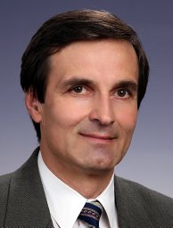 György Balla合作论文数Faculty of Medicine, University of Debrecen;H-4012 Debrecen, Hungary;Faculty of Medicine, University of Debrecen20
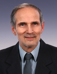 Jozsef Balla合作论文数Div. of Nephrology, Faculty of Medicine, University of Debrecen;Department of Internal Medicine, University of Debrecen;Department of Internal Medicine, Internal Medicine Clinic, University of Debrecen Clinical Centre20
 Méhes Gábor合作论文数Department of Pathology, Faculty of Medicine, University of Debrecen, Debrecen, 4032,
Hungary11
Méhes Gábor合作论文数Department of Pathology, Faculty of Medicine, University of Debrecen, Debrecen, 4032,
Hungary11 Hendrik Zoltán合作论文数9
 David Petho合作论文数Kalman Laki Doctoral Sch Biomed & Clin Sci, Univ Debrecen8Gáll Tamás合作论文数Medical and Health Science Centre, University of Debrecen6
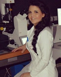 Katalin Éva Sikura合作论文数ELKH UD Vasc Biol & Myocardial Pathophysiol Res G, Hungarian Acad Sci5
 Viktória Jeney合作论文数Medical and Health Science Center, University of Debrecen5Melinda Oros合作论文数Laboratory of Neurobehavior Genetics, The Nathan S. Kline Institute for Psychiatric Research5Abolfazl Zarjou合作论文数Department of Medicine, University of Debrecen5Niké Posta合作论文数4
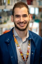 Andreas Patsalos合作论文数Johns Hopkins All Childrens Hosp, Johns Hopkins Univ4
 Eva Csosz合作论文数Medical and Health Science Center, University of Debrecen4Beke Lívia合作论文数4Matt Whiteman合作论文数Institute of Biomedical and Clinical Science, College of Medicine and Health, University of Exeter4
 Gergo Kallo合作论文数4
 Szilard Poliska合作论文数Department of Biochemistry and Molecular Biology, Faculty of Medicine, University of Debrecen4
 Csaba D. Toth合作论文数Department of Mathematics, California State University Northridge;Department of Computer Science, Tufts University4
 Laszlo Nagy合作论文数University of Debrecen;Institute for Fundamental Biomedical Research, Johns Hopkins All Children’s Hospital;Johns Hopkins Center for Metabolic Origins of Disease;Division of Endocrinology, Diabetes and Metabolism, Department of Medicine, School of MedicineJohns Hopkins University;Department of Biological Chemistry, School of Medicine, Johns Hopkins University3
 Anupam Agarwal合作论文数Harvard-Thorndike Laboratory and the Department of Medicine, Beth Israel Deaconess Medical Center and Harvard Medical School;Cardiovascular and Metabolic Division, GlaxoSmithKline Pharmaceuticals3
 Peter Nagy合作论文数Department of Molecular Immunology and Toxicology, National Institute of Oncology, Hungary;Debrecen University;Department of Pathology, University of Otago3Mark E. Wood合作论文数3Mónika Nyitrai合作论文数2
 Zsolt Combi合作论文数2Roberta Torregrossa合作论文数2Erzsébet Zavaczki合作论文数2
 Paolo Arosio合作论文数Biochemical Engineering Laboratory, Institute for Chemical and Bioengineering, Department of Chemistry and Applied Biosciences, ETH Zurich2Attila Fintha合作论文数2
 Tamas Szerafin合作论文数2Annamaria Nagy合作论文数2
 Miguel Che Parreira Soares合作论文数Instituto Gulbenkian de Ciencia2Becs Gergely合作论文数2Péter Sótonyi合作论文数2
 Peter Gergely合作论文数Second Department of Medicine, Semmelweis University2Agnes Gyetvai合作论文数Medical and Health Science Center Laboratory of Immunology, University of Debrecen2
 Eva Katona合作论文数University of Szeged;Department of Image Processing and Computer Graphics2Ágnes Szappanos合作论文数2
 John W. Eaton合作论文数Departments of Medicine, and Pharmacology & Toxicology, School of Medicine, University of Louisville2Ibolya Furtos合作论文数2Sándor Olvasztó合作论文数1Eszter Zöld合作论文数1Beáta Nagy合作论文数1Anita Vasas合作论文数1
 Frida Rosenblum合作论文数1József Tözsér合作论文数1Hs Jacob合作论文数1Balazs Lukucz合作论文数1Szabolcs Balla合作论文数1Dorottya Garai合作论文数1




