ADC is a potential post treatment imaging biomarker in colorectal liver metastasis however measurements are affected by respiratory motion. This is compounded by increased statistical uncertainty in ADC measurement with decreasing tumour volume. In this prospective study we applied a retrospective motion correction method to improve the image quality of 15 tumour data sets from 11 patients. We compared repeatability of ADC measurements corrected for motion artefact against non-motion corrected acquisition of the same data set. We then applied an error model that estimated the uncertainty in ADC repeatability measurements therefore taking into consideration tumour volume. Test-retest differences in ADC for each tumour, was scaled to their estimated measurement uncertainty, and 95% confidence limits were calculated, with a null hypothesis that there is no difference between the model distribution and the data. An early post treatment scan (within 7 days of starting treatment) was acquired for 12 tumours from 8 patients. When accounting for both motion artefact and statistical uncertainty due to tumour volumes, the threshold for detecting significant post treatment changes for an individual tumour in this data set, reduced from 30.3% to 1.7% (95% limits of agreement). Applying these constraints, a significant change in ADC (5th and 20th percentiles of the ADC histogram) was observed in 5 patients post treatment. For smaller studies, motion correcting data for small tumour volumes increased statistical efficiency to detect post treatment changes in ADC. Lower percentiles may be more sensitive than mean ADC for colorectal metastases.
Apparent Diffusion Coefficient (ADC) is a potential quantitative imaging biomarker for tumour cell density and is widely used to detect early treatment changes in cancer therapy. We propose a strategy to improve confidence in the interpretation of measured changes in ADC using a data-driven model that describes sources of measurement error. Observed ADC is then standardised against this estimation of uncertainty for any given measurement. 20 patients were recruited prospectively and equitably across 4 sites, and scanned twice (test-retest) within 7 days. Repeatability measurements of defined regions (ROIs) of tumour and normal tissue were quantified as percentage change in mean ADC (test vs. re-test) and then standardised against an estimation of uncertainty. Multi-site reproducibility, (quantified as width of the 95% confidence bound between the lower confidence interval and higher confidence interval for all repeatability measurements), was compared before and after standardisation to the model. The 95% confidence interval width used to determine a statistically significant change reduced from 21.1 to 2.7% after standardisation. Small tumour volumes and respiratory motion were found to be important contributors to poor reproducibility. A look up chart has been provided for investigators who would like to estimate uncertainty from statistical error on individual ADC measurements.
This study describes post-processing methodologies to reduce the effects of physiological motion in measurements of apparent diffusion coefficient (ADC) in the liver. The aims of the study are to improve the accuracy of ADC measurements in liver disease to support quantitative clinical characterisation and reduce the number of patients required for sequential studies of disease progression and therapeutic effects. Two motion correction methods are compared, one based on non-rigid registration (NRA) using freely available open source algorithms and the other a local-rigid registration (LRA) specifically designed for use with diffusion weighted magnetic resonance (DW-MR) data. Performance of these methods is evaluated using metrics computed from regional ADC histograms on abdominal image slices from healthy volunteers. While the non-rigid registration method has the advantages of being applicable on the whole volume and in a fully automatic fashion, the local-rigid registration method is faster while maintaining the integrity of the biological structures essential for analysis of tissue heterogeneity. Our findings also indicate that the averaging commonly applied to DW-MR images as part of the acquisition protocol should be avoided if possible.
Many medical image analysis algorithms make assumptions concerning the image formation process, the structure of the intensity histogram, or other statistical properties of the input data. Application of such algorithms to image data that do not fit these assumptions will produce unreliable results. This paper describes a technique for the automatic identification of images that do not have histogram structure consistent with that expected. The approach is based upon a component analysis followed by statistical testing. Experiments validate its use in the identification of quantisation problems and unexpected image structure. It is intended that this test will form one component of a quality control assessment, to aid in the use of sophisticated statistical image analysis software by non-expert users.
This work assesses the reproducibility of ADC measurement for data acquired across several clinical sites within IMI QuIC-ConCePT project. We present a model for expected ADC reproducibility which takes account of the initial ADC distribution within tumours and the volume of measurement. We show that the accuracies of ADC currently achieved are on average 7.5% but that better measurements are generally associated with increasing the measurement volume. Our analysis generates methods which are capable of predicting the reproducibility of individual tumours, and is therefore suitable for guiding the region of interest selection process. Overall performance of reproducibility for the ‘on scanner’ averaged acquisition (protocol A) is found to be currently insufficient for detection of change in individuals, but recent results regarding the expected improvement using ‘off scanner’ averaging (protocol B) would suggest the possibility of patient specific adjustment of therapy. Real tumour data for protocol B data is now required in order to confirm this expected benefit and such data is planned to be acquired as part of the upcoming BOS2 trial.
Purpose: To construct an appropriate phantom for quality control use in diffusion‐weighted imaging (DWI), to establish ground truth for measurement of apparent diffusion coefficient (ADC) and to characterize measurement linearity across a relevant physiological range of ADC. Methods: Aqueous solutions containing the polymer polyvinylpyrrolidone (PVP) were mixed at concentrations of 0, 10, 20, 30, 40 and 50% by mass PVP. These solutions were placed in 20 mL vials, arranged in concentric inner and outer circles, with a central water vial, and were fixed in a spherical phantom with a diameter of 194 mm, designed to fit into commercially‐available MRI head coils. Two prototype phantoms were constructed, and underwent inter‐site comparison in the US and EU. The phantoms were filled with an ice‐water bath to ensure stable temperature; 0 °C temperature was verified by use of a thermocouple before and after scans. The phantoms were scanned using b‐values of 0, 500 and 900 s/mm 2 at several sites, using coronal and/or axial orientations and scan planes. Results: ADC values ranged from 0.12 to 1.12 × 10 ‐3 mm 2 /s, and exhibited a high degree of reproducibility across different scanners and imaging sites (coefficient of variations (CoV) ranged from 1.1 to 2.2% for 0 to 40% PVP, with 50% PVP at 11.3%). Little difference in ADCs was seen between inner and outer ring vials of the same PVP concentration (average CoV< 5% across vials, 10.3% for 50% PVP). Conclusion: The range of ADCs covers a relevant physiological range, most notably in brain white matter. The ADCs of water vials were in excellent agreement with literature values of the diffusion coefficient of water at 0 °C (1.1 × 10 ‐3 mm 2 /s). The phantom provides a much needed quality control tool for DWI, and provides ground truth with the diffusion coefficient of water at 0 °C.
Background: In previous work, the authors described a software package for the digitisation of 3D landmarks for use in geometric morphometrics. In this paper, we describe extensions to this software that allow semi-automatic localisation of 3D landmarks, given a database of manually annotated training images. Multi-stage registration was applied to align image patches from the database to a query image, and the results from multiple database images were combined using an array-based voting scheme. The software automatically highlights points that have been located with low confidence, allowing manual correction.Results: Evaluation was performed on micro-CT images of rodent skulls for which two independent sets of manual landmark annotations had been performed. This allowed assessment of landmark accuracy in terms of both the distance between manual and automatic annotations, and the repeatability of manual and automatic annotation. Automatic annotation attained accuracies equivalent to those achievable through manual annotation by an expert for 87.5% of the points, with significantly higher repeatability.Conclusions: Whilst user input was required to produce the training data and in a final error correction stage, the software was capable of reducing the number of manual annotations required in a typical landmark identification process using 3D data by a factor of ten, potentially allowing much larger data sets to be annotated and thus increasing the statistical power of the results from subsequent processing e. g. Procrustes/principal component analysis. The software is freely available, under the GNU General Public Licence, from our web-site (www.tina-vision.net).
Our aim is to develop methodologies for the determination of ADC, suitable for use in patient management and drug trials. In this paper we demonstrate improvements in ADC measurement which at the same time maintain the biological structures essential for analysis of tissue heterogeneity. Two motion correction methods are compared, one based upon non-rigid registration using freely available open source algorithms and the other a purpose designed local rigid registration. Performance of these methods is evaluated using metrics computed from regional ADC histograms. While the nonrigid registration method has the advantages of being applicable on the whole volume and in a fully automatic fashion, the local rigid registration method is much faster and also provides advantages with regard to data smoothness by avoiding interpolation and sub-sampling. Our study also shows that the averaging commonly applied to DW-MR images as part of the acquisition protocol should be avoided if at all possible.
The aim of this document is to define the quality assurance (QA) methods for scanners using the ice-water phantom and the corresponding software developed within the IMI QuIC-ConCePT project, for the upcoming ‘BOS2’ trials. The outlined methods are what we consider to constitute the best use of currently available data following our recent papers [1] [2] in which we studied ADC (apparent diffusion coefficient) measurements corresponding to five phantom tubes obtained from four scanners located at four different sites. Scanners are required to gain approval based on their phantom data quality in order to proceed to the clinical trial. The idea is to obtain one phantom data-set from each site before site activation and one week prior to clinical scans. Scanners used in our recent study resulted in about 6% average measurement variability in healthy liver data. These results and those from phantom studies, have been used here to set criteria for QA pass/fail. To approve the quality of each new scanner, several metrics of image quality will be automatically extracted from phantom data-sets. A simple statistical analysis then provides a three step procedure for the pass or fail decision. The full process is described below with example data. For the current QA we concentrate on signal to noise and absolute quantitation, ignoring issues of fat suppression and inhomogeneity which will need to be addressed separately (where possible).
BACKGROUND:The introduction and statistical formalisation of landmark-based methods for analysing biological shape has made a major impact on comparative morphometric analyses. However, a satisfactory solution for including information from 2D/3D shapes represented by 'semi-landmarks' alongside well-defined landmarks into the analyses is still missing. Also, there has not been an integration of a statistical treatment of measurement error in the current approaches.RESULTS:We propose a procedure based upon the description of landmarks with measurement covariance, which extends statistical linear modelling processes to semi-landmarks for further analysis. Our formulation is based upon a self consistent approach to the construction of likelihood-based parameter estimation and includes corrections for parameter bias, induced by the degrees of freedom within the linear model. The method has been implemented and tested on measurements from 2D fly wing, 2D mouse mandible and 3D mouse skull data. We use these data to explore possible advantages and disadvantages over the use of standard Procrustes/PCA analysis via a combination of Monte-Carlo studies and quantitative statistical tests. In the process we show how appropriate weighting provides not only greater stability but also more efficient use of the available landmark data. The set of new landmarks generated in our procedure ('ghost points') can then be used in any further downstream statistical analysis.CONCLUSIONS:Our approach provides a consistent way of including different forms of landmarks into an analysis and reduces instabilities due to poorly defined points. Our results suggest that the method has the potential to be utilised for the analysis of 2D/3D data, and in particular, for the inclusion of information from surfaces represented by multiple landmark points.
The aim of this study was to investigate the extent to which measurements made in an ADC (apparent diffusion coefficient) calibration phantom could be shown to correlate with accuracies of measurements made in normal liver. Ideally, a strong correlation might be used in the future in order to perform an assessment of scanner performance based entirely on the phantom. Several metrics of image quality were automatically extracted from the phantom and ROI’s (regions of interest) were identified manually for human livers. Analysis was performed in two stages, firstly looking at correlations between metrics measured in the phantom in order to gain an understanding of reliability, and secondly looking at the reproducibility of human data. Correlations in phantom measurements help to identify which of the possible summary statistics are likely to be best measured and meaningful. This has allowed us to refine our definitions of the parameters which are most useful for summarising phantom data. It has also allowed us to identify phantom design weaknesses which might be improved. Repeatability in humans sets a limit for realistic clinical performance and also highlights potential problems in current methodology.
When multiple scanners are used to acquire diffusion MR image data from the same patient (even on the same day), there is no guarantee that identical settings will result in comparable images of a specific organ and region. Despite this, for clinical use, any software developed to compute the apparent diffusion coefficient (ADC) must be expected to give equivalent results. What is needed is a phantom study with appropriate design which supports a calibration, so that appropriate settings for equivalent diffusion measurements can be defined among different scanners. Ideally the calibration process needs to be fully automatic, so that it can be used by non-experts in a clinical setting. We intend to developed software which automatically locates five cylinders in an ice water phantom (designed for the QuIC-ConCePT project) and then measures the different diffusion values in the cylinders. The location algorithm uses an object recognition process which culminates in robust Likelihood estimation of position and orientation, computed using probabilistic Hough Transforms. This process estimates cylinder locations to an accuracy of a few pixels, even in the presence of field inhomogeneity and significant spatial distortion. Our assessment of performance includes quantification of possible errors due to; data inaccuracy, distortion due to poor shimming, signal to noise, field inhomogeneity and image clutter (i.e. ice). Results indicate that reliable localisation can be obtained using these methods for realistic clinical settings.
This paper addresses the problem of silhouette-based human action modelling and recognition independently of the camera point of view. Action recognition is carried out by comparing a 2D motion tem- plate, built from observations, with learned models of the same type cap- tured from a wide range of viewpoints. All these 2D motion templates, are projected into a new subspace by means of the Kohonen Self Orga- nizing feature Map (SOM). A specific SOM is trained for every action, grouping viewpoint (spatial) and movement (temporal) in a principal manifold. This approach enables the interpolation of data dif- ferent and, at the same time, to establish motion correspon- dences between viewpoints without considering a mapping to a complex 3D model. Every new 2D motion template gives a distance to the map, related to the probability that motion feature belongs to that particular action. Action recognition is accomplished by a Maximum Likelihood (ML) classifier over all specific-action SOMs. We demonstrate this ap- proach on two challenging video sets: one based on real actors making 11 complex actions and another one based on virtual actors performing 20 dierent actions.
Aims The aim of this document is to make explicit the various factors which would be expected to influence a quantitative assessment of diffusion measurement changes for clinical assessment. In order to do this we must make some assumptions regarding the nature of the study, the data acquisition and the analysis. We will assume; • That quantitative measurements will be in the form of a regional averaged ADC, and that a method which allows reproducible identification of the required region (typically of 100 voxels of independent ADC measurements) in biological data has been specified. • That the acquisition is to be made on a scanner using a standard protocol, with matched phantom data. • That the expected percentage change in measured ADC following treatment can be predicted from prior understanding of the study. For convenience we will group the expected level of change into 3 classes, selected according to results found in the literature to be typical of what might be required for future studies 1. (Type A) A study seeking to establish no observable difference in ADC (for example a fasting study) which requires the most precise estimates available (10% of ADC). (Type B) A study seeking to confirm a typical change in biological behaviour which is expected to be significant (and of known direction) but not complete (30% of ADC). (Type C) A study where the expected response is so strong that all structure in tissue is destroyed and the resulting diffusion coefficient is equivalent to water (50% of ADC). Changes of ADC value beyond this are expected to be accompanied by such drastic changes in anatomical MR images that using a diffusion measurement to assess change is unnecessary. The intention now is to be able to claim that for a given class of study (A,B or C), a percentage level change of the required amount will be measureable in an individual subject using a specific MR scanner. We must therefore be able to infer the expected level of accuracy from data obtained from the phantom. We will do this by considering fractional changes in ADC precision which arise from the various forms of MRI degradation.
Our intention is to utilise the statistical methods we have developed, for analysis of shape data with an-isotropic measurement covariances, as a tool to monitor manual and automatic placement of landmarks. We wish to make quantitative assessments of measurement accuracy, and also identify potential errors in markup at the level of ≤ 5% of the data sample. This document outlines the mechanism we have used to extend 2D shape rotation analysis, and the extraction of corrected anisotropic measurement covariances (Tina Memo 2010-009), to 3D. The methods are demonstrated in the analysis of 3D mouse mandible data, both as a test of the theory/software implementation and as an illustration of use for the identification of outlier landmarks.
We propose a shape analysis system based upon the description of landmarks with measurement covariance, which extends statistical linear modelling processes to 'pseudo landmarks' for scientific studies. We discuss the properties of our approach and how measurement covariances can be considered characteristic of the local shape. Our formulation includes corrections for parameter bias, induced by the degrees of freedom within the linear model. The method has been implemented and tested on measurements from fly wing, hand and face data. We use these data to explore possible advantages and disadvantages over the use of standard Procrustes/PCA analysis. In the process we show how appropriate weighting provides more efficient use of the available landmark data(1).
In this paper we propose a method for automatic placement of landmark points in 3D volumes, for use in morphological studies, which addresses the issue of quantitative estimation of measurement error. We are given a sample 3D volume of image slices (and a number of manually annotated landmark points) and develop an algorithm that is capable of estimating landmark locations in a similar but novel 3D volume. Problems are found which arise from the local minima generated when constructing likelihood functions using shifted noisy image patches, which must be addressed during both localisation and covariance estimation. The method is tested using Monte-Carlo in order to evaluate the quantitative validity of error estimates.
Conventional methods for shape analysis, based upon Procrustes and PCA, seem incapable of dealing with ‘non-landmark’ features, meaning measured positions not associated with well defined locations. This is due to an assumption of homogeneous errors, associated with an attempt to extract linear models with biologically meaningful descriptions. We propose a shape analysis system based upon the description of landmarks with measurement covariance which will extend the modelling process to ‘pseudo-landmarks’ such as boundaries and surfaces. We discuss the properties of our approach and how these covariances can be considered characteristic of the local shape. The method has been implemented and tested on measurements from fly wing, hand and face data. We use these data to explore possible advantages and disadvantages over the use of Procrustes/PCA.
This paper describes a body of multicamera humanaction video data with manually annotated silhouette datathat has been generated for the purpose of evaluatingsilhouette-based human action recognition methods. Itprovides a realistic challenge to both the segmentationand human action recognition communities and can act asa benchmark to objectively compare proposed algorithms.The public multi-camera, multi-action dataset is animprovement over existing datasets (e.g. PETS, CAVIAR,soccerdataset) that have not been developed specificallyfor human action recognition and complements otheraction recognition datasets (KTH, Weizmann, IXMAS,HumanEva, CMU Motion). It consists of 17 action classes,14 actors and 8 cameras. Each actor performs an actionseveral times in the action zone. The paper describes thedataset and illustrates a possible approach to algorithmevaluation using a previously published action simplerecognition method. In addition to showing an evaluationmethodology, these results establish a baseline for otherresearchers to improve upon.
 Edwin Hancock合作论文数Department of Computer Science, University of York;Beijing Advanced Innovation Centre for Big Data and Brain Compuring, Beihang University26
 Neil Thacker合作论文数Division of Informatics, Imaging and Data Sciences, School of Health Sciences, Faculty of Biology, Medicine and Health, The University of Manchester13
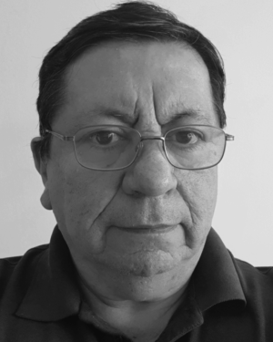 Sergio A. Velastin合作论文数Department of Computer Science and Engineering, Universidad Carlos III de Madrid Leganés7
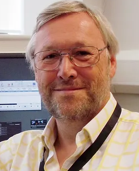 Alan Jackson合作论文数University of Manchester6David L. Morris合作论文数4Francisco Martinez-Contreras合作论文数3R Pathak合作论文数3Paul Bromiley合作论文数Division of Informatics, Imaging and Data Sciences, School of Health Sciences, Faculty of Biology, Medicine and Health, The University of Manchester3
 Paolo Remagnino合作论文数Department of Computer Science, Durham University;Wolfson Research Institute for Health and Wellbeing, Durham University3Nandita M. Desouza合作论文数3
 T. J. Ellis合作论文数Digital Imaging Research Centre;Kingston University;Digital Imaging Research Centre, Kingston University3
 Carlos Orrite合作论文数Centro Politecnico Superior, University of Zaragoza3
 Elias Herrero Jaraba合作论文数Aragon Institute for Engineering Research, University of Zaragoza3
 Stefan Klein合作论文数2D. Morris合作论文数2
 Diethard Tautz合作论文数Department for Evolutionary Genetics, Max Planck Institute for Evolutionary Biology2Anja C. Schunke合作论文数2
 Jean-Marie Guyader合作论文数2Joost Kuijer合作论文数1B. Van Beers合作论文数1Jingduo Tian合作论文数1Atsushi Shimada合作论文数1
 Alastair J. Martin合作论文数Dept Radiol & Biomed Imaging, Univ Calif San Francisco1Heerschap Arend合作论文数1
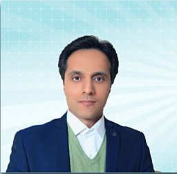 Amiri Houshang合作论文数1
 Philippe Garteiser合作论文数1
 Michael A Boss合作论文数Electromagnetics Division, National Institute of Standards and Technology1
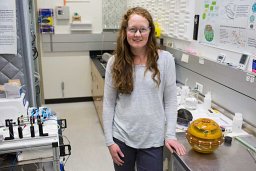 Keenan Kathryn E合作论文数1
 Mukherjee Pratik合作论文数1
 David Collins合作论文数The Royal Marsden NHS Foundation Trust;Division of Radiotherapy and Imaging, Institute of Cancer Research1
 John Waterton合作论文数Division of Informatics, Imaging & Data Sciences, School of Health Sciences, Faculty of Biology, Medicine and Health, University of Manchester1
 Antonio Robles-Kelly合作论文数School of Information Technology, Deakin University1
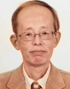 Rin-Ichiro Taniguchi (谷口倫一郎)合作论文数Laboratory For image and Media Understanding, Kyushu University1
 Will Smith合作论文数Department of Computer Science, University of York1
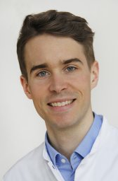 Dennis M Hedderich合作论文数Institut fur Diagnostische und Interventionelle Neuroradiologie, Neuro-Kopf-Zentrum, Technical University of Munich1Sabrina Doblas合作论文数1Thorsten Persigehl合作论文数1
 Thomas Chenevert合作论文数Department of Radiology, Medical School, University of Michigan;Michigan Institute for Data & AI in Society, University of Michigan;Rogel Cancer Center, University of Michigan1Sanchit Singh合作论文数1Mark P Saunders合作论文数1




