Objective: Temporal lobe epilepsy (TLE) is the most common focal epilepsy syndrome and remains medically refractory in a substantial proportion of patients. Dual pathology, defined as the coexistence of hippocampal sclerosis with an additional neocortical lesion, is an important cause of surgical failure when not adequately recognized. This study aimed to evaluate the clinical characteristics and postoperative seizure outcomes of patients with dual pathology compared with those with isolated mesial temporal sclerosis. Methods: We retrospectively reviewed 125 patients who underwent surgery for TLE between January 2005 and February 2023. Thirty-one patients with dual pathology, defined as hippocampal sclerosis accompanied by a neocortical tumor, were included. A control group consisted of 34 age-matched patients with isolated mesial temporal sclerosis. Clinical features, seizure characteristics, surgical procedures, postoperative outcomes assessed using the Engel classification, and complications were analyzed. Results: The mean age was similar between the dual pathology and control groups. However, the age at seizure onset was significantly later in patients with dual pathology (26.5 +/- 15.9 years vs. 9.2 +/- 7.8 years; p<0.001). Generalized tonic-clonic seizures were more frequent in the dual-pathology group, whereas focal seizures with impaired awareness predominated in patients with isolated mesial temporal sclerosis. Engel class I seizure freedom was achieved in 61.3% of patients with dual pathology and in 67.6% of controls, with no significant difference between groups. Postoperative complication rates were comparable. Conclusion: Despite differences in seizure characteristics and age at seizure onset, postoperative seizure outcomes in patients with dual pathology were comparable to those in patients with isolated mesial temporal sclerosis when both the mesial temporal structures and the associated neocortical lesion were adequately resected. Dual pathology should be considered in patients with TLE who present with mesial temporal sclerosis accompanied by a neocortical tumor, particularly in those with a relatively late age at seizure onset.
Seizure semiology and electroencephalograph (EEG) are very important for determining seizure type, hemisphere lateralization, or localization. Clinical symptoms of focal seizures, as well as findings at the onset or end of a focal to bilateral tonic–clonic seizure (FBTCS), are highly informative for lateralization. This study aimed to investigate the relationship of asymmetric last clonic jerk in patients with temporal or extratemporal lobe epilepsy with pathologies, localization, lateralization, or other semiological findings detected in neuroimaging or neuro psychometric tests and its positive predictive value for the detection of hemisphere lateralization based on seizure onset ictal EEG activation. 44 patients with asymmetric last clonic jerks (aLCJ) who were followed up in our VEM unit were randomized 1:1 with epilepsy patients without. In patients with ipsilateral automatism and contralateral posture or gustatory and olfactory hallucinations aLCJ was less or absent. In patients with unilateral tonic activity, aLCJ was more common. The positive predictive value of aLCJ for ictal EEG activation lateralization was 86.36%. In conclusion, asymmetric last clonic beat is valuable for lateralization of FBTCS and should be considered. Its presence strongly and reliably lateralizes to the side of seizure onset.
Video-elektroensefalografik izleme (VEM), nöbetlerin davranışsal ve elektroensefalografik (EEG) aktivite süresini objektif olarak değerlendirmek için altın standarttır. Bugüne kadar nöbet süreleri farklı hasta gruplarında klinik olarak veya EEG ile değerlendirilmiştir. Bu çalışma, VEM ile değerlendirilen fokal başlangıçlı bilateral tonik-klonik nöbet (FBTKN) tanısı olan epilepsi hastalarında, demografik, klinik ve nörogörüntüleme bulguları ile nöbet süresi arasındaki ilişkiyi ortaya koymayı amaçlamaktadır. Nisan 2005 ve Ocak 2024 tarihleri arasında merkezimizde FBTKN tanısı alan rastgele seçilmiş 58 hastanın tıbbi öyküleri, nörogörüntülemeler, VEM kayıtlarından elde edilen klinik ve iktal EEG bulguları retrospektif olarak analiz edildi. En kısa fokal aktivite frontal lob epilepsisinde ve en uzun jeneralize aktivite parietooksipital lob epilepsisindeydi. Fokal aktivite süresi frontal ve eksternal kapsül lokalizasyonlu lezyonlarda daha kısa, mezial temporal lokalizasyonda daha uzundu. Fokal aktivite süresi meziyal temporal sklerozda daha uzundu. Jeneralize aktivite süresi ensefalomalazi ve polimikrogiride daha uzun, kortikal displazide ise daha kısaydı. Lezyonlara kortikal atrofi eşlik ettiğinde fokal aktivite süresi daha kısaydı. Anti nöbet ilaç türü ile nöbet süreleri arasında herhangi bir korelasyon yoktu. Nöbet süreleri semiyolojik bulgulara, radyolojik özelliklere ve epilepsi sendromlarına göre değişebilir. Farklılıkları klinisyen için epilepsi sendromu türü, status olasığı, semiyolojik eşlik eden bulgular hakkında bilgi verici olabilir.
OBJECTIVE:The present study was aimed at investigating the effects of anti-seizure medications (ASMs), patient demographic characteristics, and the seizure type and frequency on the development of congenital malformations (CMs) in the infants of pregnant women with epilepsy (PWWE).METHODS:PWWE followed up at the neurology outpatient clinic of 21 centers between 2014 and 2019 were included in this prospective study. The follow-up of PWWE was conducted using structured, general pregnant follow-up forms prepared by the Pregnancy and Epilepsy Study Committee. The newborns were examined by a neonatologist after delivery and at 1 and 3 months postpartum.RESULTS:Of the infants of 759 PWWE, 7.2% had CMs, with 5.6% having major CMs. Polytherapy, monotherapy, and no medications were received by 168 (22.1%), 548 (72.2 %), and 43 (5.7 %) patients, respectively. CMs were detected at an incidence of 2.3% in infants of PWWE who did not receive medication, 5.7% in infants of PWWE who received monotherapy, and 13.7% in infants of PWWE who received polytherapy. The risk of malformation was 2.31-fold (95% confidence interval (CI): 1.48-4.61, p < .001) higher in infants of PWWE who received polytherapy. Levetiracetam was the most frequently used seizure medication as monotherapy, with the highest incidence of CMs occurring with valproic acid (VPA) use (8.5%) and the lowest with lamotrigine use (2.1%). The incidence of CMs was 5% at a carbamazepine dose <700 mg, 10% at a carbamazepine dose ≥700 mg, 5.5% at a VPA dose <750 mg, and 14.8% at a VPA dose ≥750 mg. Thus the risk of malformation increased 2.33 times (p = .041) in infants of PWWE receiving high-dose ASMs.SIGNIFICANCE:Birth outcomes of PWWE receiving and not receiving ASMs were evaluated. The risk of CMs occurrence was higher, particularly in infants of PWWE using VPA and receiving polytherapy. The incidence of CMs was found to be lower in infants of PWWE receiving lamotrigine.
Objective: Status epilepticus (SE) is a serious neurological emergency that can has high morbidity and mortality rates and requires prompt diagnosis and treatment. There are different etiologies and the prognosis varies multifactorially. The aim of this study was to reveal the etiological causes, clinical features and mortality rates of patients diagnosed with SE at our center.Methods: The records of 234 patients with a diagnosis of SE over the age of 18 who were followed up and treated at our center between 01.01.2015-01.01.2022 were evaluated retrospectively. Using the hospital information operating system database, we identified people hospitalized with an International Classification of Diseases 10th Revision code G41 for SE as the primary diagnosis. Demographic information, clinical characteristics, and discharge results were obtained from medical records.Results: One hundred-twenty (51.3%) female and 114 (48.7%) male patients were evaluated. The top 3 most common etiologic causes were: discontinuation of anti-seizure treatments without advice (n=82), cerebrovascular events (n=50), and meningitis or encephalitis (n=39). Motor seizures were detected in 183 (78.2%) patients, and non-motor seizures were detected in 51 (21.8%) patients. Seizures were suppressed by first-line treatment in 24 patients and by second -line treatments in 135 patients. Seventy-five patients whose seizures could not be suppressed were accepted as refractory SE and 9 died. The mean age of all patients was 55, and 63 of the patients died.Conclusion: In this study, clinical and demographic features, the etiological causes and in the hospital mortality rates of SE followed in a single center in the Turkish population were determined. The most common causes of patients diagnosed with SE were discontinuation of anti-seizure treatments without our recommendation, cerebrovascular diseases and central nervous system infections, respectively. In our center, no relationship was found between age and mortality. The in-hospital mortality rate was 3.9% for all patients (n=234) and 12% for patients with refractory SE (n=75).
Introduction. Ulegyria results from perinatal hypoxic-ischemic brain injury in term infants. The specific mushroom-shaped configuration of ulegyria results from small atrophic circumvolutions at the bottom of a sulcus underlying an intact gyral apex. Clinically, ulegyria is generally associated with epilepsy. Here, we aimed to delineate the characteristics of patients with ulegyria and the epileptic seizures they experience. Material and methods. Medical records including radiology and pathology reports, video-electroencephalographic (EEG) analysis, operative notes, hospital progress and outpatient clinic notes were reviewed retrospectively in a total of 10 ulegyria patients. Results. Patients ages ranged between 24 and 58 years (mean, 32 ± 9.8 years). Past medical history was confirmed for neonatal asphyxia in 2 (20%). Neurological examination was remarkable for spastic hemiparesis in 1 (10%) patient with perisylvian ulegyria and for visual field deficits in 2 patients (20%) with occipital ulegyria. Ulegyria most commonly involved the temporoparietal region (n = 5, 50%) followed by the perisylvian area (n = 2, 20%). Except the one with bilateral perisylvian ulegyria, all patients had unilateral lesions (n = 9, 90%). Hippocampal sclerosis accompanied ulegyria in 2 patients (20%). All patients experienced epileptic seizures. Mean age at seizure onset was 8.8 ± 5.4 years (range, 2-20 years). Interictal scalp EEG and EEG-video monitoring records demonstrated temporoparietal and frontotemporal activities in 5 (50%) and 2 (20%) patients, respectively. The seizures were successfully controlled by antiepileptic medication in 8 patients (n = 8, 80%). The remaining 2 patients (%20) with concomitant hippocampal sclerosis required microsurgical resection of the seizure foci due to medically resistant seizures. Discussion. Ulegyria is easily recognized with its unique magnetic resonance imaging characteristics and clinical presentation in the majority of cases. It is highly associated with either medically resistant or medically controllable epileptic seizures. The treatment strategy depends on the age at onset and extends of the lesion that has a significant impact on the severity of the clinical picture.
Epilepsy is a chronic neurological disease characterized by spontaneous and repeative seizures resulting in abnormal and excessive electrical discharge in cortical neurons [1]. In adults, epilepsy is the most common neurological disease after cerebrovascular diseases. It is important to be careful about the distinctive diagnosis of cases that are similar to clinical similarity but not epileptic feature [2]. Generally nonepileptic seizures (pseudoseizure); collected under two headings, depending on psychogenic and physiological reasons . Psychogenic nonepileptic seizures (PNES) is more common and the distinctive diagnosis is more difficult. PNES cases constitute 15-30% of patients who are refractory to pharmacological treatment who apply to epilepsy centers [3]. In a study of Bora et al, there was a 67 PNES cases in the case of 440 resistant epilepsy (15.2%) and all of these patients have been started the antiseizure medications in earlier centers reported [4].
BACKGROUND:Gelastic seizures are extremely rare, short-lasting, unprovoked, and uncontrollable laughing attacks. We conducted this retrospective evaluation to determine whether these symptoms, manifesting in different forms, such as cheerful laughter, laughing, smiling, and sobbing had any value in terms of etiology or localization.METHODS:A total of 31 patients who exhibited bouts of laughing or crying and who were under follow-up between 2000 and 2019 at tertiary epilepsy centers were included in the study. Laughing seizures were divided into three groups in terms of semiology (i.e., laughter with mirth, laughter without mirth, and smile). Dacrystic seizures were accompanied by some gelastic seizures and were divided into two groups in terms of semiology (i.e., weeping loudly [motor and voice-sobbing] and crying).RESULTS:Of the 27 patients with laughing seizures, 12 had seizures that manifested with smiling, 7 had seizures that manifested with laughing and mirth, and 8 had seizures that manifested with laughter without mirth. Dacrystic-gelastic seizures were observed in four patients, among whom 2 patients had crying and laughter without mirth and 2 patients had weeping loudly and laughter without mirth episodes.CONCLUSION:Gelastic and dacrystic seizures often suggest hypothalamic hamartomas, in the literature. This rare ictal behavior can originate from different cortical locations and lesions of a different nature. However, we found that gelastic seizures with smiling were a more homogenous group with regard to location in the temporal lobe, which we aimed to show by evaluating the patients included in this study.
Introduction: Epileptic seizures occur in approximately 35%–40% of patients with cerebral venous thrombosis (CVT). The relationship between parenchymal lesions and epileptic seizures in CVT has been investigated, but the most associated types of parenchymal lesions have not been determined. This study, therefore, aimed to identify high-risk groups. Methods: A total of 159 patients were diagnosed as having CVT between 2015 and 2021 at our tertiary center. The risk factors for epileptic seizures after CVT were determined. Results: A total of 159 patients who were diagnosed with having CVT, 109 (68.5%) females and 50 (31.5%) males, were included in this study. The mean ages of the women and men were 41.20 ± 14.15 years and 43.60 ± 16.30 years, respectively. We found that superior sagittal sinus involvement (P = 0.019), sigmoid sinus involvement (P = 0.010), cortical vein involvement (P < 0.001), parenchymal lesion (P < 0.001), and the postpartum period (P = 0.003) increased the risk of epileptic seizures. When the significant variables associated with epileptic seizures in the patients were analyzed using binary logistic regression, the most significant variable was found to be the presence of parenchymal lesions. Conclusion: We found that the most significant variable for epileptic seizures after CVT was parenchymal lesions. Juxtacortical hemorrhages and nonhemorrhagic venous infarcts were the most common causes of epileptic seizures. CVT is a heterogeneous group of diseases caused by multiple aetiologies and may show ethnic and racial differences. For this reason, more precise information can be obtained with multi-center prospective studies in our population.
Reproductive functional disorders and endocrine disorders are common in epileptic patients, particularly in patients with temporal lobe epilepsy (TLE). Pituitary size has been measured in patient populations with several diseases, but not in those with TLE so far. We compared the pituitary gland height and the morphology of its superior margin between patients with TLE and age- and sex- matched controls on magnetic resonance imaging (MRI). We found a smaller pituitary gland in patients with TLE compared to controls without any change of the morphology of its superior margin. The pituitary gland seems to be a site to check on MRI when evaluating a patient with TLE. The implications of this finding related to etiopathogenesis and clinical practice have been discussed.
Epilepsy develops related to a complex genetic heredity as many diseases in society. Lafora disease (LD) is an autosomal recessive inheritance. It is localized at EPM2A gen 6q23-25 and encodes tyrosine phosphatase (Laforin protein). About 80% of the patients have mutations in this gene. In a case of LD, rapid and progressive dementia and frequent occipital seizures are clinical symptoms. For definitive diagnosis, through genetical study, EPM2A and EPM2B genes should be analyzed. A male at the age of 18, with a medical history of meningitis and seizures with high temperature. Starting from the age of 10, there have been symptoms such as generalized tonic-clonic (GTC) seizures, startles in the whole body, and forgetfulness. In genetical tests, homozygote deletion of adenine nucleotide in the position of 468 at codon 156 and guanine nucleotide in the position of 469 at codon 157 is found. In other words, there has been dinucleotide deletion which is compatible with LD. A 20-year-old male was examined because of such symptoms as forgetfulness, myoclonia, hallucinations, and GTC clonic seizures. He was diagnosed with LD because of the heterozygote transformation of CCC to CTC at codon 111. Even though genetic disorders have many different reasons, it is advised that every society should have their own advanced studies on gene mutation. In Turkish cases, both of these genes were found mutated, each in different various studies.
Objectives: Temporal lobe epilepsy (TLE) is the most common seizure type in adults. Recent studies showed that 28–58% of TLE patients had a previous history of complex febrile convulsions (CFC). We compared the hippocampal volumes and volumes of amygdaloid body and widths of fornix and mammillary bodies on magnetic resonance imaging (MRI) of TLE patients with and without history of CFC. Methods: MRI scans of 42 subjects retrospectively examined. The amount of atrophy in hippocampus, amygdaloid body, fornix and mammillary bodies were determined by two formulas depending on the mean values of the controls. Results: We found no difference between TLE patients with a history of CFC and TLE patients without such a history in terms of all the quantitative measurements results (p>0.05) except the absolute right-left hippocampus volume and fornix % difference rate (p<0.01, p<0.05 respectively). Conclusion: Forniceal atrophy was more prominent in the TLE group of patients with previous CFC history when compared to those patients without a CFC history. The CFCs should not be underestimated in the childhood, as they are associated with more atrophy in the particular brain structures in patients with TLE.
METHODS This study included 100 patients with epilepsy (60 females, 40 males) between 13 and 66 years of age who applied to the Epilepsy Polyclinic of Uludağ University Faculty of Medicine. Patients with symptomatic epilepsy, secondary headaches and mental retardation were excluded. The study was approved by the Uludağ University Faculty of Medicine Ethics Committee with decision number: 2005-18/29 and written informed consent forms were obtained from all patients.
ABSTRACT Background: It has been reported that 10 to 30% of patients sent to epilepsy centers with a diagnosis of refractory epilepsy are diagnosed with psychogenic non-epileptic seizure (PNES). A wide variety of provocative methods are used to assist PNES diagnosis. Objective: To investigate the effect of seizure induction on the diagnosis and prognosis of PNES. Methods: We retrospectively examined 91 patients with PNES complaints in our video-EEG laboratory. Intravenous saline was administered to all patients for induction of seizures. Results: Saline injection was performed in 91 patients referred to our EEG lab with PNES initial diagnosis, 57 of whom were female and 34 male. Saline injection triggered an attack in 82 patients (90%). Conclusions: In this study we have concluded that provocative methods are practical, cheap and, most of all, effective for patient diagnosis. In clinical practice, explaining the diagnosis is the first and most important step of the treatment, and careful patient-doctor communication has a positive impact on patient prognosis.
Introduction The purpose of the present study is to examine the frequency of headaches based on their relationship with seizures in epileptic patients as well as types of these headaches, and their clinical characteristics. Methods 100 patients with epilepsy (60 female, 40 male), who applied to the epilepsy outpatient clinic of Faculty of Medicine of Uludağ University, were included in the study after accepting their consent forms. Patients with symptomatic epilepsy, secondary headaches and mental retardation were excluded from the study. Patients with epilepsy were divided into two groups as the patients with or without headaches associated with seizure. In addition, according to their temporal relationships with seizures, headaches were grouped as preictal, ictal and postictal headaches and the characteristics of headaches associated with seizure were examined and the patients with and without headache associated with seizure were compared in terms of their demographic and clinical features. Results In this study, the prevalence of headache associated with seizure was found as 42%. Headaches associated with seizure were more frequent in the postictal period and they were mostly characterized as migraine-like headache. According to the seizure periods, 22 (52.3%) of the patients experienced pain during every seizure period. It was determined that preictal headache was frequently migraine-like compared to postictal headache and this headache was more frequently accompanied by aura. Conclusion Headache and epilepsy are the most frequent paroxysmal neurological conditions. However, because the symptoms of epilepsy are more remarkable, and its clinical presentation has a more dramatic picture, additional neurological conditions may be overlooked. Since both epilepsy and headache symptoms decrease the quality of life, it is important to treat both conditions. Examination of the correlation between these two situations can guide the physicians for selecting the treatment type, as well as helping them to improve the quality of life.
Objective: In this study, we aim to share the data of patients who were followed-up and treated with a diagnosis of juvenile myoclonic epilepsy (JME), and to draw attention to the difficulties in diagnosis and the problems that may occur in treatment. Method: In this study, seizure types, demographic and EEG characteristics of 75 patients with JME were retrospectively analyzed in our tertiary care center. Results: Of the total 75 cases, 48 patients (64%) were female and 27 patients (36%) were male. The overall female/male ratio was 1.7/1. The age of onset of seizures ranged from 6 to 24 years old. According to seizure types, all patients had myoclonic seizures, 65 patients (86%) had generalized tonic clonic seizures and 17 patients (22.6%) had absence seizures. Of the cases, 13 patients (17.3%) had febrile convulsions, 4 patients (5.3%) had a history of febrile convulsions in their families and 10 patients (13.3%) had a family history of epilepsy. For 63 (84%) patients, seizures were under control with valproic acid alone. When the patients EEGs were examined, 55 patients (73.3%) had generalized epileptiform activity, 11 patients (14.7%) had focal abnormaly and 9 patients (12%) had no abnormality. It was determined that the diagnosis of JME was not established at the onset of the disease and the seizures were not under control for 40% of the patients who were admitted to our outpatient clinic from different centers. Conclusion: Physicians should be very careful in the diagnosis of JME and the presence of myoclonia and absence seizures should be questioned in all patients presenting with generalized tonic-clonic seizures between 8-20 years of age in polyclinic practice.
Objectives: Sleep respiratory disorders, which are more common in epilepsy patients, are treatable diseases. We have discussed the diagnosis of sleep apnea syndrome after PSG of epileptic patients with visible apnea and snoring, in light of the findings in the literature. Methods: For this study, from 1120 patients with epilepsy, 32 of them who were diagnosed with OSA in the PSG test were selected. Thirty-two patients with epilepsy that snoring and apnea who consulted sleep center. Patients with simple snoring and upper airway resistance were not selected. Epworth Sleep Scales was applied to all patients and all patients were taken. Results: In this study, the findings showed that the ratio of OUAS in patients with epilepsy was 2.9%. 21 of the 32 cases were male and 11 were female. The mean age was 53 (42-69) years. 72% had partial epilepsies and 28% generalized. The Apne-hypopnea index was 28 (14-48). PAP treatment was performed after the diagnosis of sleep apnea syndrome. Conclusion: Sleep respiratory disorders are more common in patients with epilepsy than the rest of the population and they are treatable diseases. The other study revealed that the frequency of OSAS among epilepsy was 10,2%. In this study, the findings showed that the ratio of OSAS in patients with epilepsy was 2.9%. This a risk for OSAS and the medication used for the treatment should be selected appropriately. Clinical screening of OSAS in settings of epileptic patients may be needed to diagnose to find out it maybe a potential and modifiable risk factor for epilepsy.
Video EEG monitorizasyon (VEM) ünitelerinde hastalara iki elektrot aracığıyla eş zamanlı EKG monitörizasyonu yapılmaktadır. EKG kaydının olması, EEG artefaktlarının epileptik deşarjlardan ayırımının yanı sıra, interiktal ya da periiktal dönemde ortaya çıkabilicek kardiyak aritmileri de gösterebilmektedir. Bu aritmilerin ayrıca ani beklenmeyen ölümlere (SUDEP: Sudden unexpexcted death of epilepsy) yol açabileceği düşünülmektedir. Bu çalışmada VEM ünitelerinde takip edilen hastaların eş zamanlı EEG-EKG kayıtları retrospektif değerlendirilerek aritmilerin tanımlanması ve ilişkili olabilecek faktörlerin belirlenmesi araştırılması amaçlandı. Mart 2014 ile Şubat 2016 arasında VEM ünitesinde takip edilen hastaların EEG-EKG kayıtları ve nöbet semiyolojileri retrospektif olarak incelendi. Yaş, cinsiyet, nöbet sınıflandırması, nöbet sayısı, aritmi varlığı, ortaya çıkış zamanı ve tiplendirmesi yapılarak, bunların birbiri ile ilişkisi değerlendirildi. Çalışmaya dahil edilen 165 hastanın %45,4 ‘ü (n:75) kadın, %54,5 ‘i (n:90) erkekti. Kadın hastaların yaş ortalaması 34±3 iken erkeklarin yaş ortalaması 49± 5 idi. Tüm bu hastaların EEG- EKG ve nöbet bulguları değerlendirildi. Hastaların %77’si (n:127) fokal, %23’ü (n:38) jeneralize epilepsi hastasıydı. Kayıt edilen toplam 370 fokal nöbetin %62,9’u (n:233) temporal, %24,8’i (n:92) frontal, %9 (n:35) parietoksipital kaynaklıydı. Temporal lob kaynaklı nöbetlerin %35’inde (n:82), frontal lob nöbetlerin %50’sinde (n:46) parietoksipital nöbetlerin %11’inde (n:4) iktal taşikardi saptandı. Tespit edilen 79 jeneralize nöbetin %87’sinde (n:69) iktal taşikardi tespit edildi. Temporal lob kaynaklı nöbetlerin %5,6’sında (n: 13), frontal nöbetlerin %2,2’sinde (n:2) ve primer jeneralize nöbetlerin ise %1’inde (n:8) iktal bradikardi saptandı. Temporal epilepsili 1 (%0,43) hastada ise iktal asistoli tespit edildi. Çalışmamızda epilepside ortaya çıkabilecek ritm bozukluklarının sıklık, nöbet semiyolojisi ve EEG ile ilişkisinin belirlenmesi amaçlanmıştır.
 Aylin Bican Demir合作论文数55
 Bahattin Hakyemez合作论文数Department of Radiology, Uludag University15Ozlem Taskapilioglu合作论文数9
 Sevda Erer合作论文数Medical Faculty, Uludag University9
 Mehmet Zarifoglu合作论文数Medical Faculty Department of Neurology, Uludag University7
 Cengiz Akkaya合作论文数Üniversitesi Tip Fakültesi;Psikiyatri Anabilim Dali;Üniversitesi Tip Fakültesi, Psikiyatri Anabilim Dali7PINAR UZUN USLU合作论文数6
 Cemile Haki合作论文数Yuksek Ihtisas Training & Res Hosp, Univ Hlth Sci6
 Meral Seferoglu合作论文数Bursa Yuksek Ihtisas Training & Res Hosp, Univ Hlth Sci5
 Mustafa Bakar合作论文数Fac Med, Bursa Uludag Univ5
 Ozlem Akdogan合作论文数Fac Med, Corum Hitit Univ5
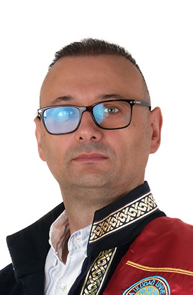 Gokhan Ocakoglu合作论文数Faculty of Medicine, Bursa Uludağ University5
 Vahap Ozan Kotan合作论文数5Guven Ozkaya合作论文数Faculty of Medicine, Uludağ University5
 A. Sivaci合作论文数4Furkan Saridas合作论文数4Erhan Bilir合作论文数4Ahmet Bekar合作论文数School of Medicine, Uludag University4
 G. Atasayar合作论文数3
 Yasemin Dinc合作论文数Fac Med, Bursa Uludag Univ3
 Sahsine Tolunay合作论文数Medical Faculty, Uludag University Bursa3
 Seher Naz Yeni合作论文数Cerrahpasa Medical Faculty, Istanbul University3Kaan Yucel合作论文数Department of Psychiatry and Behavioural Neurosciences, McMaster University3Necdet Karli合作论文数Faculty of Medicine, Uludag University3Kezban Aslan合作论文数3
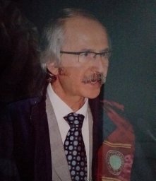 Mufit Parlak合作论文数Medical Faculty, Uludag University3
 Çi̇gdem Özkara合作论文数Department of Neurology, Cerrahpasa Faculty of Medicine, Istanbul University;Department of Internal Medicine, Cerrahpasa Faculty of Medicine, Istanbul University3Hacer Bozdemir合作论文数3Faruk Turan合作论文数3Sukran Yurtogullari合作论文数Medical Faculty, Uludag University3
 Nerses Bebek合作论文数Istanbul Medical Faculty, Istanbul University3Candan Gurses合作论文数The Institute of Experimental Medicine, Istanbul University3Ebru Aykutlu合作论文数2Gizem Mesut合作论文数2mine ayse altun emirza合作论文数2Dursun Ceylan合作论文数2E Ogul合作论文数2Aygul Gunes合作论文数2
 Sakir Delil合作论文数Faculty of Medicine, Istanbul University Cerrahpasa2Demir Aylin Bican合作论文数Faculty of Medicine, Uludag University2
 Zeynep Kotan合作论文数2
 Sibel Velioglu合作论文数Medical Faculty, Karadeniz Technical University2
 Serap Saygi合作论文数2
 Kadriye Agan合作论文数Department of Neurology, Marmara University Hospital2
 Betül Baykan (Baykal)合作论文数Department of Neurology, Faculty of Medicine, Istanbul University;EMAR Medical Center2
 Gulnihal Kutlu合作论文数2Ender Carkungoz合作论文数2
 Selcuk Kirli合作论文数Medical Faculty, Uludag University2Berrin Aktekin合作论文数2




