Resonance Raman spectroscopy offers a mechanism for the noninvasive measurement of in vivo and in situ hemoglobin oxygen saturation (HbO(2)Sat) in living tissue. Clinically informative signals can be provided by resonance enhancement with deep violet excitation. It is notable that fluorescence does not significantly degrade the quality of the signals. During the controlled hemorrhage and resuscitation of rats, signal intensity ratios of oxy- vs deoxyhemoglobin from sublingual mucosa correlated with co-oximetry values of blood withdrawn from a central venous catheter. The spectroscopic application described here has potential as a noninvasive method for the diagnosis of clinical shock and guidance of its therapy.
Since skeletal muscle represents up to 40% of body mass and suffers severe reductions in blood flow during HH, we examined the microcirculatory consequences of prolonged hypotension due to hemorrhage in the rat spinotrapezius muscle. An aortic transducer was implanted for continuous measurement of cardiac output. Systemic hemodynamic and respiratory variables were measured 7 days after the implantation. Functional capillary density, microvascular diameter, arteriolar SO2 and hemoglobin concentration ([Hb]) were measured using intravital microscopy. Measurements were made before and up to 4 h after HH. Additional bleeding or lactated Ringer's infusion were used to maintain mean arterial pressure at 40 mmHg. Controls were subjected to the same procedures except HH. Fifty-three percent of rats survived ≥ 3 h (S); others were considered nonsurvivors (NS). There was a positive correlation (r= 0.83, p ≤ 0.05) between microvascular SO2 and respiratory rate in S. Significant differences between S and NS were obtained for arteriolar SO2 (lower in S, p ≤ 0.05). Microvascular [Hb] was correlated with the systemic [Hb] (r= 0.93, p ≤ 0.001). Small arterioles constricted more in S than in NS, while large arterioles dilated in S. The diameter findings may explain why S showed higher total peripheral resistance levels for a longer period of time during HH. Our findings suggest that respiratory compensations and microvascular changes are essential for survival of prolonged HH. Support: DOD and NIH HL079087.
Clinicians now realize the limitations of the physical examination in detecting compensated shock states, the severity of uncompensated states, and in determining the adequacy of resuscitation in order to prevent subsequent post-traumatic multisystem organ failure and death. A renewed interest has developed in interrogating the state of oxygen transport at the end-organ level in the trauma patient. Although used as a research tool and now clinically to monitor cerebral oxygenation during complex cardiovascular and neurosurgery, near infrared absorption spectroscopy (NIRS) is being more aggressively investigated and now marketed clinically as a noninvasive means to assess tissue oxygenation in the trauma patient at the end organ level. This paper will describe the principles of NIRS and the basis for its proposed use in the trauma patient to assess tissue oxygenation. This includes its known limitations, current controversies, and what will be needed in the future to make this technology a part of the initial and ongoing assessment of the trauma patient. The ultimate goal of such techniques is to prevent misassessment of patients and inadequate resuscitation, which are believed to be major initiators in the development of multisystem organ failure and death.
Noninvasive measurements of PO2 and SO2 were made using intravital microscopy. Resonance Raman spectroscopy was used to determine SO2 in vitro and in vivo. Scattered Raman light from laser‐excited regions was directed to a spectrometer connected to a cooled CCD detector. The intensities of oxy‐ and deoxy‐ heme spectral markers were used to determine the SO2 by Raman (R_SO2). Phosphorescence quenching microscopy was employed to measure PO2 using a pulsed 532‐nm laser and Pd‐MTCPP oxygen probe. Since the exposure of oxygenated red blood cell (RBC) to laser light may result in photo‐induced displacement of O2 from Hb, the relationship between this photodamage and the RBC velocity was evaluated in vitro. During acquisition of Raman spectra, blood was circulated through glass capillaries at RBC velocities from 0.1 to 5.0 mm/s. Photodissociation was estimated by the difference between R_SO2 and a CO‐oximeter SO2 (O_SO2). There was a negative correlation between the RBC velocity and the R_SO2 / O_SO2 difference. When SO2 and acquisition time were held constant, decreasing the laser energy lowered the extent of photodamage.
We assessed the systemic effects of exchanges with blood or hemoglobin (Hb) raffimer under conditions of critical oxygen delivery (Do(2)crit). We compared Do(2)crit in animals receiving Hb-based oxygen carrier (HBOC; Hemolink), fresh blood (collected <24 h), or stored blood (10 days) before hemodilution. Rats were randomized to control, blood, or HBOC isovolemic exchange. Oxygen consumption was measured by using expired gas (o(2)a) and blood (o(2)b) samples, whereas whole-body oxygen delivery (Do(2)) was calculated from cardiac output and arterial oxygen content. After exchange, rats were subjected to stepwise isovolemic hemodilution. Blood pressure, gases, acid-base status, glucose, Hb oxygen saturation, heart rate, and total peripheral resistance were also measured. We found that 1) HBOC-treated rats showed an increased mean arterial blood pressure and total peripheral resistance throughout the hemodilution, 2) Do(2)crit calculated with o(2)a or o(2)b gave identical results, 3) Do(2)crit was not different between animals receiving blood and those receiving HBOC, 4) the terminal Hb concentration (1.8 +/- 0.1 g/dL) and Do(2) (5 +/- 1 mL . min(-1) . kg(-1)) were similar for all animals, and 5) most oxygen transport and biochemical variables changed similarly during hemodilution. The data suggest that tolerance to Do(2)crit is not altered by 50% replacement of native Hb by stored blood or Hb raffimer.
A system is described for in vivo noninvasive measurements of hemoglobin oxygen saturation (HbO2Sat) at the microscopic level. The spectroscopic basis for the application is resonant Raman enhancement of Hb in the violet/ultraviolet region, allowing simultaneous identification of oxy- and deoxyhemoglobin with the same excitation wavelength. The heme vibrational bands are well known, but the technique has never been used to determine microvascular HbO2Sat in vivo. A diode laser light (power: 0.3 mW) was focused onto sample areas 15-30 microm in diameter. Raman spectra were obtained in backscattering geometry by using a microscope coupled to a spectrometer and a cooled detector. Calibration was performed in vitro by using glass capillaries containing blood at several Hb concentrations, equilibrated at various oxygen tensions. HbO2Sat was estimated using the Raman band intensities at 1,360 and 1,375 cm(-1). Glass capillary path length and Hb concentration had no effect on HbO2Sat estimated from Raman spectra. In vivo observations were made in blood flowing in microvessels of the rat mesentery. The Hb Raman peaks observed in oxygenated and deoxygenated blood were consistent with earlier Raman studies that used Hb solutions and isolated cells. The method allowed HbO2Sat determinations in the whole range of arterioles, venules, and capillaries. Tissue transillumination allowed diameter and erythrocyte velocity measurements in the same vessels. Raman microspectroscopy offers distinct advantages over other currently used techniques by providing noninvasive and reliable in vivo determinations of HbO2Sat in thin tissues as well as in solid organs and tissues, which are unsuitable for techniques requiring transillumination.
Studies are needed to provide a rigorous examination of the relevance of monitored variables during prolonged hemorrhagic hypotension (HH). This study was designed to investigate the parameters that describe biochemical and O2 transport patterns in animals subjected to HH. Systemic parameters that could differentiate survivors from nonsurvivors were identified. An aortic flow probe was implanted in rats (n = 21) for continuous measurement of cardiac output. Experiments were performed 6-9 days after surgery. Rats were bled to a mean arterial pressure of 40 mmHg and kept at that level using Ringer-lactate solution. Arterial and venous blood pressures, gases, acid-base status, glucose, lactate, electrolytes, hemoglobin, O2 saturation, heart and respiratory rates, total peripheral resistance, and O2 delivery and consumption were measured before hemorrhage, soon after 40 mmHg was reached, and 0.5, 1, 2, 3, and 4 h later. Fifty-three percent of rats survived > or =3 h (survivors); others were considered nonsurvivors. Nonsurvivors showed a significantly greater degree of metabolic acidosis than survivors. Arterial PO2, respiratory rate, O2 saturation, O2 content, glucose, and pH were significantly higher in survivors. The rate of Ringer-lactate infusion, arterial K+, and PCO2 were lower in survivors. Arterial K+ and respiratory rate were the only parameters significantly different between survivors and nonsurvivors at all time points during HH. Arterial levels of K+ showed the clearest distinction between survivors and nonsurvivors and may explain the sudden death experienced by animals during HH. The data suggest that early respiratory and metabolic compensations are essential for survival of prolonged HH.
We tested the hypotheses that continuous total peripheral resistance (TPR) measurements are superior to intermittent data collection and that variables related to TPR can be used to distinguish between survivors and nonsurvivors (NS), respectively, of prolonged hemorrhagic hypotension (HH). One week after a transit-time ultrasound probe was implanted on their ascending aortas, 21 rats were subjected to 4 h of HH at 40 mmHg. Measurements were made before and up to 4 h after initiation of HH. Additional bleeding or Ringer L-lactate (RL) infusion was used to maintain HH. TPR was continuously measured online using recordings of blood flow and arterial pressure. Approximately 67% of the rats survived > or =3 h; others were considered NS. Data collected at 30-min intervals failed to detect the maximum value of TPR (TPRmax). The times to reach TPRmax were similar for survivors and NS and were strongly correlated with the bleeding end points and with the RL infusion-onset times. However, survivors showed higher TPRmax values than NS (P <0.005) and had a significantly longer period than NS during which TPR was above baseline level (116 +/- 20 vs. 51 +/- 10 min). In conclusion, 1) the transit-time ultrasound technique at high sampling rate allowed continuous and accurate real-time monitoring of TPR, 2) the bleeding end point and RL infusion-onset times may be used as surrogates of the time to TPRmax, 3) TPRmax of survivors and NS could be detected only using a continuous TPR measurement, and 4) differences between survivors and NS could be revealed by the continuous TPR curve.
Systemic variables were evaluated with respect to O(2) delivery to test the hypothesis that critical O(2) delivery and critical Hb can be estimated by multiple variables collected simultaneously. Rats were subjected to transfusion with either fresh or stored blood and then subjected to stepwise isovolemic hemodilution. Critical levels were measured by the dual-regression method from plots of systemic variables against O(2) delivery and Hb. Delivery was calculated from cardiac index and arterial O(2) content. We found that 1) after hemodilution, O(2) delivery changed in a nonlinear relationship with Hb; 2) critical delivery calculated using 30 different systemic variables was not statistically different from each other; 3) critical delivery and critical Hb were correlated but were not different between animals receiving fresh or stored blood; and 4) similar critical levels were found using a single variable from several animals and using several variables from the same subject. The best variables to estimate critical delivery were lactate, bicarbonate, base excess, O(2) extraction ratio, expired CO(2), pulse pressure, cardiac index, and systolic pressure. The data suggest that a multivariable analysis of critical delivery may help determine the physiological oxygenation boundary at the whole body level. This may assist in finding therapeutic triggers on an individual basis using systemic markers of the transition from aerobic to anaerobic metabolism.
We have developed a system with two epi-illumination sources, a DC-regulated lamp for transillumination and mechanical switches for rapid shift of illumination and detection of defined areas (250-750 microm(2)) by fluorescence and phosphorescence videomicroscopy. The system permits investigation of standard microvascular parameters, vascular permeability as well as intra- and extravascular PO2 by phosphorescence quenching of Pd-meso-tetra (4-carboxyphenyl) porphine (PORPH). A Pechan prism was used to position a defined region over the photomultiplier and TV camera. In order to validate the system for in vivo use, in vitro tests were performed with probes at concentrations that can be found in microvascular studies. Extensive in vitro evaluations were performed by filling glass capillaries with solutions of various concentrations of FITC-dextran (diluted in blood and in saline) mixed with different amounts of PORPH. Fluorescence intensity and phosphorescence decay were determined for each mixture. FITC-dextran solutions without PORPH and PORPH solutions without FITC-dextran were used as references. Phosphorescence decay curves were relatively unaffected by the presence of FITC-dextran at all concentrations tested (0.1 microg/ml to 5 mg/ml). Likewise, fluorescence determinations were performed in the presence of PORPH (0.05 to 0.5 mg/ml). The system was successfully used to study macromolecular extravasation and PO2 in the rat mesentery circulation under controlled conditions and during ischemia-reperfusion.
We evaluated systemic and microvascular effects of hypertonic NaCl solution on normovolemic and hemorrhaged animals. Forty-three Wistar rats (186 +/- 4 g, mean +/- SEM) were anesthetized with pentobarbital and cannulated for mean arterial pressure (MAP), heart rate (HR), and mean pulse pressure (MPP) monitoring and blood withdrawal. Diameters of 126 arterioles and 88 venules of the exteriorized mesentery were studied by using intravital microscopy. Microvascular blood flow was calculated from diameter and red blood cell velocity measurements. The protocol consisted of 15 min control, 30 min hypotension (MAP = 52.9 +/- 0.9 mmHg, hemorrhaged vol. = 17.1 +/- 0.7 mL/kg) and 60 min post-infusion of either normal (0.9%) or hypertonic saline (7.5%, 4 mL/kg). Normovolemic animals showed no systemic or microvascular effects of hypertonic saline. Hemorrhagic hypotension resulted in HR fall that was not changed after infusions. Hypertonic infusion reversed MPP decrease during hypotension but only partially restored MAP and microvascular blood flow. Venules did not change diameter during protocols. During hypotension, 24% of arterioles displayed vasomotion (38% of the rats) with low- and high-frequency components present in 74% and 87% of arterioles, respectively. Arterioles with vasomotion during hypotension had larger control diameters (28.9 +/- 2.0 mum) and contracted more (30.8 +/- 4.1%) than arterioles without vasomotion (18.7 +/- 1.2 mum and 8.1 +/- 1.5%, respectively). Mean arteriolar diameter did not change after infusions, After hypertonic solution, the number of vessels showing vasomotion increased 80%, frequency of vasomotion was unchanged, and amplitude increased. These findings may help to explain some of the mechanisms underlying resuscitation effects of hypertonic infusions during hemorrhagic hypotension.
Although vasomotion has been considered a feature of the microvascular bed under physiological conditions, it has also been observed following hypotension in several tissues. In this work, 158 mesenteric microvessels of 36 rats were investigated quantitatively in normovolemic and hemorrhaged animals, focussing on diameter changes, particularly vasomotion incidence and characteristics. The femoral arteries of Wistar rats (body weight BW = 188 +/- 23 g, mean +/- SD) anesthetized with pentobarbital were cannulated for arterial pressure (AP) monitoring and blood withdrawal. The protocol consisted of 15 min control and 30 min of hemorrhagic hypotension (AP = 52 +/- 5 mmHg, hemorrhaged vol. = 17 +/- 4 ml/kg BW). During control normovolemic conditions, analysis of mesenteric microcirculation using intravital videomicroscopy revealed neither arteriolar nor venular vasomotion. During hemorrhagic hypotension (HH) microvascular blood flow reduced to 25% of control. While venules did not show diameter changes during HH, arterioles contracted to 85 +/- 20% of control and arteriolar vasomotion appeared in 42% of the animals and 27% of the arterioles. The amplitude of arteriolar diameter change during HH relative to mean diameter and to control diameter averaged 65 +/- 24% (range: 32-129%) and 41 +/- 10% (range: 25-62%), respectively. Vasomotion analysis showed two major frequency components: 1.7 +/- 0.8 and 7.0 +/- 5.2 cycles/min. Arterioles showing vasomotion had a mean control diameter larger than the remaining arterioles and showed the largest constriction during HH. We conclude that hemorrhagic hypotension does not change venular diameter but induces arteriolar constriction and vasomotion in rat mesentery. This activity is expressed as slow waves with high amplitude and fast waves with low amplitude, and is dependent on vessel size.
Previous studies from our laboratory have shown that the average arteriolar pO2 in the hamster skinfold preparation is lower than arterial systemic pO2. In the present work we tested the hypothesis that there is a longitudinal gradient of pO2 along precapillary vessels. Experiments were performed in Syrian golden hamsters bearing a dorsal skin chamber. The oxygen-dependent quenching of phosphorescence of palladium-porphyrin complexes was used to measure intravascular pO2 in the microcirculation. Arterioles were classified in four orders according to their position in the network, first-order vessels being the entrance points. Simultaneous determinations of diameter (D), red blood cell velocity, and systemic blood gases were also made. There was a significant fall of pO2 between vessels of different orders. First-order arterioles (mean D = 64 microns) had pO2 of 51.8 +/- 9.8 mm Hg (mean +/- SD) which was equivalent to approximately equal to 73% of the arterial systemic pO2. Within the arteriolar network, further decreases of intravascular pO2 were measured, leading to a pO2 of 34.0 +/- 7.9 mm Hg in terminal arterioles (mean D = 7.7 microns). In some vessels pO2 was measured in different positions of the same arteriole. The average longitudinal arteriolar oxygen saturation gradient was 3.4 +/- 0.4 delta %/mm (range 0.8-7.2). A significant and positive correlation was found between pO2 and microhemodynamic parameters when arterioles were grouped according to their order. This relation was not significant for venules which showed a mean pO2 of 30.8 +/- 10.8 mm Hg. Tissue pO2 averaged 24.6 +/- 5.8 mm Hg. We conclude that: (1) There is an oxygen loss in arterial vessels larger than 100 micrograms in diameter, (2) arteriolar pO2 in this preparation depends on the position of the vessel within the network, (3) a substantial portion of oxygen delivery to the hamster skin is provided by the arteriolar network, and (4) only a small pO2 gradient exists between terminal arterioles and venules, suggesting that the contribution of the capillary network to tissue oxygenation is relatively small.
Simultaneous measurements of intravascular and interstitial oxygen partial pressure (PO2) in any tissue have not previously been reported, despite the importance of oxygen in health and in disease. This is due to the limitations of current techniques, both invasive and noninvasive. We have optically measured microscopic profiles of PO2 with high spatial resolution in subcutaneous tissue and transplanted tumors in mice by combining an oxygen-dependent phosphorescence quenching method and a transparent tissue preparation. The strengths of our approach include the ability to follow PO2 in the same location for several weeks and to relate these measurements to local blood flow and vascular architecture. Our results show that (i) PO2 values in blood vessels in well-vascularized regions of a human colon adenocarcinoma xenograft are comparable to those in surrounding arterioles and venules, (ii) carbogen (95% O2/5% CO2) breathing increases microvascular PO2 in tumors, and (iii) in unanesthetized and anesthetized mice PO2 drops to hypoxic values at < 200 microns from isolated vessels but drops by < 5 mmHg (1 mmHg = 133 Pa) in highly vascularized tumor regions. Our method should permit noninvasive evaluations of oxygen-modifying agents and offer further mechanistic information about tumor pathophysiology in tissue preparations where the surface of the tissue can be observed.
Topological and geometrical characteristics of the anastomotic arteriolar network in cat sartorius muscle were studied. The vessels were dilated and filled with gelatin-ink solution and the muscle cleared with methylsalicylate. The analysis was done on 11 muscles and included 2297 vascular segments and 772 vascular loops classified according to their position within the network. On average, each muscle had 644 transverse arterioles arising from the anastomotic vessels. The length of vascular segments close to feeding arteries was greater than those located in the central region, while the number of transverse arterioles per unit length of arcade vessel showed an opposite tendency. Most vessel orders had similar diameters, except for the segments at the periphery of the muscle, which were significantly larger. No correlation was found between vessel length and diameter. Vascular loops located in the central part of the network were smaller, as assessed by area, perimeter, number of segments, segment length and number of branches. The variability of the parameters between muscles was smaller than variability within each muscle. We concluded that, in addition to the parameters previously reported, quantitative descriptions of anastomotic networks may be enhanced by considering certain topological aspects of microvessels. The separation of segments and loops according to the position in the network may reveal differences between muscles of different sizes and functions which would not be detected if the vessels were considered as a single group.
Microvascular parameters as well as respiratory and cardiac rates were measured in the freshwater catfish Hypostomus cf. punctatus. Unanesthetized animals were kept in a box with flowing water. Microcirculation of the pectoral fin was studied using a closed-circuit television system coupled to a microscope. In the first order venules, diameter and red blood cell (RBC) velocity averaged (mean ± SD) 37.2 ± 14.5 μm and 0.63 ± 0.40 mm/sec, respectively. Second order venules had values of 22.8 ± 5.6 μm and 0.45 ± 0.32 mm/sec. Arterioles (diameter = 4.7 ± 4.0 μm) emerging from the soft rays had RBC velocities ranging from 0.04 to 2.31 mm/sec with a mean of 1.25 ± 0.87 mm/sec. Capillary diameter and RBC velocity averaged 6.0 ± 1.8 μm and 0.15 ± 0.09 mm/sec, respectively. Some observations were also made on the secondary system of blood vessels.
 Eliete Bouskela合作论文数Universidade do Estado do Rio de Janeiro9
 Luciana N. Torres合作论文数Departamento de Patologia - FMVZ-USP8
 PC JOHNSON合作论文数7
 Kevin R. Ward合作论文数Department of Biomedical Engineering, College of Engineering, University of Michigan-Ann Arbor7
 Robert Barbee合作论文数Department of Physiology, Virginia Commonwealth University7
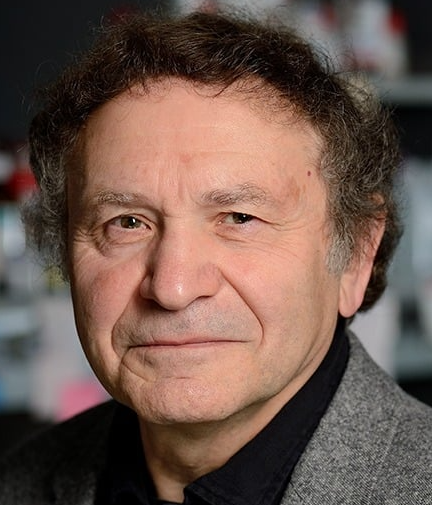 Aleksander S. Popel合作论文数Popel Systems Biology Laboratory, Department of Biomedical Engineering, School of Medicine, Johns Hopkins University5
 Randall N. Pittman合作论文数Department of Pharmacology, School of Medicine, University of Pennsylvania5E BOUSKELATORRES合作论文数4
 Roland Pittman合作论文数Department of Cellular, Molecular and Genetic Medicine, School of Medicine Physiology and Biophysics, Virginia Commonwealth University4James Terner合作论文数4V.Martuchelli Filho合作论文数4F CYRINO合作论文数4M. H. Tiba合作论文数3
 Marcos Intaglietta合作论文数Department of Bioengineering, Jacobs School of Engineering, University of California, San Diego;Institute of Engineering in Medicine, University of California, San Diego3Ls Neves合作论文数2Fzga Cyrino合作论文数2Daniel Contaifer合作论文数2
 Rao R Ivatury合作论文数Department of Surgery, Virginia Commonwealth University2Bruce D. Spiess合作论文数2H Kerger合作论文数2Ma Boegehold合作论文数1K. R. Ward合作论文数1MMD BREITENBACH合作论文数1Sd House合作论文数1
 Egberto Moura合作论文数1K. Ward合作论文数1Carmen Cabanelas Pazos-Moura合作论文数Carlos Chagas Filho Institute of Biophysics, Federal University of Rio de Janeiro1
 Roland N Pittman合作论文数1
 Fan Yuan合作论文数Department of Biomedical Engineering, Pratt School of Engineering, Duke University1
 Michael Leunig合作论文数Schulthess Klinik1
 Rakesh K. Jain合作论文数Harvard Medical School, Harvard University;Edwin L. Steele Laboratories for Tumor Biology, Department of Radiation Oncology, Massachusetts General Hospital1Matthew A. Boegehold合作论文数Department of Medicine, West Virginia University School of Medicine1




