Soret-excited resonance Raman spectra (λex 413.1 nm) were acquired for manganese(III) and gold(III) tris(pentafluorophenyl)corrole, each as four different isotopomeric samples: natural abundance, fully pyrrole-15N-substituted, fully meso-13C-substituted, and fully pyrrole-15N-meso-13C-substituted. The spectra were modeled with density functional theory-based vibrational analyses, which in general did an excellent job of reproducing both the absolute frequencies and isotope shifts. The results led to the assignment and visualization of approximately 10 prominent Raman bands. A key finding was that the bands could be categorized into two broad classes: Class A, exhibiting large 15N isotope shifts, assignable to vibrations with predominant Cα-N character, and Class B, exhibiting large meso-13C isotope shifts, assignable to vibrations with predominant Cα-Cmeso character. Preliminary evidence suggests that the class A bands may serve as core size markers, while class B bands may correlate with the innocence or otherwise of the corrole macrocycle.
BACKGROUND:The knowledge of hemoglobin oxygen saturation (SO2) and tissue oxygenation is critical to identify the presence of shock and therapeutic options. The resonance vibrational enhancement of hemoglobin allows measurement of oxy- and deoxy species of hemoglobin and resonance Raman spectroscopy (RRS-StO2) has been successfully used to measure aggregate microvascular oxygenation. We tested the hypothesis that noninvasive oxygen saturation measured by RRS-StO2 could serve as surrogate of systemic central venous SO2.METHODS:In anesthetized rats, measurements of RRS-StO2 made in oral mucosa, skin, muscle, and liver were compared with measurements of central venous SO2 using traditional multi-wavelength oximetry. Various oxygenation levels were obtained using a stepwise hemorrhage while over 100 paired blood samples and Raman-based measurements were performed. The relationships between RRS-StO2 and clinically important systemic blood parameters were also evaluated. RRS-StO2 measurements were made in 3-mm diameter tissue areas using a microvascular oximeter and a handheld probe.RESULTS:Significant correlations were found between venous SO2 and RRS-StO2 measurements made in the oral mucosa (r = 0.913, P < 0.001), skin (r = 0.499, P < 0.01), and liver (r = 0.611, P < 0.05). The mean difference between sublingual RRS-StO2 and blood sample SO2 values was 5.4 ± 1.6%. Sublingual RRS-StO2 also correlated with lactate (r = 0.909, P < 0.01), potassium (r = 0.757, P < 0.01), and pH (r = 0.703, P < 0.05).CONCLUSIONS:Raman-based oxygen saturation is a promising technique for the noninvasive evaluation of oxygenation in skin, thin tissues, and solid organs. Under certain conditions, sublingual RRS-StO2 measurements correlate with central venous SO2.
Schwermetallcorrole sind von großem Interesse als optische Sensoren, Nah-IR-Farbstoffe, Leuchtstoffe, OLEDs und Tumortherapeutika, allerdings ist die Insertion von 5d-Metallen in Corrole schwierig. A. Ghosh et al. beschreiben in ihrer Zuschrift auf S. 14639 ff. die oxidative Metallierung von meso-Triarylcorrolen mit [Os3(CO)12]/NaN3 als Route zu Nitridoosmium(VI)-corrolen. Diese Komplexe weisen ausgeprägt gewölbte Makrocyclen auf und sind durch multiple Os-Nitrido-Streckschwingungsfrequenzen charakterisiert.
BACKGROUND The ability to monitor the patient of hemorrhage noninvasively remains a challenge. We examined the ability of resonance Raman spectroscopy to monitor tissue hemoglobin oxygenation (RRS-StO2) during hemorrhage and compared its performance with conventional invasive mixed venous (SmvO2) and central venous (ScvO2) hemoglobin oxygen saturation as well as with near-infrared spectroscopy tissue hemoglobin oxygenation (NIRS-StO2). METHODS Five male swine were anesthetized and instrumented followed by hemorrhage at a rate of 30 mL/min for 60 minutes. RRS-StO2 was continuously measured from the buccal mucosa, and NIRS-StO2 was continuously measured from the forelimb. Paired interval measures of SmvO2, ScvO2, and lactate were made. Pearson correlation was used to quantify the degree to which any two variables are related. Receiver operating characteristic (ROC) area under the curve values were used for pooled data for RRS-StO2, NIRS-StO2, SmvO2, and ScvO2 to compare performance in the ability of tissue oxygenation methods to predict the presence of an elevated arterial blood lactate level. RESULTS Sequential RRS-StO2 changes tracked changes in SmvO2 (r = 0.917; 95% confidence interval [CI], 0.867–0.949) and ScvO2 (r = 0.901; 95% CI, 0.828–0.944) during hemorrhage, while NIRS-StO2 failed to do so for SmvO2 (r = 0.283; 95% CI, 0.04919–0.4984) and ScvO2 (r = 0.142; 95% CI, −0.151 to 0.412). ROC curve performance of oxygenation measured to indicate lactate less than or greater than 3 mM yielded the following ROC area under the curve values: SmvO2 (1.0), ScvO2 (0.994), RRS-StO2 (0.972), and NIRS-StO2 (0.611). CONCLUSION RRS-StO2 seems to have significantly better ability to track central oxygenation measures during hemorrhage as well as to predict shock based on elevated lactate levels when compared with NIRS-StO2.
Heavy-element corroles are of great interest as optical sensors, near-IR dyes, phosphors, organic light-emitting diodes, and anticancer compounds. Insertion of 5d metals into corroles, however, is often a difficult and unpredictable process. Against this backdrop, oxidative metalation of meso triarylcorroles with [Os3 (CO)12 ]/NaN3 in refluxing 1:2 diethylene glycol monomethyl ether/glycol has provided a convenient and relatively high-yielding route to nitridoosmium(VI) corroles, three of which could be characterized with single-crystal X-ray structure analysis.
In this work we demonstrate the coupling of the photothermal effects of gold nanostructures of controlled size and shape with graphene oxide nanosheets dispersed in water. The enhanced photothermal effects can be tuned by controlling the shape and size of the gold nanostructures, which result in a remarkable increase in the heating efficiency of the laser-induced size reduction of gold nanostructures. The Raman spectra of the Au-graphene nanosheets provide direct evidence for the presence of more structural defects in the graphene lattice induced by laser irradiation of graphene oxide nanosheets in the presence of Au nanostructures. The large surface areas of the laser-reduced graphene oxide nanosheets with multiple defect sites and vacancies provide efficient nucleation sites for the ultrasmall gold nanoparticles with diameters of 2-4 nm to be anchored to the graphene surface. This defect filling mechanism decreases the mobility of the ultrasmall gold nanoparticles and, thus, stabilizes the particles against the Ostwald ripening process, which leads to a broad size distribution of the laser-size-reduced gold nanoparticles. The Au nanostructures/graphene oxide solutions and the ultrasmall gold-graphene nanocomposites are proposed as promising materials for photothermal therapy and for the efficient conversion of solar energy into usable heat for a variety of thermal, thermochemical, and thermomechanical applications.
We report the development of a facile process for the synthesis of hydrogen-terminated graphene using the laser vaporization-controlled condensation (LVCC) method. The method allows rapid deoxygenation of bulk graphite oxide (GO) in an atmosphere of helium or a helium-hydrogen mixture to produce hydrogen-terminated graphene (HTG) nanosheets without the need for any chemical reducing agents or solvents. Direct laser vaporization/ionization (LVI) of bulk GO in a vacuum coupled with a beam expansion of a carrier gas produces the carbon cluster series C-n with n in the range 4-30, in addition to hydrogenated carbon ions with a significant enhancement in the ion intensity of the n-even ions suggesting the formation of hydrogen-capped polyyne chains corresponding to the H-(C C)(n)H--(+) and H-(C C)(n)(-) formulas. In contrast, LVI of bulk graphite under identical experimental conditions generates mainly the carbon duster series C-n without any significant hydrogenation in both the positive and negative ion modes. The results confirm that the LVCC method of bulk GO produces HTG nanosheets which can be ionized and dissociated in a vacuum to produce the observed hydrogen-capped polyyne chains. These species could be deposited from the gas phase to form one-dimensional conducting molecular wires for a variety of potential applications in nanoelectronics, sensors, and devices.
We have previously developed a methodology to measure hemoglobin (Hb) oxygen saturation using Raman spectroscopy in vivo (AJP 289:H488, 2005; JAP 104:1809, 2008). However, the technique has not yet been used in sickle cell disease (SCD). We have established a colony of transgenic (Tg) SCD mice. This strain no longer expresses mouse Hba and Hbb, but express human sickle hemoglobin (HbS). It mimics the genetic, hematologic and histopathologic features found in humans with SCD. Whole blood samples were obtained from anesthetized Tg mice and normal control mice. Raman spectra from these samples were obtained in vitro using the exciting wavelengths in the range of 405 ‐ 532 nm emitted by a tunable laser. The scattered light was sampled at 90 degrees using a 0.5 m spectrometer and a nitrogen cooled CCD detector. In order to induce HbS polymerization, Raman spectra were collected from blood samples exposed to high and low oxygen environments. The findings suggest that strong Raman signals can be obtained from normal and SCD blood, opening the possibility for in vivo noninvasive measurements of hemoglobin Hb and HbS saturation in the microcirculation. The resonance Raman technique should provide additional worthwhile microscopic information in the study of the abnormal hemoglobins in vivo.SupportVCU.
We have developed a facile and scalable chemical reduction method assisted by microwave irradiation for the synthesis of chemically converted graphene sheets and metal nanoparticles dispersed on the graphene sheets. The method allows rapid chemical reduction of exfoliated graphite oxide (GO) using a variety of reducing agents in either aqueous or organic media. It also allows the simultaneous reduction of GO and a variety of metal salts thus resulting in the dispersion of metallic and bimetallic nanoparticles supported on the large surface area of the thermally stable 2D graphene sheets.
The resonant Raman enhancement of hemoglobin (Hb) in the Q band region allows simultaneous identification of oxy- and deoxy-Hb. The heme vibrational bands are well known at 532 nm, but the technique has never been used to determine microvascular Hb oxygen saturation (So(2)) in vivo. We implemented a system for in vivo noninvasive measurements of So(2). A laser light was focused onto areas of 15-30 microm in diameter. Using a microscope coupled to a spectrometer and a cooled detector, Raman spectra were obtained in backscattering geometry. Calibration was performed in vitro using blood at several Hb concentrations, equilibrated at various oxygen tensions. So(2) was estimated by measuring the intensity of Raman signals (peaks) in the 1,355- to 1,380-cm(-1) range (oxidation state marker band nu(4)), as well as from the nu(19) and nu(10) bands (1,500- to 1,650-cm(-1) range). In vivo observations were made in microvessels of anesthetized rats. Glass capillary path length and Hb concentration did not affect So(2) estimations from Raman spectra. The Hb Raman peaks observed in blood were consistent with earlier Raman studies using Hb solutions and isolated cells. The correlation between Raman-based So(2) estimations and So(2) measured by CO-oximetry was highly significant for nu(4), nu(10), and nu(19) bands. The method allowed So(2) determinations in all microvessel types, while diameter and erythrocyte velocity could be measured in the same vessels. Raman microspectroscopy has advantages over other techniques by providing noninvasive and reliable in vivo So(2) determinations in thin tissues, as well as in solid organs and tissues in which transillumination is not possible.
Ultrafast laser spectroscopy techniques are used to measure the low-frequency vibrational coherence spectra and nitric oxide rebinding kinetics of Caldariomyces fumago chloroperoxidase (CPO). Comparisons of the CPO coherence spectra with those of other heme species are made to gauge the protein-specific nature of the low-frequency spectra. The coherence spectrum of native CPO is dominated by a mode that appears near 32-33 cm(-1) at all excitation wavelengths,with a phase that is consistent with a ground-state Raman-excited vibrational wavepacket. On the basis of a normal coordinate structural decomposition (NSD) analysis, we assign this feature to the thiolate-bound heme doming mode. Spectral resolution of the probe pulse ("detuned" detection) reveals a mode at 349 cm-1, which has been previously assigned using Raman spectroscopy to the Fe-S stretching mode of native CPO. The ferrous species displays a larger degree of spectral inhomogeneity than the ferric species, as reflected by multiple shoulders in the optical absorption spectra. The inhomogeneities are revealed by changes in the coherence spectra at different excitation wavelengths. The appearance of a mode close to 220 cm(-1) in the coherence spectrum of reduced CPO excited at 440 nm suggests that a subpopulation of five coordinated histidine-ligated hemes is present in the ferrous state at a physiologically relevant pH. A significant increase in the amplitude of the coherence signal is observed for the resonance with the 440 nm subpopulation. Kinetics measurements reveal that nitric oxide binding to ferric and ferrous CPO can be described as a single-exponential process, with rebinding time constants of 29.4 +/- 1 and 9.3 +/- 1 ps, respectively. This is very similar to results previously reported for nitric oxide binding to horseradish peroxidase.
The usefulness of the 532 nm laser excitation to estimate hemoglobin oxygen saturation (SO2) based on Raman spectra was investigated. Noninvasive microscopy and Raman spectroscopy were used to determine SO2 in vitro and in vivo. Up to six peaks could be used to estimate SO 2 by Raman spectroscopy, corresponding to three oxidation state marker bands of hemoglobin (Hb), designated as n4, n10 and n19. The estimated SO2 using Raman correlated well with SO2 measured via co-oximetry, allowing SO2 to be determined in the physiological range. The system was successfully used to determine local SO2 of rats and mice.
ADVERTISEMENT RETURN TO ISSUEPREVBook ReviewNEXTMetal Ions in Life Sciences, Volume 3: The Ubiquitous Roles of Cytochrome P450 Proteins Edited by Astrid Sigel, Helmut Sigel (University of Basel, Switzerland), and Roland K. O. Sigel (University of Zürich, Switzerland). John Wiley & Sons, Ltd: Chichester. 2007. xxvi + 652 pp. $360. ISBN 978-0-470-01672-5.James Terner and Avram GoldView Author Information Virginia Commonwealth University The University of North Carolina at Chapel HillCite this: J. Am. Chem. Soc. 2007, 129, 51, 16279–16280Publication Date (Web):November 14, 2007Publication History Published online14 November 2007Published inissue 1 December 2007https://pubs.acs.org/doi/10.1021/ja076984ahttps://doi.org/10.1021/ja076984abook-reviewACS PublicationsCopyright © 2007 American Chemical SocietyRequest reuse permissionsArticle Views257Altmetric-Citations2LEARN ABOUT THESE METRICSArticle Views are the COUNTER-compliant sum of full text article downloads since November 2008 (both PDF and HTML) across all institutions and individuals. These metrics are regularly updated to reflect usage leading up to the last few days.Citations are the number of other articles citing this article, calculated by Crossref and updated daily. Find more information about Crossref citation counts.The Altmetric Attention Score is a quantitative measure of the attention that a research article has received online. Clicking on the donut icon will load a page at altmetric.com with additional details about the score and the social media presence for the given article. Find more information on the Altmetric Attention Score and how the score is calculated. Share Add toView InAdd Full Text with ReferenceAdd Description ExportRISCitationCitation and abstractCitation and referencesMore Options Share onFacebookTwitterWechatLinked InRedditEmail Other access optionsGet e-Alertsclose SUBJECTS:Detoxification,Genetics,Hydrocarbons,Ions,Peptides and proteins Get e-Alerts
Raman spectroscopy provides non-invasive in vivo and in situ hemoglobin oxygen saturation (SO2) measurements of tissue. Clinically informative Raman signals arise from resonance enhancement provided by excitation in the deep violet. In order to obtain Raman spectra from tissues of live human subjects, we used a handheld device coupled with optical fibers for laser excitation and signal collection. The technique opens the possibility of noninvasive measurements of SO2 that can prove clinically valuable, avoiding procedures such as central venous catheterization.
Resonance Raman spectroscopy offers a mechanism for the noninvasive measurement of in vivo and in situ hemoglobin oxygen saturation (HbO(2)Sat) in living tissue. Clinically informative signals can be provided by resonance enhancement with deep violet excitation. It is notable that fluorescence does not significantly degrade the quality of the signals. During the controlled hemorrhage and resuscitation of rats, signal intensity ratios of oxy- vs deoxyhemoglobin from sublingual mucosa correlated with co-oximetry values of blood withdrawn from a central venous catheter. The spectroscopic application described here has potential as a noninvasive method for the diagnosis of clinical shock and guidance of its therapy.
We have employed rapid scan stopped-flow spectroscopy to examine whether the mechanism of oxidative dehalogenation catalyzed by C. fumago chloroperoxidase (CCPO) involves two consecutive one-electron steps or a single two-electron oxidation. First, we optimized the formation of CCPO compound I (CCPO-I) [Fe(IV)=O/porphyrin radical] and CCPO compound II (CCPO-II) [Fe(IV)=O] for use in double mixing rapid scan stopped-flow experiments. Reaction of CCPO-I with 2,4,6-trichlorophenol (TCP) quickly yielded CCPO-II. Reaction of CCPO-II, a one-electron oxidant, with TCP rapidly regenerated the ferric resting state of the enzyme. The rates of the reaction of both CCPO-I and -II with TCP are first-order with respect to [TCP]. In the absence of organic substrate, CCPO-I is slowly reduced to CCPO-II and then the ferric state. The ability of both CCPO-I and -II to carry out the oxidative dehalogenation reaction is consistent with a mechanism involving two consecutive one-electron oxidations. In contrast, reaction of CCPO-I with thioanisole generated the ferric enzyme with no evidence of CCPO-II, consistent with a single two-electron oxidation by insertion of an oxygen atom. The relative stability of CCPO-I and -II has allowed us to differentiate between one- and two-electron substrate oxidations using rapid scan stopped-flow techniques.
A system was developed for in vitro studies using flowing blood. The system (priming volume: 5–10 ml) includes a computer-controlled pump, gas mixer, oxygenator and interconnected tubing that allows the flowing blood to be exposed to various O2 and CO2 tensions, yielding controlled levels of hemoglobin (Hb) O2 saturation (SO2). Therefore, blood sampled at different O2 partial pressures (PO2) can be used to obtain Hb O2 dissociation curves and to determine the Hb O2 affinity. SO2 and PO2 can also be measured optically using Raman micro-spectroscopy and phosphorescence quenching, respectively. In SO2 determinations, scattered Raman light from laser-excited regions is directed to a spectrometer connected to a cooled CCD detector. The intensities of up to 6 Raman peaks can be used, corresponding to oxidation state marker bands of Hb, designated as ν4, ν10 and ν19. Since the blood flow and the pressure drop are measured, alterations in blood viscosity are continuously recorded. Changes in blood cell morphology and interactions between blood cells and endothelial cells can be evaluated at each oxygenation level using video-microscopy. The system allows critical physiological variables to be studied using blood sampled from animals and patients. If the medical history of a patient is known, it is possible to correlate the clinical findings with the information obtained using this system and his/her own blood. Support: ONR and NIH
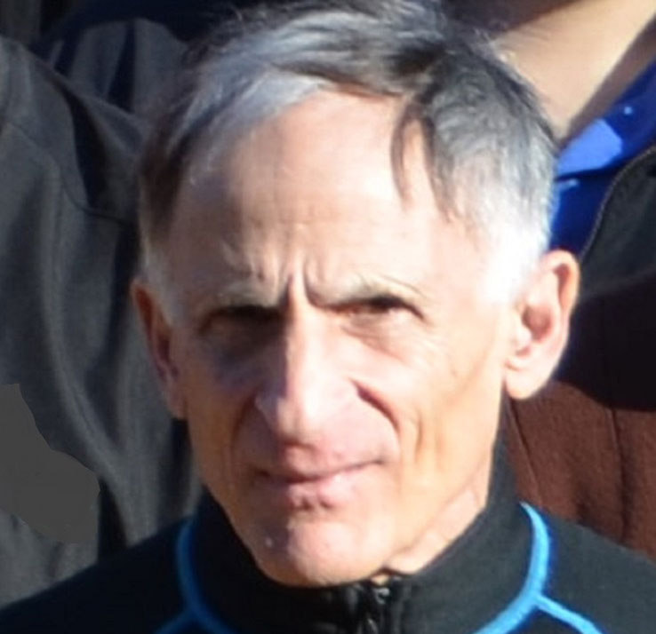 Avram Gold合作论文数Department of Environmental Sciences and Engineering, Gillings School of Global Public Health, The University of North Carolina at Chapel Hill25
 Alfred X. Trautwein合作论文数Institute of Physics, Medical University Lübeck20
 Kevin R. Ward合作论文数Department of Biomedical Engineering, College of Engineering, University of Michigan-Ann Arbor16
 Aj Sitter合作论文数15K Jayaraj合作论文数15Raymond Weiss合作论文数14
 Rachel Narehood Austin合作论文数Department of Chemistry, Barnard College, Columbia University13
 Eckhard Bill合作论文数Max Planck Institute for Chemical Energy Conversion13
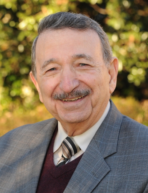 Mostafa El-Sayed合作论文数El-Sayed Group, Laser Dynamics Laboratory, School of Chemistry & Biochemistry, College of Sciences, Georgia Institute of Technology13Dominique Mandon合作论文数13Cm Reczek合作论文数11
 Thomas G. Spiro合作论文数Department of Chemistry, University of Washington11
 Ivo P. Torres Filho合作论文数Department of Anesthesiology, Virginia Commonwealth University11
 Robert Barbee合作论文数Department of Physiology, Virginia Commonwealth University10Vaithianathan Palaniappan合作论文数DEPT CHEM, UNIV CALIF RIVERSIDE10
 Luciana N. Torres合作论文数Departamento de Patologia - FMVZ-USP9Am Sullivan合作论文数8Jean Fischer合作论文数8
 Roland Pittman合作论文数Department of Cellular, Molecular and Genetic Medicine, School of Medicine Physiology and Biophysics, Virginia Commonwealth University7M.M. Fitzgerald合作论文数6Cm Hosten合作论文数6
 Volker Schunemann合作论文数Institut für Physik, Medizinische Universität Lübeck6
 Philippe Ochsenbein合作论文数6Cl Hsieh合作论文数5Markus Muther合作论文数5M. Nagumo合作论文数4Ip Torres合作论文数4De Reed合作论文数4
 Thomas L. Poulos合作论文数Department of Molecular Biology and Biochemistry, University of California, Irvine4
 Rao R Ivatury合作论文数Department of Surgery, Virginia Commonwealth University4Khadija Ayougou合作论文数4MF NICOL合作论文数3A CAMPION合作论文数3Daryoosh Vakhshoori合作论文数3Ar Burns合作论文数3Fm Hawkridge合作论文数3Wt Henshaw合作论文数3S Sun合作论文数3
 Roland N Pittman合作论文数3
 Karin Kuhnel合作论文数3
 Abhik Ghosh合作论文数Department of Chemistry, UiT The Arctic University of Norway3
 Ilme Schlichting合作论文数Department of Biosciences, Heidelberg University;Department of Physics and Astronomy, Heidelberg University;Department of Biomolecular Mechanisms, Max Planck Institute for Medical Research3Munirathinam Sundaramoorthy合作论文数Department of Biochemistry;Molecular Biology;University of Kansas Medical Center3
 M. Samy El-Shall合作论文数Department of Chemistry, College of Humanities and Sciences, Virginia Commonwealth University3Karupiah Jayaraj合作论文数3Louise M. Ball合作论文数3M. H. Tiba合作论文数2




